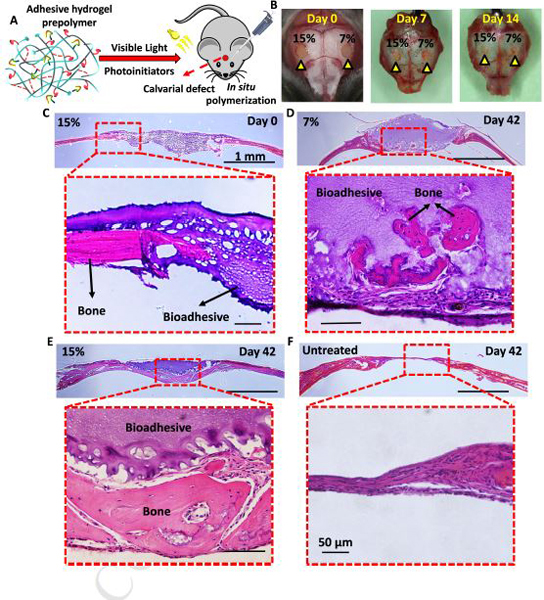
Figure 5. In vivo evaluation of bioadhesive hydrogels using a mouse calvarial defect model.

(A) Schematic diagram of in situ application of bioadhesive hydrogels in a mouse calvarial defect model. (B) 7% and 15% bioadhesive hydrogels were delivered to artificially created bone defects in mouse calvaria (yellow arrowheads), and photopolymerized for 1 min using a commercially available dental curing light. 7 and 14 days after implantation, samples remained in place, without any sign of detachment. (C) Histological evaluation (H&E staining) of the 15% (w/v) bioadhesives at day 0 post implantation. Representative H&E images for (D) 7% (w/v) and (E) 15% (w/v) bioadhesive treatment, and (F) untreated sample after 42 days post implantation.
