



Abstract
Background
Congenital heart disease (CHD) is a common birth defect originating from both environmental and genetic factors. An overabundance of copy number variations (CNVs) affecting cardiac‐related genes has previously been detected in individuals with CHD.
Objective
To evaluate if the presence of CNVs in the 22q11.2 region, and to determine whether GATA4, NKX2‐5, TBX5, BMP, and CRELD1 genes contributed toward the pathogenesis of isolated incidences of CHDs in southwest China.
Methods
In total 167 patients from southwest China with sporadic CHD were studied, including 121 patients with ventricular septal defect (VSD), 24 with atrial septal defect (ASD), 12 with tetralogy of fallot (TOF), six VSD cases with TOF, two cases with patent ductus arteriosus (PDA), and two VSD cases with ASD. 22q11.2, GATA4, NKX2‐5, TBX5, BMP4, and CRELD1 regions were screened using MLPA and copy number variation sequencing (CNV‐Seq).
Results
A 2.5‐2.8 Mb deletion in the 22q11.2 region was identified in 5 patients with CHD. Two of these patients were diagnosed with VSD, while two had VSD and ASD, and the other had TOF. 5 patients correspond to the same classical DiGeorge syndrome. A 0.86 Mb duplication in the 22q11.2 region was identified in a PDA patient, whom was without extracardiac symptoms.
Conclusion
These data suggest that copy number variation in the 22q11.2 region is common in CHD patients in southwest China. Regardless of the presence or absence of extracardiac symptoms, results also indicate that it is necessary to perform prenatal screening for CHD.
Keywords: 22q11 region, congenital heart disease, copy number variations, MLPA
1. INTRODUCTION
Congenital heart disease (CHD) is a common birth defect resulting from both environmental and genetic causes, which affects 0.85%‐1% newborns1 globally. Studies have revealed that 11.9% and 7.4% of cases are associated with monogenetic and cytogenetic anomalies, respectively. Collectively, these account for an estimated 20% of all CHD cases,2 while the etiology for the remaining 80% is still unclear. Recently, copy number variations (CNVs), which constitute either gross DNA deletions or duplications, were identified as being critical dosage‐sensitive genome for cardiac development and have also emerged as important contributors to CHDs. Examples of this include the TBX1 deletion at 22q11.2, the GATA4 deletion at 8p23.1, and the NKX2‐5 deletion at 5q35.1.3, 4 An increasing assortment of CNVs affecting cardiac‐related genes have been detected in individuals with CHDs. Recent population studies indicate that the 22q11.2 deletion is the most common microdeletion syndrome, occurring in 1 per 5950 live births and nearly 0.5%‐1.9% of all CHDs,5 usually arising de novo, which accounts for 34% of truncus arteriosus and 16% of tetralogy of fallot (TOF) cases.6 Previous studies have identified that GATA4, NKX2.5, and TBX5 contribute to monogenic forms of CHD,7, 8 while BMP4 and CRELD1 are closely related to the development of CHD. Inactivation of BMP4 within the myocardium results in neonatal lethality results in severe defects in septation and valve diseases such as ASD and VSD.9 CRELD1 gene can cause CHD in Down syndrome–atrioventricular septal defect (AVSD) and also in sporadic forms of AVSD without any syndromic features.10 However, combined mutation screening revealed that congenital heart defects are rarely caused by mutations in these genes.11 Thus far, no study has been conducted to investigate the role of NKX2.5, TBX5, BMP4, and CRELD1 CNVs in Chinese children with sporadic congenital heart disease, we analyzed a cohort of 167 patients with sporadic cardiac anomalies using multiplex ligation‐dependent probe amplification (MLPA) and copy number variation sequencing (CNV‐Seq).
2. METHODS
2.1. Materials and methods
The study cohort enrolled 167 patients after an ultrasound diagnosis with unselected CHD at the Liuzhou Maternal and Child Healthcare Hospitals, Guangxi, southwest China between 2012 and 2016. All the patients in this study had no history of CHD in first‐degree relatives. Congenital cardiac malformations were diagnosed by echocardiography and subsequently confirmed during surgery when performed. Extra‐cardiac findings were extracted from medical records and included dysmorphic features, major anomalies, non‐cardiac medical problems, and deficiencies in growth or developmental delay. This study was approved by the Liuzhou maternal and children healthcare hospital ethics review board on human subjects. All the patients were assessed by MLPA and CNV‐seq.
2.2. MLPA analysis
Genomic DNA was extracted from peripheral blood by phenol/chloroform extraction and quality determined with a NanoDrop 2000 UV–vis spectrophotometer (ThermoFischer Scientific, Waltham, MA, USA). MLPA reactions were performed with a SALSA‐P311A kit (MRC Holland, Amsterdam, the Netherlands) according to the manufacturer's instructions on an Applied Biosystems Veriti PCR system (ThermoFischer Scientific). The P311‐A1 kit includes probes for all seven GATA4 exons; upstream and downstream regions of GATA4′ the two NKX2‐5 exons; eight of the ten TBX5 exons; the five BMP4 exons; three of the 11 exons (exons 1, 3 and 10) of CRELD1; and three probes for chromosomal region 22q11 (DiGeorge syndrome). The MLPA products were analyzed on an ABI 3500Dx sequencer (ThermoFischer Scientific). MLPA data were collected with Gene Mapper (ThermoFischer Scientific) and analyzed with Coffalyser software (MRC Holland), as per manufacturer's recommendations. Sequence deletion or duplication was considered when a 35%‐50% variation in the relative peak area of the amplification product of the respective probe was obtained.
2.3. Next generation sequencing
Copy number variation sequencing was performed with next generation sequencing (NGS) as previously described.12 We constructed DNA libraries by end ligation of oligonucleotide adaptors to 50 ng of fragmented DNA and PCR amplified molecules. Adaptors included a 9‐bp barcode, and libraries were subjected to massive parallel sequencing using the HiSeq2500 platform (Illumina, San Diego, CA, USA). Unmapped reads, duplicate reads, and reads with a low mapping scores were filtered out by the Wheel‐Burrows algorithm, allowing perfect and unique mapping of high quality reads (2.8‐3.2 million) against the hg19 reference genome. Mapped reads were progressively allocated along the length of each chromosome to 20 kb sequencing bins, and copy number (CN) data analysis performed using previously described algorithms. For reporting CNVs, stringent CN ranges of 2.9‐3.1 for a duplication and 0.9‐1.1 for a deletion were applied.
3. RESULTS
A cohort of 167 patients with sporadic CHD were enrolled. CHD phenotypes included ventricular septal defect (VSD, n = 121), atrial septal defect (ASD, n = 24), TOF, (n = 12), VSD combined with TOF (n = 6), patent ductus arteriosus (PDA, n = 2), and VSD combined with ASD (n = 2). The ages of the patients ranged from 2 months to 17 years, while the mean age was 2.8 years.
Among these 167 CHD patients, no imbalances were detected in any of the GATA4, NKX2‐5, TBX5, BMP4, or CRELD1 exons. Associated with the 22q11 region, the MLPA assay detected a heterozygous deletion in the CDC45, GP1BB, and DGCR8 genes (Figure 1). Of the 5 patients with a 22q11 region deletion, two were diagnosed with VSD and ASD, one had VSD, and one other was diagnosed with TOF. The specific type of cardiac defect and hemodynamic status identified in the 6 patients is shown in Table 1. ASD in Patient 1 and 2 was Ostium secundum type, while VSD in Patient 1‐4 was perimembranous type.
Figure 1.
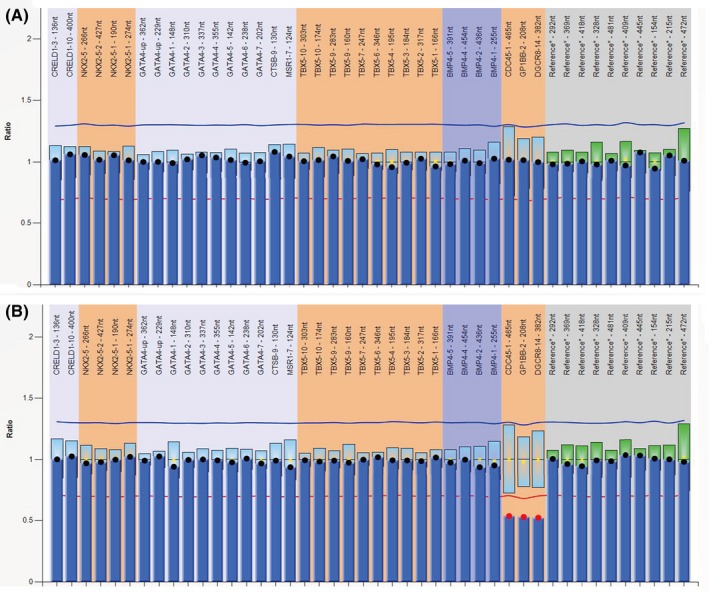
Multiplex ligation‐dependent probe amplification (MLPA) analysis. A, Normal control. B, Positive for deletion of the CDC45, GP1BB, and DGCR8 probes located in 22q11 region (red dots)
Table 1.
Specific type and hemodynamic status of cardiac defect identified in the 6 patients
| No. | Gender/age | Cardiac phenotype | Specific type | Defect size (mm) | SpO2 (%) | |
|---|---|---|---|---|---|---|
| Preoperation | Postoperation | |||||
| Patient 1 | F/7 y | ASD | Ostium secundum | 2 × 2 | 100 | 100 |
| VSD | Perimembranous | 6 × 6 | ||||
| Patient 2 | F/8 m | ASD | Ostium secundum | 6 × 5 | 95 | 99 |
| VSD | Perimembranous | 8 × 8 | ||||
| Patient 3 | F/8 y | VSD | Perimembranous | 13 × 13 | 98 | 98 |
| Patient 4 | F/6 y | VSD | Perimembranous | 12 × 12 | 98 | 98 |
| Patient 5 | M/6 m | TOF | – | – | 97 | 99 |
| Patient 6 | F/6 y | PDA | – | – | 98 | 99 |
ASD, atrial septal defect; PDA, patent ductus arteriosus; TOF, tetralogy of fallot; VSD, ventricular septal defect.
MLPA results from each of these 5 patients were confirmed as all patient samples were sequenced on a NGS platform. However, NGS results of one of the 167 patients indicated the presence of a duplication of the 22q11 region in which MLPA results for the same patient were reported as normal. Estimated breakpoints in the 22q11.21 region of GRCh37/hg19 were 18900001 and 21400000 for the 2.5 Mb interstitial deletion in Patient 1 (Figure 2A), 18880001 and 21460000 for the 2.58 Mb interstitial deletion in Patient 2 (Figure 2B), 18900001 and 21460000 for the 2.56 Mb interstitial deletion in Patient 3 (Figure 2C), 18920001 and 21800000 for the 2.88 Mb interstitial deletion in Patient 4 (Figure 2D), 18900001 and 21460000 for the 2.56 Mb interstitial deletion in Patient 5 (Figure 2E), and 20940001 and 21800000 for the 0.86 Mb interstitial duplication in Patient 6 (Figure 2F). The five low copy repeats (LCR) A‐D involved in the deletions were detected in Patients 1‐5, which were responsible for the majority of 22q11.21 deletion syndromes, while the Patient 6 had a LCR C‐D duplication on chromosome 22q11.21, overrides about 20% of the 22q11 duplication syndrome, and this CNV has not been previously described in DECIPHER, UCSC, and DGV database. The genes listed below are located within the duplication, and their positions in detail are shown in Table S1. Further study indicated that the CNV of 0.86 Mb duplication was de novo. These six CHD patients, Patients 1‐5 correspond to the same classical DiGeorge syndrome, and involved the TBX1 gene, and the Patient 6 had not previously presented with extracardiac symptoms. Results are shown in Table 2. First‐degree relatives of the patients in this study had no history of CHD and negative for the abnormal CNV.
Figure 2.
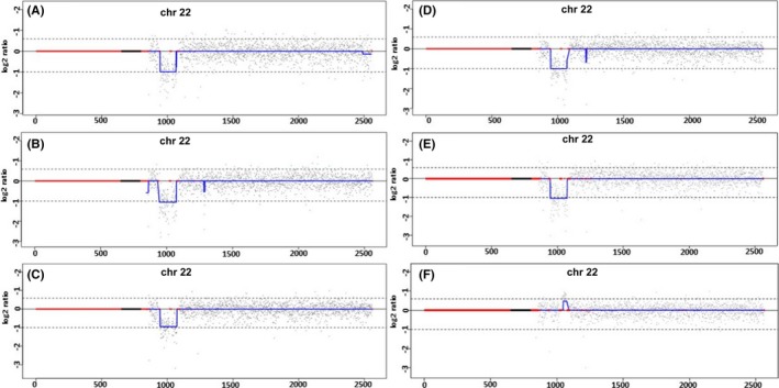
Next generation sequencing analysis on chromosome 22. A, The 2.5 Mb 22q11 deletion (Patient 1); B, The 2.58 Mb 22q11 deletion (Patient 2); C, The 2.56 Mb 22q11 deletion (Patient 3); D, The 2.88 Mb 22q11 deletion (Patient 4); E, The 2.56 Mb 22q11 deletion (Patient 5); F, The 0.86 Mb 22q11 duplication (Patient 6)
Table 2.
Distribution of 6 patients regarding the type of CHD and the results detected by CNV‐seq
| Gender/age | Cardiac phenotypes | Other anomalies | CNV‐Seq (size) | LCR | Inheritance | |
|---|---|---|---|---|---|---|
| Patient 1 | F/7 y | VSD/ASD | Developmental delay and learning disability | 22q11.2 deletion (2.5 Mb) | A‐D | De novo |
| Patient 2 | F/8 m | VSD/ASD | Microcephalus | 22q11.2 deletion (2.58 Mb) | A‐D | De novo |
| Patient 3 | F/8 y | VSD | Short stature | 22q11.2 deletion (2.56 Mb) | A‐D | De novo |
| Patient 4 | F/6 y | VSD | Delayed independent walking | 22q11.2 deletion (2.88 Mb) | A‐D | De novo |
| Patient 5 | M/6 m | TOF | Cleft palate | 22q11.2 deletion (2.56 Mb) | A‐D | De novo |
| Patient 6 | F/6 y | PDA | None | 22q11.2 duplication (0.86 Mb) | C‐D | De novo |
ASD, atrial septal defect; CNV‐Seq, copy number variation sequencing; F, female; M, male; PDA, patent ductus arteriosus; TOF, tetralogy of fallot; VSD, ventricular septal defect.
4. DISCUSSION
Genetic causes of CHDs include structural abnormalities of the chromosome and gene dosage disorders. There is accumulating evidence indicating that a number of genomic disorders are strongly associated with gene dosage and the development of CHDs.13 Determining which CNVs contribute toward disease is an issue that is worth studying. With the development of genetic testing for CHDs, CNVs play an important role in diagnosis and gene discovery.14 Characteristics of a pathogenic CNV may include abnormalities in a known disease‐associated regions, or gene dosage irregularities.15 These may therefore be observed in individuals with cardiovascular malformations.
As molecular targets for CHD, MLPA was used to screen the 22q11.2 region, and GATA4, NKX2‐5, TBX5, BMP4, and CRELD1 genes. The presence of CNVs was confirmed by NGS sequencing. In the 167 patients with isolated incidences of CHD, normalized single probes for the GATA4, NKX2‐5, TBX5, BMP4, and CRELD1 genes fell within the standard range, while six CNVs were identified. Among the CNVs, five 22q11.2 deletions and one 22q11.2 duplication was observed.
The 22q11.2 deletion syndrome is the second most common cause of CHD globally16 and was the most prevalent abnormality in this study having been found in ~3.0% (5/167) of all samples. This is close to the 2.56% found by Campos et al.17 An abundance of LCRs present in the 22q11 region caused non‐allelic homologous recombination during meiosis, which resulted in the observed deletions and duplications.18 There were eight LCR blocks (LCR22‐A to LCR22‐H) in the 22q11 region, but only LCR22‐A to LCR22‐D were implicated in the 22q11.2 deletion syndrome. Over 90% of the patients in this study shared a deletion between the LCR22‐A and LCR22‐D blocks.19 In the present study, Patients 2‐5 possessed a 22q11.2 rearrangement and carried the ~2.5 Mb LCR22A‐D deletion. According to literature, VSD, ASD, and TOF are the most common heart defects associated with chromosomal anbnormalities.20 Interestingly, our results corroborate this observation as Patients 2‐5 presented with VSD. Patient 2 additionally presented with ASD. Patient 1 carried the 2.5 Mb LCR22A‐C deletion in the 22q11 region, and presented with VSD and ASD.
Unlike the 22q11 deletion, duplications in this region are rarely reported.21 Less is known about the highly variable phenotype linked to these duplications,22 but it appears to be associated with elevated rates of ASD and delays in language and psychomotor development.23 In the present study, we detected a 0.86 Mb de novo duplication in the 22q11.2 region (chr22: 20940001‐21800000), in a 6‐year‐old girl (Patient 6) with PDA. The duplication's range from LCR22‐C to LCR22‐D was identified by CNV‐seq. This is the first report of a patient with PDA presenting with a duplication in the 22q11.2 region. However, some limitations must be noted. Firstly, language spoken by the patient was not assessed. Secondly, this duplication is frequently inherited and we failed to verify its presence in the family. Further investigation into this finding is thus required.
The 167 patients used in this study were initially characterized by CNV‐seq and MLPA. Using MLPA, five of the patients (Table 1) had 22q11 deletion, and the other 162 patients were negative. Further detection of CNV‐seq verified the accuracy of the results. Five of the previously identified 22q11 deletion were detected by this method, and no false‐positive results were yielded in the remaining 162 samples, indicating 100% sensitivity and specificity. And MLPA could become a commonly worldwide used methodology for diagnosis of several genomic disorders as the DiGeorge syndrome.
5. CONCLUSION
In summary, our results suggest that deletions in the 22q11.2 region are common in CHD cases in southwest China. Regardless of the presence or absence of extracardiac symptoms, results also indicate that it is necessary to perform prenatal screening for CHD. Although limited in sample size, there is no evidence from this study that CNVs in the GATA4, NKX2‐5, TBX5, BMP4, and CRELD1 genes play a role in fetal CHD in southwest China.
Supporting information
Li Z, Huang J, Liang B, et al. Copy number variations in the GATA4, NKX2‐5, TBX5, BMP4 CRELD1, and 22q11.2 gene regions in Chinese children with sporadic congenital heart disease. J Clin Lab Anal. 2019;33:e22660 10.1002/jcla.22660
Funding information
The project was supported by the Guangxi Scientific Research Technology Development Project (Guigongke 1598011‐8), Liuzhou Science and Technology Development Program (2016G020219), and Guangxi Zhuang Autonomous Region Health Department (Z20170545).
Zhetao Li and Jiwei Huang contributed equally to this work and should be considered co‐first authors
REFERENCES
- 1. van der Linde D, Konings EEM, Slager MA, et al. Birth prevalence of congenital heart disease worldwide. J Am Coll Cardiol. 2011;58(21):2241‐2247. [DOI] [PubMed] [Google Scholar]
- 2. Pierpont ME, Basson CT, Benson DJ, et al. Genetic basis for congenital heart defects: current knowledge: a scientific statement from the American Heart Association Congenital Cardiac Defects Committee, Council on Cardiovascular Disease in the Young: endorsed by the American Academy of Pediatrics. Circulation. 2007;115(23):3015‐3038. [DOI] [PubMed] [Google Scholar]
- 3. Kruszka P, Addissie YA, McGinn DE, et al. 22q11.2 deletion syndrome in diverse populations. Am J Med Genet A. 2017;173(4):879‐888. [DOI] [PMC free article] [PubMed] [Google Scholar]
- 4. Glessner JT, Bick AG, Ito K, et al. Increased frequency of de novo copy number variations in congenital heart disease by integrative analysis of SNP‐array and exome sequence data. Circ Res. 2014;115(10):884‐896. [DOI] [PMC free article] [PubMed] [Google Scholar]
- 5. Russell MW, Chung WK, Kaltman JR, Miller TA. Advances in the understanding of the genetic determinants of congenital heart disease and their impact on clinical outcomes. J Am Heart Assoc. 2018;7(6):e006906. [DOI] [PMC free article] [PubMed] [Google Scholar]
- 6. McDonald‐McGinn DM, Tonnesen MK, Laufer‐Cahana A, et al. Phenotype of the 22q11.2 deletion in individuals identified through an affected relative: cast a wide FISHing net!. Genet Med. 2001;3:23‐29. [DOI] [PubMed] [Google Scholar]
- 7. Sarkozy A, Conti E, Neri C, et al. Spectrum of atrial septal defects associated with mutations of NKX2.5 and GATA4 transcription factors. J Med Genet. 2005;42:e16. [DOI] [PMC free article] [PubMed] [Google Scholar]
- 8. Mori AD, Bruneau BG. TBX5 mutations and congenital heart disease: Holt‐Oram syndrome revealed. Curr Opin Cardiol. 2004;19(3):211‐215. [DOI] [PubMed] [Google Scholar]
- 9. Qian B, Mo R, Da M, Peng W, Hu Y, Mo X. Common variations in BMP4 confer genetic susceptibility to sporadic congenital heart disease in a Han Chinese population. Pediatr Cardiol. 2014;35(8):1442‐1447. [DOI] [PMC free article] [PubMed] [Google Scholar]
- 10. Alcántara‐Ortigoza MA, De Rubens‐Figueroa J, Reyna‐Fabian ME, et al. Germline mutations in NKX2‐5, GATA4, and CRELD1 are rare in a Mexican sample of Down syndrome patients with endocardial cushion and septal heart defects. Pediatr Cardiol. 2015;36(4):802‐808. [DOI] [PubMed] [Google Scholar]
- 11. Granados‐Riveron JT, Pope M, Bu'Lock FA, et al. Combined mutation screening of NKX2‐5, GATA4, and TBX5 in congenital heart disease: multiple heterozygosity and novel mutations. Congenit Heart Dis. 2012;7(2):151. [DOI] [PMC free article] [PubMed] [Google Scholar]
- 12. Shanshen E, Rosenberg J, Van Bergen AH. Identification of novel congenital heart disease candidate genes using chromosome microarray. Pediatr Cardiol. 2017;23:1‐12. [DOI] [PubMed] [Google Scholar]
- 13. Azhar M, Ware SM. Genetic and developmental basis of cardiovascular malformations. Clin Perinatol. 2016;43(1):39. [DOI] [PMC free article] [PubMed] [Google Scholar]
- 14. Costain G, Silversides CK, Bassett AS. The importance of copy number variation in congenital heart disease. NPJ Genom Med. 2016;1:16031. [DOI] [PMC free article] [PubMed] [Google Scholar]
- 15. Lander J, Ware SM. Copy number variation in congenital heart defects. Curr Genet Med Rep. 2014;2(3):168‐178. [Google Scholar]
- 16. Ware SM, Jefferies JL. New genetic insights into congenital heart disease. J Clin Exp Cardiol. 2012;S8:003. [DOI] [PMC free article] [PubMed] [Google Scholar]
- 17. Campos CMR, Zanardo EA, Dutra RL, Kulikowski LD, Kim CA. Investigation of copy number variation in children with conotruncal heart defects. Arq Bras Cardiol. 2015;104(1):24‐31. [DOI] [PMC free article] [PubMed] [Google Scholar]
- 18. Edelmann L, Pandita RK, Morrow BE. Low‐copy repeats mediate the common 3‐Mb deletion in patients with velo‐cardio‐facial syndrome. Am J Hum Genet. 1999;64(4):1076‐1086. [DOI] [PMC free article] [PubMed] [Google Scholar]
- 19. Bertini V, Azzarà A, Legitimo A, et al. Deletion extents are not the cause of clinical variability in 22q11.2 deletion syndrome: does the interaction between DGCR8 and miRNA‐CNVs play a major role? Front Genet. 2017;8:47. [DOI] [PMC free article] [PubMed] [Google Scholar]
- 20. Bellucco FTDS, Belangero SIN, Farah LMS, et al. Investigating 22q11.2 deletion and other chromosomal aberrations in fetuses with heart defects detected by prenatal echocardiography. Pediatr Cardiol. 2010;31(8):1146. [DOI] [PubMed] [Google Scholar]
- 21. Lin A, Crk C, Vajdi A, et al. Mapping 22q11.2 gene dosage effects on brain morphometry. J Neurosci. 2017;37(26):6183. [DOI] [PMC free article] [PubMed] [Google Scholar]
- 22. Wentzel C, Fernström M, Ohrner Y, et al. Clinical variability of the 22q11.2 duplication syndrome. Eur J Med Genet. 2008;51(6):501. [DOI] [PubMed] [Google Scholar]
- 23. Wenger TL, Miller JS, Depolo LM, et al. 22q11.2 duplication syndrome: elevated rate of autism spectrum disorder and need for medical screening. Mol Autism. 2016;7(1):1‐16. [DOI] [PMC free article] [PubMed] [Google Scholar]
Associated Data
This section collects any data citations, data availability statements, or supplementary materials included in this article.
Supplementary Materials