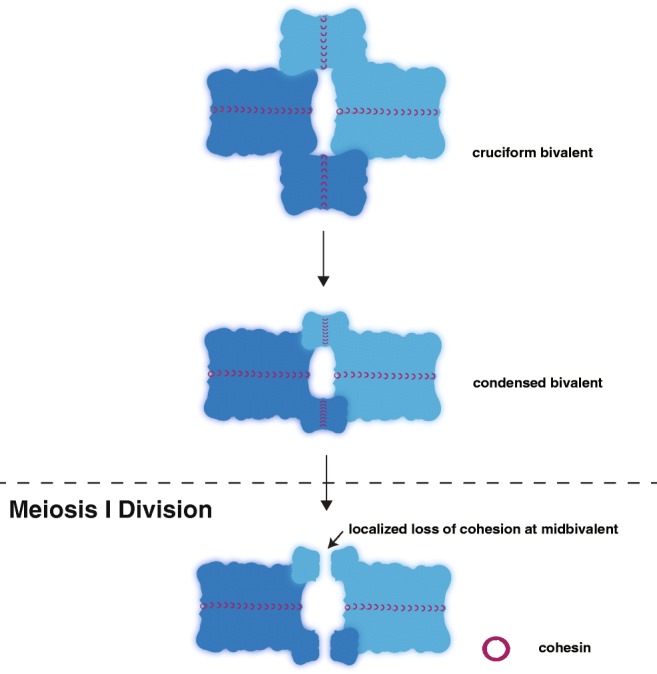
Fig. 5.

Diakinesis. The homologous chromosomes (dark and light blue) are rearranged during diakinesis to form a cruciform bivalent structure. This bivalent structure is then condensed by a group of proteins likely including cohesins, condensins, and DNA topoisomerases. At the meiosis I division, cohesin (maroon rings) is locally lost distal to the crossover site at the midbivalent allowing the homologs to separate. The residual cohesin is maintained between the sister chromatids until the second meiotic division when it is removed to allow the sisters to segregate (not depicted)
