Abstract
Cerebral cavernous malformations (CCMs) is a microvascular disorder in the central nervous system. Despite tremendous efforts, the causal genetic mutation in some CCM patients has not be identified, raising the possibility of an unknown CCM locus. The CCM2/MGC4607 gene has been identified as one of three known genes causing CCMs. In this report, we defined a total of 29 novel exons and 4 novel promoters in CCM2 genomic structure and subsequently identified a total of 50 new alternative spliced isoforms of CCM2 which eventually generated 22 novel protein isoforms. Genetic analysis of CCM2 isoforms revealed that the CCM2 isoforms can be classified into two groups based on their alternative promoters and alternative start codon exons. Our data demonstrated that CCM2 isoforms not only are specific in their subcellular compartmentation but also have distinct cellular expression patterns among various tissues and cells, indicating the pleiotropic cellular roles of CCM2 through their multiple isoforms. In fact, the complexity of the CCM2 genomic structure was reflected by the multiple layers of regulation of CCM2 expression patterns. At the transcriptional level, it is accomplished by alternative promoters, alternative splicing, and multiple transcriptional start sites and termination sites; while at the translational level, it is carried out with various cellular functions with a distinguishable CCM2 protein group pattern, specified abundance and composition of selective isoforms in a cell and tissue specific fashion. Through experimentation, we discovered a unique phosphotyrosine binding (PTB) domain, namely atypical phosphotyrosine binding (aPTB) domain. Some long CCM2 isoform proteins contain both classes of PTB domains, making them a dual PTB domain-containing protein. Both CCM1 and CCM3 can bind competitively to this aPTB domain, indicating CCM2 as the cornerstone for CCM signaling complex (CSC).
Subject terms: Gene expression profiling, Comparative genomics, Stroke, Genetics of the nervous system
Introduction
Cerebral cavernous malformations (CCMs) are malformed microvasculatures predominantly in the central nervous system, with prevalence in approximately 0.1–0.5% of the population and represent 10% of all vascular malformations1. The pathology of CCMs can be characterized by distended capillary cavities enclosed by a thin layer of endothelial cells (EC) lacking the support of intervening parenchyma, making lesions susceptible to stroke. CCMs are located primarily in the brain but can also be found in the other parts of the body such as spinal cord, skin, and rarely the retina. Familial forms of CCM is caused by loss of function mutations in one of three known CCM genes, KRIT1/CCM1, MGC4607/Malcavernin/CCM2, and PDCD10/CCM3. Despite tremendous efforts, there are still CCM patients with mutations that have not yet been defined, suggesting the possible existence of an unknown CCM locus.
CCM2 has a strong interaction with both CCM1 and CCM3 proteins, suggesting all three proteins can form a complex, termed CCM signaling complex (CSC), which might be involved in integrin mediated signaling. Subsequently, CSC interacts with other cellular components to mediate intracellular and cell/extracellular matrix (ECM) signaling2–6. Both in-vitro and in-vivo studies showed that through their interaction7, CCM proteins influence the angiogenic performance of vascular ECs by regulating β1-integrin-mediated signaling cascades8. Obstructed microvasculature in CCM1 and CCM2 (Ccm1/2) animal models5 is the provenance of various phenotypic expressions such as enlarged heart9, dilated axial primitive vessels10,11, and blood stasis in and around the heart, thus demonstrating the importance of CSC complex in angiogenesis5,12,13.
In this study, we aimed to define the CCM2 gene, in both transcriptional and translational levels. We found that there are multiple alternative promoters, alternative splicing, and multiple transcriptional start sites and termination sites in the genomic structure of CCM2 gene, which play significant roles during the transcriptional events, generating various CCM2 RNA isoform species with apparently distinct biological functions. These CCM2 isoforms were further confirmed at the protein level, leading us to identify a novel PTB domain in CCM2 protein, which helps us better understand the complexity of CSC and its associated cellular factors which contribute to the angiogenic events and underlining molecular and cellular etiology in the pathogenesis of CCMs.
Results
Identification of new exons, alternative spliced exons, and new isoforms in CCM2
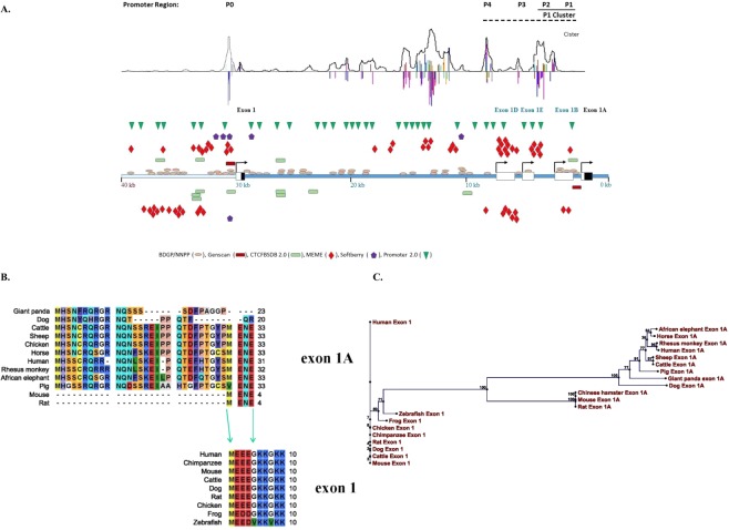
A total of 31 exons, including 8 new exons and 13 new alternative spliced exons derived from existing exons, were identified in the CCM2 gene. Most of the newly identified exons were located near or at the 5′end, just downstream of the original bonafide start codon exon (exon1); as alternative transcription start exons with their own promoters, only two of them (6A, 6B) resided in the middle of CCM2 genomic structure (Table 1, Fig. 1A). Interestingly among newly identified alternative transcription start exons, only one was found harboring another start codon, which makes it a novel alternative start codon exon (exon1A) with its own distinct promoter (Fig. 1A). We then identified a total of 50 isoforms of CCM2 using two sets of full-length CCM2 gene primers with genomic analysis tools. In addition, 11 new isoforms without exon1 or exon1A were identified. Almost all of these new exons and partial sequences of isoforms have been reported in NCBI EST/ExAC databases, reaffirming their cellular existence (Tables 1, 2). These new CCM2 isoforms are categorized into three groups based on their alternative promoters and alternative start codon exons; A group has a start codon in exon1 with the original promoter (P0) and B group in exon1A with a novel promoter (P1) downstream of exon 1, while C group in other exons are uncertain (Table 2, Fig. 1A). Both A group and B group have a notable biological significance based on their cellular abundance in various tissues (Figs 2A, 4A, Suppl. Fig. 1). Considering the shared identical open-reading frame (ORF) of some CCM2 isoforms, a total of 32 CCM2 isoforms were eventually confirmed with different coding schemes, indicating the complexity of CCM2 isoform regulation at transcription level (Table 2).
Table 1.
Identification of new exons and alternative spliced exons (as) of CCM2 gene.
| Exons | Genomic position | Hits in EST | ||
|---|---|---|---|---|
| Start | End | Length(bp) | Database (%) | |
| exon 1* | 45039933 | 45039962 | 30 | 89 |
| exon 1A | 45067304 | 45067396 | 93 | 61 |
| exon 1B | 45066608 | 45066865 | 258 | 5 |
| exon 1Bas1 | 45066608 | 45066869 | 262 | 2 |
| exon 1Bas2 | 45066608 | 45066821 | 214 | 2 |
| exon 1C | 45069043 | 45069108 | 66 | 2 |
| exon 1D | 45064095 | 45064371 | 277 | 2 |
| exon 2 | 45077852 | 45078025 | 174 | 100 |
| exon 2as1 | 45077852 | 45078019 | 168 | |
| exon 2A | 45099802 | 45099907 | 106 | 2 |
| exon 2B | 45102692 | 45102814 | 123 | 0 |
| exon 3 | 45103517 | 45103600 | 84 | 100 |
| exon 4 | 45104062 | 45104245 | 184 | 100 |
| exon 4as1 | 45104062 | 45104097 | 36 | |
| exon 4as2 | 45104062 | 45104151 | 90 | |
| exon 5 | 45108042 | 45108178 | 137 | 100 |
| exon 6 | 45109425 | 45109560 | 136 | 100 |
| exon 6A | 45111349 | 45111471 | 123 | 13 |
| exon 6B | 45109988 | 45110129 | 142 | 2 |
| exon 7 | 45112325 | 45112382 | 58 | 100 |
| exon 7as1 | 45112325 | 45112356 | 32 | |
| exon 8 | 45113059 | 45113170 | 112 | 100 |
| exon 9 | 45113869 | 45114007 | 139 | 100 |
| exon 9as1 | 45113841 | 45114007 | 167 | 1 |
| exon 10** | 45115376 | 45115656 | 281 | 100 |
| exon 10as1 | 45115500 | 45115757 | 258 | |
| exon 10as2 | 45115573 | 45115757 | 185 | |
| exon 10as3 | 45115721 | 45115757 | 37 | |
| exon 10as4 | 45115376 | 45115440 | 65 | 100 |
| exon 10as5 | 45115652 | 45116068 | 417 | 100 |
| exon 10as6 | 45115432 | 45115757 | 326 | 100 |
Genomic location of exons and alternative slicing sites of CCM2 based on the Genome Reference Consortium Human Genome Build 37/Human Genome Assembly 19 (GRCh37/hg19). Both underlined exons 1 and 1A contain a bonafide start codon with different promoters. Newly identified exons are highlighted with bold letter, whereas the alternatively spliced exons were italicized (as). The genomic location of initial and end sites and exact length of each exon are detailed in the table. The maximum number of blast hit for each exon in human EST database is set at 100, reaching this limit is considered as the most abundant in CCM2 cDNA/EST, while some blanks in this category indicate that the alterative spliced isoform are located within its coordinated exons. Exon 1 is the foremost 5′ end among all exons along the genomic sequence, *or **indicates exons containing alternative transcriptional start sites; ***containing the alternative termination sites.
Figure 1.
Genomic structure, conservation, and variability among alterative start-codon exons and promotors of CCM2. (A) The complex promoter regions of human CCM2 locus were defined with bioinformatics (promoter prediction software from top to bottom: Cister, promotor2.0, Softberry, MEME, CTCFBSDB, BDGP/NNPP and Genscan as indicated by different colors). Symbols on top of DNA templates are on positive strand, below are on negative strand. The single promoter for the original bonafide start-codon exon, exon 1, simply lies immediately upstream of the transcription start site for exon 1, as P0. The promoter region for exon 1A is much more complicated. Although a seemingly weak promoter, P1 lies immediately upstream of its transcription-start site; in addition, there are three relatively strong promoters (P2-P4) upstream adjacent to P1 promoter. Three exons (exon 1B, 1D, 1E) with the transcription start site driven by these three promoters (P2-P4), respectively, usually skip exon 1A (coding exon), presumably to down-regulate the transcription level of group B CCM2 isoforms. Genomic structure of 5′ region of the human CCM2 locus is schematically summarized in this map. Noncoding region within a transcription-start exon labeled as white box while coding region within the exon labeled as black box. (B) Multiple-alignment between two alterative start codon exons (exon 1 and exon 1A) across species reveals a vertebrate-specific exon 1 and a mammalian-specific exon 1A and their evolutional relationship. Exon 1A is evolutionarily evolved from exon1 with its C-terminus homolog to the N-terminus of exon1. (C) Phylogenetic relationships between exon 1A and exon 1 among CCM2 isoforms across species based on neighbor joining (NJ) method which hypothesizes a stochastic process in different lineages during evolution.
Table 2.
Identification and nomenclature of alternatively spliced CCM2 isoforms.
| Isoform identifier | Containing exons | Missing exons | Peptide Length (aa) | cDNA/EST Clones |
|---|---|---|---|---|
| CCM2-A | ||||
| CCM2-100 | 1, 2, 3, 4, 5, 6, 7, 8, 9, 10 | 444 | BX337677 | |
| CCM2-101 | 1, 3, 4, 5, 6, 7, 8, 9, 10 | 2 | 386 | BM922546 |
| CCM2-102 | 1, 3, 4, 7, 8, 9, 10 | 2, 5, 6 | 295 | |
| CCM2-103 | 1, 2, 8, 9, 10 | 3, 4, 5, 6, 7 | 142* | |
| CCM2-104 | 1, 2, 7, 8, 9, 10 | 3, 4, 5, 6 | 142* | |
| CCM2-105 | 1, 2, 3, 4, 6, 7, 8, 9, 10 | 5 | 142* | |
| CCM2-106 | 1, 4, 5, 6, 7, 8, 9, 10 | 2, 3 | 358 | |
| CCM2-107 | 1, 6, 7, 8, 9, 10 | 2, 3, 4, 5 | 251 | |
| CCM2-108 | 1, 7, 8, 9, 10 | 2, 3, 4, 5, 6 | 142* | |
| CCM2-1209 | 1, 8, 9, 10 | 2, 3, 4, 5, 6, 7 | 142 | |
| CCM2-111 | 1, 5, 6, 7, 8, 9, 10 | 2, 3, 4 | 142* | |
| CCM2-115 | 1, 2, 5, 6, 7, 8, 9, 10 | 3, 4 | 142* | BG385641 |
| CCM2-116 | 1, 2, 6, 7, 8, 9, 10 | 3, 4, 5 | 309 | BI752347 |
| CCM2-117 | 1, 2, 3, 4, 7, 8, 9, 10 | 5, 6 | 353 | BG421932 |
| CCM2-118 | 1, 2, 3, 4, 5, 7, 8, 9, 10 | 6 | 207 | BX439329 |
| CCM2-119 | 1, 2, 3, 4, 5, 6, 8, 9, 10 | 7 | 142* | BX462850 |
| CCM2-120 | 1, 3, 4, 6, 7, 8, 9, 10 | 2, 5 | 142* | CX872822 |
| CCM2-129 | 1, 7, 8, 9, 10 | 2, 3, 4, 5, 6 | 142* | |
| CCM2-130 | 1, 1C, 2, 3, 4, 6, 7, 8, 9, 10 | 5 | 142* | BX460017 |
| CCM2-131 | 1, 1C, 2, 7, 8, 9, 10 | 3, 4, 5, 6 | 142* | |
| CCM2-132 | 1, 1C, 2, 5, 6, 7, 8, 9, 10 | 3, 4 | 142* | |
| CCM2-300 | 1, 2B, 3, 4, 5, 6, 7, 8, 9, 10 | 2 | 142* | BI523801 |
| CCM2-401 | 1, 3, 4as1, 10as1 | 2, 5, 6, 7, 8, 9 | 81 | |
| CCM2-402 | 1, 2, 3, 4as2, 10as2 | 5, 6, 7, 8, 9 | 153 | |
| CCM2-403 | 1, 2as1, 10as3 | 3, 4, 5, 6, 7, 8, 9 | 95 | |
| CCM2-404 | 1, 2, 3, 4, 7, 8, 9, 10as4, 10as5 | 5, 6 | 334 | H7C6075-68 |
| CCM2-600 | 1, 2, 3, 4, 5, 6, 6A, 7, 8, 9, 10 | 485 | BG722235 | |
| CCM2-601 | 1, 3, 4, 5, 6, 6A, 7, 8, 9, 10 | 2 | 427 | CF145136 |
| CCM2-602 | 1, 2, 3, 4, 6, 6A, 7, 8, 9, 10 | 5 | 142* | |
| CCM2-603 | 1, 3, 4, 6, 6A, 7, 8, 9, 10 | 2, 5 | 142* | H7C6075-63 |
| CCM2-604 | 1, 5, 6, 6A, 7, 8, 9, 10 | 2, 3, 4 | 142* | CF145056 |
| CCM2-606 | 1, 4, 5, 6, 6A, 7, 8, 9, 10 | 2, 3 | 400 | |
| CCM2-607 | 1, 6, 6A, 7, 8, 9, 10 | 2, 3, 4, 5 | 293 | |
| CCM2-610 | 1, 2, 3, 4, 5, 6, 6B, 8, 9, 10 | 7 | 473 | |
| CCM2-B | ||||
| CCM2-200 | 1A, 2, 3, 4, 5, 6, 7, 8, 9, 10 | 1 | 465 | BX363852 |
| CCM2-201 | 1A, 3, 4, 5, 6, 7, 8, 9, 10 | 2 | 407 | BI766123 |
| CCM2-202 | 1A, 3, 4, 5, 8, 9, 10 | 2, 6, 7 | 148 | BI906707 |
| CCM2-203 | 1A, 2, 3, 4, 7, 8, 9, 10 | 5, 6 | 374 | H7C6075-81 |
| CCM2-204 | 1A, 2, 4, 5, 6, 7, 8, 9, 10 | 3 | 437 | H7C6075-86 |
| CCM2-205 | 1A, 7, 8, 9, 10 | 2, 3, 4, 5, 6 | 142* | |
| CCM2-206 | 1A, 2, 3, 4, 5, 6, 6A, 7, 8, 9, 10 | 1 | 506 | BQ073060 |
| CCM2-207 | 1A, 2, 3, 5, 6, 6B, 8, 9, 10 | 7 | 494 | BX415190 |
| CCM2-208 | 1A, 2, 7, 8, 9, 10 | 3, 4, 5, 6 | 142* | |
| CCM2-209 | 1A, 8, 9, 10 | 2, 3, 4, 5, 6, 7 | 178 | |
| CCM2-210 | 1A, 3, 4, 7, 8, 9, 10 | 2, 5, 6 | 316 | |
| CCM2-211 | 1A, 5, 6, 7, 8, 9, 10 | 2, 3, 4 | 142* | |
| CCM2-212 | 1A, 6, 7, 8, 9, 10 | 2, 3, 4, 5 | 272 | |
| CCM2-213 | 1A, 2, 3, 4, 5, 6, 7as1, 10as4 | 8, 9 | 354 | |
| CCM2-214 | 1A, 3, 4, 5, 6, 7as1, 10as4 | 2, 8, 9 | 296 | |
| CCM2-215 | 1A, 3, 4, 7as1, 10as4 | 2, 5, 6, 8, 9 | 205 | |
| CCM2-216 | 1A, 2, 3, 4, 6A, 7, 8, 9, 10 | 5, 6 | 415 | |
| CCM2-C | ||||
| CCM2-220 | 1B, 3, 4, 5, 6, 8, 9, 10 | 2, 7 | 142* | |
| CCM2-221 | 1B, 2, 8, 9, 10 | 3, 4, 5, 6, 7 | 142* | |
| CCM2-222 | 1B, 3, 4, 5, 6, 7, 8, 9, 10 | 2 | 142* | |
| CCM2-223 | 1Bas2, 3, 8, 9, 10 | 2, 4, 5, 6, 7 | 179 | BI907152 |
| CCM2-224 | 1B, 7, 8, 9, 10 | 2, 3, 4, 5, 6 | 142* | |
| CCM2-225 | 1B, 2, 7, 8, 9, 10 | 3, 4, 5, 6 | 142* | |
| CCM2-226 | 1B, 5, 6, 7, 8, 9, 10 | 2, 3, 4 | 142* | |
| CCM2-227 | 1B, 1A, 5, 6, 8, 9, 10 | 2, 3, 4 | 142* | |
| CCM2-230 | 1D, 2, 3, 4, 5, 6, 7, 8, 9, 10 | 142* | CV572216 | |
| CCM2-240 | 1B, 2, 3, 4, 5, 6, 7, 8, 9, 10 | 142* | DA983503 | |
| CCM2-241 | 1B, 6, 7, 8, 9, 10 | 2, 3, 4, 5 | 142* | |
Novel CCM2 isoforms identified can be further validated through EST database search. A specific isoform identifier (IsoID) is assigned for each isoform based on the information of the exon composition. CCM2 isoforms are classified into three major groups based on their alternative promoters and alternative start codon exons: CCM2-A (exon1), CCM2-B (exon1A), and CCM2-C (Undetermined). Among identified isoforms, CCM2-100 isoform is original “bonafide” CCM2 with ten coding exons, while CCM2-1209 isoform is a short protein isoform with a common C-terminal peptide sequence of HH domain (Harmonin homology) shared by many isoforms (such as 109, 209 etc). The genomic structure of the representative isoforms of CCM2 is further validated by their existing correlative cDNA/EST clones in NCBI database. *At peptide length column indicates the isoform sharing the identical open-reading frame with isoform CCM2-1209 with various length of 5′ UTRs.
Figure 2.
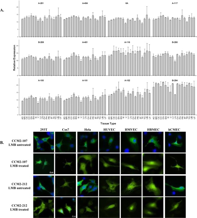
Relative expression profiling of endogenous CCM2 isoforms among various tissues. (A) The relative expression (2−∆CT) of CCM2 isoforms measured by qPCR in various tissues is presented with bar plots. Allele-specific real-time quantitative PCR (qPCR) assays were performed in triplicates (n = 3) and represented with means and standard deviations (M ± SD) of the relative expressions. Adrenal gland (AG), Breast (BR), Cervix (CE), Colon (CO), Endometrium (EN), Esophagus (ES), kidney (KI), Liver (LI), Lung (LU), Lymph node (LN), Ovary (OV), Pancreas (PA), Prostate (PR), Stomach (ST), Testis (TE), Thyroid gland (TG), Urinary bladder (UB), and Uterus (UT). (B) Subcellular localization of CCM2 isoform pairs, CCM2-107 and CCM2-212, in various immortalized cell lines: Immortalized Human Embryonic Kidney cells (293 T), Immortalizing Monkey Kidney Fibroblast cells (COS7), Immortalized Human Cervical Cancer cells (HeLa); and several primary/immortalized human endothelial cell lines: Human Umbilical Vein Endothelial Cells (HUVEC), Human Microvascular Endothelial Cells (HMVEC), Human Brain Microvascular Endothelial Cells (HBMEC), and Immortalized Human Cerebral Microvascular Endothelial Cells, hCMEC/D3 cells (hCMEC). Isoform CCM2-107s is seen predominantly in the cytoplasm in every cell line but accumulates in the nucleus after treatment with leptomycin (LMB treated). However, isoform CCM2-212 behaves differently and is seen predominantly in the cytoplasm in some cells, but distributed evenly in both the nucleus and cytoplasm before leptomycin treatment (LMB untreated), indicating its differentiated cellular compartmentation, possible nuclear function, and potential association with new cellular partners. Scale bars represent 25 µm and are located in CCM2-107 LMB treated 293 T panel and CCM2-212 LMB treated Cos7 panel; all images were acquired using the same microscope and magnitude and processed identically to each other.
Figure 4.
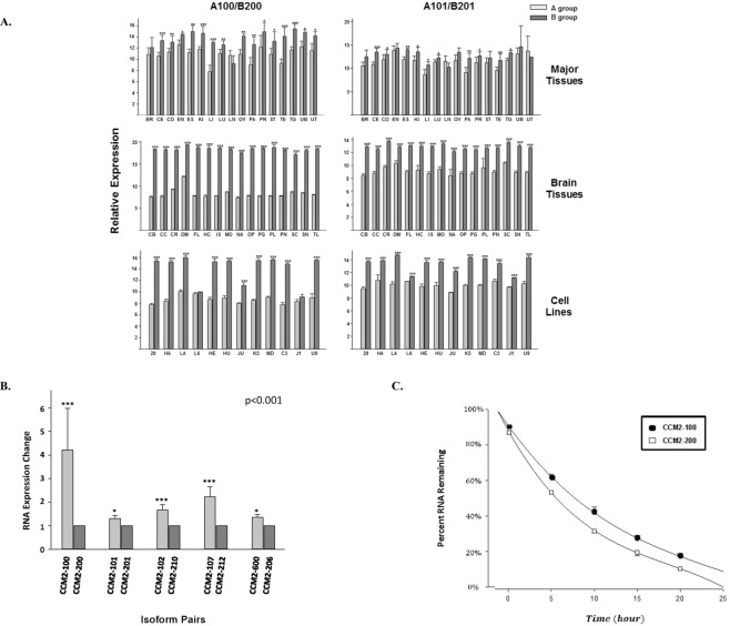
The relative expression level and cellular stability between A and B group isoforms. (A) Comparison of endogenous expression levels between CCM2 isoform pairs among various tissues. The relative mRNA expression levels of paired-CCM2 isoforms (2−∆CT) were presented with bar plots, in which light grey bars represent A group isoforms, dark grey bars represent their respective counterparts, B group isoforms. For experimental design, the left three panels represent expression levels between CCM2 isoform pairs with primer set, CCM2-A100 and CCM2-B200; the right three panels for CCM2 isoform pairs with primer set, CCM2-A101 and CCM2-B201. For tissue location and cell lines, upper two panels represent the expression levels of paired-CCM2 isoforms among major tissues (see Fig. 2A), middle two panels for various brain tissues, and lower two panels for multiple cell lines (see Suppl. Fig. 1) Middle and lower panels are further described in supplemental Fig. 1. One-way ANOVA was also performed for the comparison between A and B groups of isoforms among different tissues and cells; it was found there is a very significant difference (P < 0.001). The detailed information for isoform-qPCR primer sets is listed in Suppl. Table 2. (B) The changes in the expression levels between ectopic expressed A and B group isoform pairs. The expression levels were measured by allele-specific qPCR (V5-tag), then normalized first by internal expression control (Neo) and followed by mean values for B group isoforms, presented as fold changes. (C) Comparison of RNA decay rates between two ectopic expressed CCM2-100 (A) and CCM2-200 (B), isoform pairs from groups A and B respectively, measured with allele-specific qPCR primers (Suppl. Table 2) at five different time points (after 5, 10, 15, 20, and 25 hours). The solid circle presents A Group isoform (CCM2-100), while hollow square presents Group B isoform (CCM2-200). One-way ANOVA was also performed for the comparison between A and B groups of isoforms and found there is a very significant difference for the expression levels between A and B groups of isoforms (P < 0.001). ***, **, and *above bar indicate P =< 0.001, 0.01, and 0.05 respectively for paired t-test. For major tissue abbreviations refer to Fig. 2; for brain tissue and cell line abbreviations please refer to Suppl. Fig. 1.
Newly identified CCM2 isoforms are detected at both transcriptional and translational levels
Bioinformatics data indicate the pleiotropic roles of CCM2 isoforms
All CCM2 isoforms were analyzed and their functional domains were defined by NCBI conserved domains database using genomic analysis tools (Suppl. Table 3). Noteworthy, CCM2 protein isoforms share identical or similar amino acid sequences like isoforms in other genes and many of them contain a phosphotyrosine binding (PTB) domain which has already been shown to interact with the NPXY motifs (Asn-Pro-X-Tyr, X can be any amino acid) of CCM1 and β1-integrin7,14. However, the classic PTB domain was notably missing in several alternatively spliced isoforms (termed PTB-less isoforms). Furthermore, 8 additional different domains with distinct cellular functions were identified across different isoforms (Suppl. Table 3), indicating the pleiotropic roles of these alternatively spliced isoforms in various cellular events. Several new exons with transcription start sites and their own promoters (P0-P5) were identified, but only two of them contain distinct start-codon exons (exons 1 and 1A, labeled with a black box, respectively, Fig. 1A) and are regulated by their alternative promoters, P0 or P1, respectively. Transcripts starting with other exons (exon 1B, 1D, and 1E) usually generate a coding fragment of the canonical CCM2-1209 isoform, which is a common truncated C-terminal fragment containing solely the Harmonin homology (HH) domain. Our preliminary data suggest that promoter P1 might coordinate with P2 (exon 1B) to form a P1 promoter cluster (P1c) for regulation at the transcription level of B group CCM2 isoforms; additionally P3-P5 promoters may also have some influence on this P1c (Fig. 1A), indicating that the transcriptional regulation of B group CCM2 isoforms is more complicated than that of A group CCM2 isoforms.
Distinct cellular roles of two major groups of CCM2 isoforms
Multiple alignments (Fig. 1B) and phylogenetic analysis for the start-codon exons between two major groups (Fig. 1C) across species, indicated that the originally identified start codon exon 1 is highly conserved and vertebrate-specific, while the newly identified start codon exon 1A is mammalian-specific. Their evolutionary relationship indicates the mammalian-specific and more variable exon 1A likely originated from more conserved exon 1, probably through a series of genomic recombination and duplication events, based on the homologous sequence of C-terminus of exon1A to the N-terminus of exon 1 (Fig. 1B,C).
The variable nature of exon 1A was shown by the significantly shortened amino acid sequence in rodents (resulting from a 13 bp insertion in the coding region possibly during ancient DNA replication events in mouse and rat), which is further supported by coding variation in giant panda, dog, and even human, thus substantiating phylogenetic results (Fig. 1B,C). Further, the promoter region for exon 1A is much more complicated than the promoter for the original exon 1 (Fig. 1A). This finding led us to hypothesize that different isoforms of CCM2 behave differently in the different cell types and might have different functions in various biogenic events.
Differentially transcribed isoform RNAs in various tissues and cell lines
To test our hypothesis, we preformed qPCR which demonstrated that all identified CCM2 isoforms were ubiquitously expressed in all major tissues, but their expression levels varied anatomically. However, many CCM2 isoforms seem to be expressed relatively higher in reproductive tissues including Breast (BR), Cervix (CE), Endometrium (EN), Ovary (OV), Prostate (PR), Testis (TE) and Uterus (UT), compared to other tissues (Fig. 2A). Likewise, detailed gene expression profiling on various brain tissues and cell lines was also performed (Suppl. Fig. 1A,B). Upon elucidating additional data, it was observed that the CCM2 isoforms were ubiquitously distributed in various brain tissues and cell lines albeit at considerably different levels of expressions, suggesting that each CCM2 isoform may have a distinct cellular function.
Distinct cellular compartmentation for different isoforms
Efforts for a detailed subcellular localization of CCM2 has thus far been unsuccessful15, possibly due to the unknown existence of its polymorphic isoforms. To define the cellular localization of CCM2 isoforms, we first selected one isoform pair, CCM2-107 (A group) and CCM2-212 (B group), to investigate their subcellular localization. Surprisingly, we found significantly differentiated cellular localization patterns in various cell lines. CCM2-107 was predominantly found in the cytoplasm in all tested cell lines but accumulated in the nucleus following leptomycin treatment, similar to the isoform CCM2-100 and its shuttle, CCM1, as described before7,16. However, in some cell lines isoform CCM2-212 was distributed evenly in both the nucleus and cytoplasm at steady state before leptomycin treatment (Fig. 2B) which indicates that there are either some cellular factors shuttling these isoforms into the nucleus or isoforms themselves have the ability. To confirm this finding, all identified CCM2 isoform pairs were used to screen in HeLa cells (Immortalized Cervical cancer cells) and three primary human endothelial cell lines, Human Umbilical Vein Endothelial Cells (HUVEC), Human Microvascular Endothelial Cells (HMVEC), and Immortalized Human Cerebral Microvascular Endothelial Cells (hCMEC/D3) (Suppl. Fig. 2A–D), and final results were summarized in Table 3. The data from these experiments reaffirmed our previous observation that some CCM2 isoforms localize differently in different cells, probably through their association with different cellular factors or complexes. Intriguingly, there are more CCM2 isoforms retained within the nucleus at steady state in primary endothelial cell lines suggesting important and diverse cellular roles these nuclear CCM2 isoforms might play during vascular angiogenesis (Table 3, Suppl. Fig. 2).
Table 3.
Differential cellular compartmentations among different CCM2 isoforms in various cell lines.
| Cell Type | Isoform | LMB Untreated | LMB Treated | Cell Localization |
|---|---|---|---|---|
| LA | CCM2-107 | C | CN | S |
| CCM2-100 | C | CN | S | |
| CCM2-101 | C | CN | S | |
| CCM2-102 | C | CN | S | |
| CCM2-117 | C | CN | S | |
| CCM2-116 | C | CN | S | |
| CCM2-402 | CN | CN | S-N | |
| CCM2-601 | C | CN | S | |
| CCM2-206 | C | CN | S | |
| CCM2-212 | CN | CN | S-N | |
| CCM2-201 | C | CN | S | |
| CCM2-203 | C | CN | S | |
| CCM2-210 | C | CN | S | |
| CCM2-216 | C | CN | S | |
| HU | CCM2-107 | C | CN | S |
| CCM2-100 | C | CN | S | |
| CCM2-101 | CN | CN | S-N | |
| CCM2-102 | C | CN | S | |
| CCM2-117 | C | CN | S | |
| CCM2-116 | C | CN | S | |
| CCM2-402 | CN | CN | S-N | |
| CCM2-601 | C | CN | S | |
| CCM2-206 | C | CN | S | |
| CCM2-212 | CN | CN | S-N | |
| CCM2-201 | C | CN | S | |
| CCM2-203 | C | CN | S | |
| CCM2-210 | C | CN | S | |
| CCM2-216 | C | CN | S | |
| CCM2-600 | C | CN | S | |
| HE | CCM2-212 | C | CN | S |
| CCM2-100 | C | CN | S | |
| CCM2-101 | CN | CN | S-N | |
| CCM2-102 | CN | CN | S-N | |
| CCM2-117 | C | CN | S | |
| CCM2-116 | CN | CN | S-N | |
| CCM2-402 | C | C/N | S | |
| CCM2-601 | CN | CN | S-N | |
| CCM2-206 | CN | CN | S-N | |
| CCM2-212 | CN | CN | S-N | |
| CCM2-201 | CN | CN | S-N | |
| CCM2-203 | C | CN | S | |
| CCM2-210 | CN | CN | S-N | |
| CCM2-216 | CN | CN | S-N | |
| HC | CCM2-107 | C | C/N | S |
| CCM2-100 | C | C/N | S | |
| CCM2-101 | C | C/N | S | |
| CCM2-102 | C | C/N | S | |
| CCM2-117 | C | C/N | S | |
| CCM2-116 | C | C/N | S | |
| CCM2-402 | C/N | C/N | S-N | |
| CCM2-601 | C/N | C/N | S-N | |
| CCM2-206 | C | C/N | S | |
| CCM2-212 | C | C/N | S | |
| CCM2-201 | C/N | C/N | S-N | |
| CCM2-203 | C | C/N | S | |
| CCM2-210 | C | C/N | S | |
| CCM2-216 | C | C/N | S |
Subcellular localization with /without treatment of leptomycin and proposed shuttling scheme are listed in HeLa cell (LA) and 3 endothelial cell lines: HUVEC (HU), human dermal microvascular endothelial cells (HE), and immortalized human brain microvascular endothelial cells (HC). In the table, C: observed in cytoplasm; N: observed in nucleus; CN: observed in both cytoplasm and nucleus; S: shuttle between cytoplasm and nucleus mostly in cytoplasm in the steady state; S-N: shuttle between cytoplasm and nucleus evenly distributed in cytoplasm and nucleus in the steady state.
Differentially translated CCM2 isoforms in various cells
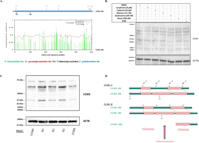
We have previously detected the cellular protein expression of CCM2 isoforms with multiple bands in Western blots (data not shown), which could be addressed with different interpretations. One major reason (or artifact) causing multiple protein bands is due to various post-translational modifications (PTMs) as indicated by our bioinformatics data (Fig. 3A). 293 T (derived from Human Embryonic Kidney 293) cells were treated with most available PTM inhibitors with no observed band shifts or intensity changes eliminating this possibility (Fig. 3B). Further, silencing CCM2 RNAs in 293 T cells significantly decreased intensity of all bands (Fig. 3C), which eliminates another potential source of artifact caused by non-specificity of the antibody. Our current data validate the belief that multiple CCM2 isoform proteins are expressed in cells although expression levels of each isoform are quite different (Fig. 3B,C).
Figure 3.
Cellular distribution and motifs/domains among different endogenous CCM2 isoform proteins. (A) the potential post-translation modification (PTM) sites were defined with prediction software by searching the longest CCM2 isoform, CCM2-206. In the upper panel, CSS (clustering and scoring strategy) was used to scan palmitoylation sites (P), farnesylation sites (F), and geranylgeranylation sites (G) while NetNGlyc 1.0 was used to predict N-glycosylation sites (NG). In the lower panel, YinOYang 1.2 was used to predict O-glycosylation sites: green bars surpassing the red threshold line have significant chance to be glycosylated at the site through O-glycosylation. Each major PTM is color coded. (B) Multiple CCM2 protein bands are not a result of post-translation modifications. Two different vehicle-controls (DMSO, EtOH), inhibitors of farnesylation (Lonafarnib, Tipifarnib, Gliotoxin) and geranylgeranylation (GGTI-298), N-linked glycosylation inhibitor (Swainsonine), and O-linked glycosylation inhibitor (benzyl-α-GalNAc, Benzyl) were used to treat 293 T cells. None of the treatments resulted in missing bands or significantly changed band density, comparable to the controls. (C) Multiple CCM2 protein bands are diminished by silencing CCM2. 293 T cells were treated with either CCM2 RNAi (siRNA-CCM2) or scrambled control (SC). Significantly decreased densities of all protein bands of CCM2 were observed consistently (two shown) in CCM2 knockdown cells, relative to SC controls. (D) Bioinformatics analysis of potential functional domains and putative linear motifs in CCM2 isoforms. (D1). Two longest isoform pairs of CCM2 from A group (100 and 600) and B group (200 and 206) were selected, to screen for intrinsic globularity with GlobPLot 2.3 and putative linear motifs with ELM, (Eukaryotic linear motifs). Structurally globular regions are considered to be composed of different secondary structures and fold types (pink), in contrast to disorder (unstructured) regions (green). All isoforms of CCM2 from A group and B group share two common globular regions: N-terminal globular region which harbors PTB domain and C-terminal region which covers HH domain (Harmonin homology). Intriguingly, a third globular region was identified, by analyzing two longest isoforms of CCM2, which have an additional newly identified 41 amino acid (aa) peptide coded by exon 6A (CCM2-600 and CCM2-206), compared to their respective paired isoforms, CCM2-100 (A) and CCM2-200 (B). The appearance of a new middle globular region might suggest an additional secondary structure and fold created in conjunction with this additional peptide. (D2). With motif prediction tool, ELM, we found five major linear protein motifs along CCM2 isoforms. (D2.1). Motif for protein degradation (red colored). 11, signal motif targeting to endoplasmic reticulum (ER) lumen; 12, signal motif targeting the protein for degradation in a cell cycle dependent manner; 13, signal motif targeting the protein for degradation by binding to the UBR-box of N-recognins; 14, S/T rich motif for SPOP/Cul3-dependant ubiquitination; 15, a degron motif, for the cyclin’s degradation; 16, LIR motif in autophagy; 17, di-Arg ER retention motif, targeting to endoplasmic reticulum (ER) lumen; 18, Sorting motif, targeting to the lysosomal-endosomal-complex. (D2.2). Motif for protein phosphorylation (pink colored). 21, canonical motif for the CDK phosphorylation site; 22, canonical motif for MAP kinases docking or phosphorylation site; 23, CK1 phosphorylation site; 24, GSK3 phosphorylation recognition site. (D2.3). Motif for proteinase cleavage (dark blue colored). 31, canonical motif for proteinase cleavage site. (D2.4). Motif for protein-protein binding (light blue colored). 41, Docking motif in calcineurin; 42, USP7 MATH domain binding motif; 43, USP7 CTD domain binding motif; 44, WW domain interaction motif. (D2.5). Motif for nucleocytoplasmic shuttling (brown colored). NLS, nuclear localization signals; NES, nuclear export signals. Most of the predicted motifs for two pairs are identical, except a few motifs which are located at the beginning of transcript (exon 1 for A group, exon 1A for B group).
Newly identified exons and isoforms suggest a novel domain in CCM2 protein
We have previously reported that despite lacking either a nuclear localization signal (NLS) or a nuclear export signal (NES) in its sequence, the original CCM2-100 isoform (canonical CCM2 protein) predominantly exists in the cytoplasm in HeLa cells at steady state, but shuttles in between the nucleus and cytoplasm7. Since all identified CCM2 isoforms share similar coding sequences with CCM2-100 isoform and contain neither a nuclear localization signal nor a nuclear export signal, most CCM2 isoforms from both groups were predicted to behave similarly as CCM2-100 isoform does within mammalian cells, which contradicts our current data (Fig. 2B, Table 3, Suppl. Fig. 2). We reinvestigated CCM2 protein domains and motifs with multiple bioinformatics tools. By comparing the two longest isoforms in each group (A, B), a PTB domain at N-terminus and an HH domain at C-terminus were found in all paired-isoforms as predicted. However, surprisingly, we not only identified the existence of both NES and NLS in A group (only NES detected in B group), we also detected a novel unknown domain in the middle of CCM2 protein that contains newly defined exon 6A (Fig. 3D), which is in line with our earlier description of an unknown domain (DUF1722) (Suppl. Table 3) predicted from bioinformatics search on both CDD and Pfam databases.
Newly identified isoform B group is dominant despite A group RNAs being more stable
Newly identified isoform B group is the dominant group of endogenously expressed isoforms
To delineate the intracellular relationship between two major groups of CCM2 isoforms, which are expressed ubiquitously in various tissues and cell lines (Fig. 2A, Suppl. Fig. 1), we examined the difference of relative expression profiling between A and B group by using qPCR with two sets of primers (A100/B200 and A101/B201, Suppl. Table 2). On careful evaluation of the data, it was found that the relative mRNA expression level was almost always higher for the newly identified mammalian-specific B group isoforms (Fig. 4A). Further, the expression level differences were even more significant in the more homogeneous tissue samples (brain) and cell lines (middle panel and lower panel, Fig. 4A), further validating this observation.
Ectopic expression levels of isoform A group is higher than that of isoform B group
To investigate the molecular mechanism of the consistently higher expression of CCM2 B group isoforms in mammalian cells, we performed qFCM assay with V5 tag-specific antibodies to precisely measure the expressed protein levels at steady state for 5 ectopic expressed isoform pairs between A/B groups (CCM2-100/CCM2-200, CCM2-101/CCM2-201, CCM2-102/CCM2-210, CCM2-107/CCM2-212, and CCM2-600/CCM2-206). In contrast to our endogenous data, flow cytometry results showed that the protein expression level of CCM2 A group isoforms was always higher than that of CCM2 B group isoforms within each pair and the overall difference was found to be statistically significant (p < 0.01) (Table 4), demonstrating the relative higher expression of A group protein isoforms at translational level. Using tag-specific (V5) and vector control (Neomycin) primer sets (Suppl. Table 2), we further performed allele-specific qPCR to examine the RNA abundance within the same 5 isoform-pairs and found very significant differences within each pair, with the overall difference also being highly statistically significant (p < 0.001) (Fig. 4B). Our qPCR data further supported our qFCM results, indicating the notion that the consistently higher protein level of A group CCM2 isoforms (Table 4) is rooted from their significantly higher expression at the transcriptional level (Fig. 4B).
Table 4.
Differential protein expression levels between A and B groups of CCM2 isoforms.
| Isoforms | Relative Expression Level | |||
|---|---|---|---|---|
| V5 | Neo | GMFI Index | Group A/B ** | |
| CCM2-100 (A) | 44.9 | 1.7 | 26.44 | 2.39 |
| CCM2-200 (B) | 4.97 | 0.45 | 11.04 | |
| CCM2-101 (A) | 22.5 | 1.1 | 20.46 | 2.40 |
| CCM2-201 (B) | 7.09 | 0.83 | 8.54 | |
| CCM2-102 (A) | 40.5 | 1.48 | 27.33 | 2.92 |
| CCM2-210 (B) | 8.33 | 0.89 | 9.36 | |
| CCM2-107 (A) | 64.1 | 2.73 | 23.48 | 2.13 |
| CCM2-212 (B) | 31 | 2.81 | 11.01 | |
| CCM2-600 (A) | 14.7 | 0.74 | 19.86 | 1.56 |
| CCM2-206 (B) | 8.27 | 0.65 | 12.72 | |
The geometric median fluorescence intensity (GMFI) is used to account for the log-normal flow data for each isoform and normalized by Neomycin (Neo) signals as GMFI index. The fold changes of expressed proteins between two groups were presented as ratio (A/B Group). One-way ANOVA was performed among all isoform pairs from A and B groups and a significant difference is defined (P < 0.01, **).
Isoform A group RNAs are more stable than isoform B group RNAs
We next measured the cellular stabilities of a representative set of ectopic expressed paired CCM2-100 (A group) and CCM2-200 (B group) isoforms at the transcriptional level. Our data demonstrated that the mRNA half-life of CCM2-100 was approximately 1.3 times longer than the CCM2-200 (Fig. 4C). This finding further corroborates with our allele-specific qPCR data that the consistently higher relative expressional levels of A group CCM2 isoforms at the transcriptional level is due to the longer half-life of their mRNAs, compared to B group CCM2 isoforms. (Fig. 4C).
CCM2 contains a novel second PTB domain
CCM2 PTB-less isoforms are able to bind to NPXY motifs
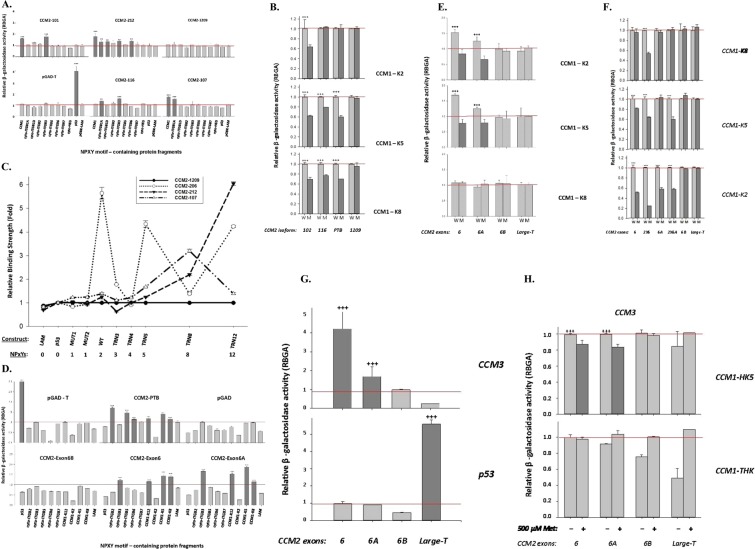
Among all CCM2 isoforms with PTB domain that we defined, three isoforms (CCM2-107, 116, and 212) do not have apparent classic PTB domain, termed PTB-less (missing exons 3, 4, and 5 coding for PTB domain) (Suppl. Table 4). We used these three isoforms to screen many proteins that contain NPXY motif through yeast-two hybrid system (Y2H). Intriguingly, these isoforms were found to bind efficiently to NPXY motifs (Fig. 5A); mutagenesis analysis further confirmed their binding to NPXY motifs in CCM1 protein (Fig. 5B). Further, constructs with increasing number of NPXY motifs (0–12) showed an overall trend of enhancing binding affinity in the PTB-less isoforms, although this trend is not linear, suggesting there is some specificity to the binding possibly associated with structure conformation (Fig. 5C). In sum, these experiments imply the existence of the second functional PTB domain in the middle of CCM2 protein.
Figure 5.
Molecular interactions defined with yeast two-hybrid system. (A) Interactions between various NPXY-motif containing protein fragments and CCM2 PTB-less isoforms (CCM2-116, CCM2-107, and CCM2-212). CCM2-101 serves as positive control, while CCM2-1209 as negative control. pGAD-T with p53 is a system control. (B) Interactions between wild type (W) and mutated (M) three NPXY motifs of CCM1 (K2, K5, and K8) and a CCM2 PTB-less isoform (CCM2-116). CCM2-102 and full-length CCM2 PTB domain serves as positive control, while CCM2-1209 as negative control. (C) Interactions between protein fragments containing various number of NPXY-motifs and CCM2 PTB-less isoforms (CCM2-107, and CCM2-212). CCM2-206 serves as positive control, while CCM2-1209 as negative control. (D) Interactions between various NPXY-motif containing protein fragments and CCM2 exons (6, 6A, and 6B). CCM2-PTB serves as positive control, while pGAD as negative control. pGAD-T with p53 is a system control. (E) Interactions between wild type (W) and mutated (M) three NPXY motifs of CCM1 (K2, K5, and K8) and CCM2 exons (6, 6A, and 6B). Large-T serves as system control. (F) Interactions between wild type (W) and mutated (M) three NPXY motifs of CCM1 (K2, K5, and K8) and CCM2 exons (6, 6A, and 6B) and duplicate forms (2 × 6 and 2 × 6A). Large-T serves as system control. (G) Interactions between CCM3 protein and CCM2 exons (6, 6A, and 6B). Large-T with p53 serves as system control. (H) Competition assays between CCM3 protein with either CCM1-HK5 (containing 1st NPXY motif) (upper panel) or CCM1-THK (containing 2nd and 3rd NPXY motif) (lower panel) binding to CCM2 exons (6, 6A, and 6B). β -galactosidase activity of each transformant was measured, normalized, and converted to relative β -galactosidase activity (RBGA). The normalized data were represented with means and standard deviations (M ± SD) generated from at least three independent assays (n = 3). RBGA+++, ++: significantly higher than that observed in any negative controls (P < 0.001, 0.01 respectively). K2, K5, and K8 are fragments containing the first, second, and third NPXY motif in CCM1 respectively. Cyto-ITGB represents cytoplasmic tails of β integrins (usually containing two NPXY motifs).
The second PTB domain of CCM2 is an atypical PTB domain with novel structure
Since our genomic and bioinformatics data indicated the second PTB domain is highly likely located within the predicted new domain in the middle of CCM2, encompassing exon 6 and 6A (Fig. 3D, Suppl. Table 3), we next tested the binding ability of NPXY motifs to the constructs containing amino acid sequences around exon 6 up till exon 7, which include exon 6, exon 6A, and exon 6B with CCM2 full-length PTB domain as a positive control and CCM2-1209 isoform containing solely HH domain (encompassing C terminal exon 7, 8, 9, and 10) as a negative control through yeast-two hybrid system. Both exons 6 and 6A but not exon 6B were found to bind to NPXY motifs (Fig. 5D), and mutated NPXY motifs in CCM1 protein lost their binding capacity to both exons 6 and 6A (Fig. 5E), while constructs with duplicated (2X) exons 6 and 6A, increase their binding capacity to NPXY motif (Fig. 5F). Interestingly, it has been previously reported that CCM3-CCM2 interaction is mediated through LD (Leucine-Aspartate Repeat) motif-like region within exon 6 of CCM217, which is the consensus sequence-determined binding18,19. After examining the binding of exons 6, 6A, and 6B to full-length CCM3, it was found that both exons 6 and 6A independently interact with CCM3 protein (Fig. 5G). Further, this interaction between CCM3 and exons 6/6A can be inhibited by CCM1 fragment HK5 (containing the 1st NPXY motif) but not CCM1 fragment THK (containing the 2nd and 3rd NPXY motifs) (Fig. 5H)2,7,20, suggesting that the first NPXY motif of CCM1 can compete binding to aPTB domain of CCM2 with CCM3. Together, these results indicate that exons 6 and 6A can independently interact with NPXY motif. The interactions between NPXY motifs were further confirmed in western blots both with PTB-less isoforms (Fig. 6A) and with exons 6 and 6A (Fig. 6B,C), validating this newly defined interaction observed in Fig. 5F.
Figure 6.
Molecular interactions defined by co-immunoprecipitation (CO-IP). (A) Various NPXY-motif containing protein fragments pulling down PTB-less CCM2 isoforms (CCM2-107, CCM2-116, and CCM2-212). CCM2-201 serves as positive control. Interactions between cytoplasmic tails of β-Integrins and CCM2 isoforms were confirmed by IP with GST beads pull-down. (B) Various NPXY-motif containing protein fragments pulled down CCM2 exons (6, 6A, and 6B) with GST beads. Full-length CCM2 PTB domain serves as positive control, while CCM2-1209 as negative control. (C) Various NPXY-motif containing protein fragments pulling down CCM2 exons (6, 6A, and 6B) with HIS beads. NPXY motifs containing protein fragments are cytoplasmic tails of β-Integrins and CCM1 or PTB cores. CCM2-1209 (CCM2 C-terminal fragment, residues 303-444, no PTB domain), CCM1 fragments, K2 (1st NPXY motif), K5 (2nd NPXY motif), and mock control. Mock control is host cell lysate. MagneGST Glutathione Particles (Promega) for GST-tagged bait proteins and dynabeads (Invitrogen) for HIS-tagged bait proteins. All target proteins were labeled with radioactive with S35.
In sum, we found that both protein fragments coded by exon 6 and exon 6A can independently interact with various NPXY motifs confirmed by Y2H (Fig. 5), Co-IP assays (Fig. 6), and characterized with BLI and ITC results (Table 5). Protein fragments coded by exon 6 and exon 6A were named as aPTB-core-N and aPTB-core-C respectively (Fig. 6C).
Table 5.
Kinetic analysis of the interactions between NPXY motif of CCM1 and two cores of novel aPTB domain.
| Method | BLI | ITC | |
|---|---|---|---|
| Affinity | KD (M) | KD (M) | |
| Ligand | CCM1-K8 | CCM1-K8 | CCM1-K2 |
| CCM2-EX6 | 4.06 × 10⁻⁶ | 1.99 × 10⁻⁶ | 6.45 × 10⁻⁶ |
| CCM2-Ex6A | 2.97 × 10⁻⁵ | NA | 6.02 × 10⁻⁵ |
| CCM2-Ex6B | NA | NA | NA |
Affinity data obtained using two different platforms (BLI and ITC) were presented. Binding affinity is presented with the equilibrium dissociation constant (KD). NA: not available due to undetectable binding.
The binding of atypical PTB domain of CCM2 to CCM1 and CCM3 is structurally determined
Our predicted tertiary structure data show that exon 6 (aPTB-core-N) is a novel structure that has not been reported before, which is shaped as a horseshoe composed of two α-helices (Fig. 7A). In contrast, exon 6B fragment does not bind to NPXY motif, due to absence of this structure. Interestingly, the initial predicted tertiary structure of exon 6A fragment seems to be a structurally immature analog of exon 6, with only one crude C-terminal α-helix, a turn and flexible N-terminus. However, in the presence of its partner exon 6 sequence, it matures to become an exon 6-like structure, a horseshoe composed with two α-helices. Both exon 6 and exon 6A linked as two horseshoes form a serine-rich four alpha helix bundle (hairpin), structurally similar to that of the homodimer of exon 6 (CCM2-exon6x2) (Fig. 7B). Interestingly, this novel four-helix bundle (45 aa/horseshoe) is structurally similar to but smaller in size than previously reported serine-rich four-helix bundle focal adhesion adaptor protein, p130-CAS (55–60 aa/ horseshoe)21,22. To determine whether the sequence or the length of amino acid chain at N-terminal exon6A could cause this structure to transition to a “mature” exon6 horseshoe structure, 4 (same length of exon 6) and 9 (5aa longer than exon 6) amino acids from C-terminus of exon6 were added to the N-terminus of exon6A respectively; however, the tertiary structure did not show significant shift to assemble the structure of a “mature” exon6, which suggests there are some issues hindering the correct prediction of exon6A as horseshoe composed with two α-helices, which we believe is the case, from bioinformatics (Fig. 7C).
Figure 7.
Structures of the novel atypical PTB domain. (A) Ribbon representations of exon 6, exon 6A and exon 6B. Both exon 6 and exon 6A share a structural similarity with a C-terminal α helix followed by a turn, but not exon 6B. (B) Predicted ribbon presentation of CCM2 exon homo-and hetero-dimers. Homodimer of exon 6 (2X exon6), homodimer of exon 6A (2X exon6A) and heterodimer of exon 6 and 6A (canonical aPTB domain, exon6 + 6A) were simulated. Surprisingly, homodimer of exon 6 (2X exon6) was found to assemble to a naturally occurring heterodimer between exon 6 and 6A (exon6 + 6A), a dual horseshoe or simple duplication of exon 6, raising the doubt of the accuracy of predicted structure of exon 6A. (C) To determine accuracy of predicted structure of exon 6/6A, 4 or 9 amino acids from exon 6 were added to C-terminus of exon6A (N4 + exon6A and N9 + exon6A respectively). N4 + exon6A is equal to exon 6 in size (45 aa), while N9 + exon6A is larger than exon 6 in size (50 aa) and contains > 20% of amino acid sequence from exon 6, suggesting that neither size nor sequencing from exon 6 influences conformation of exon 6A. Red color: c-terminus and blue color: N-terminus.
Sequence comparison showed that there is a possible LD motif-like region present in exon 6, but no such homolog can be found in exon 6A (Suppl Table 4). Therefore, the interaction between exon 6A and CCM3 can’t be mediated through LD-motif like sequence (Fig. 5G,H and Suppl Table 4). The possibility that exon 6 and exon 6A interact with CCM3 through different mechanisms seems minimum, therefore, due to the similarity in tertiary structure between exon 6 and exon 6A, and the competitive nature of both cores with the 1st NPXY motif of CCM1, we conclude that the binding between CCM2 and CCM3 is structurally determinant rather than sequence determinant (Figs 5, 7).
By analyzing these data, we conclude that the newly defined atypical PTB domain (aPTB) is composed of exon 6 and exon 6A, each approximately 45 amino acids long, and can independently bind to either CCM1 or CCM3 proteins. In fact, this binding potential can be augmented when both form homo- or hetero- dimers folding into structurally identical four-alpha helix bundle structure (Figs 5F, 7B). The homodimer of exon 6 has the better tendency than exon 6 alone to bind to the NPXY motif of CCM1, suggesting that the binding between CCM2 aPTB domain and the 1st NPXY motif of CCM1 is mediated through tertiary structure, similar to the binding between CCM2 PTB and the 2nd and 3rd NXPY motif of CCM1-canonical PTB-NPXY binding. Since, the full aPTB domain present in CCM2 isoforms is a four alpha helix bundle composed of exon 6 and exon 6A, it can be reasonably assumed that the minimum requirement for binding between aPTB of CCM2 and NPXY motif is the existence of C-terminal alpha helix and turn (highly possible including the 2 alpha helix as the horseshoe for its basic functional unit) of both cores of aPTB domain of CCM2.
Discussion
This current report may help define more CCM mutations
Upon comparing linkage results23 and mutational screening data24 of known CCM genetic loci (CCM 1–3), there is a noticeable discrepancy in the relative incidence of CCM mutations among CCM patients, raising the possibility of the existence of an additional CCM4 locus25. However, endeavors to find the new locus have thus far failed. Based on our previous RNA and protein data on CCM2, we hypothesized that the additional exons might exist within CCM2 genomic structure, contributing to this discrepancy. In this report, we have defined a total of 29 exons, including 8 new exons and 11 alternatively spliced exons derived from existing exons and 5 novel promotors in CCM2 gene. Using our designed primers (Suppl. Table 1), we screened a small group of previously defined CCM2 patients. Ironically, we still could not identify novel CCM mutations in these newly identified exons, which could be due to our small cohort of CCM patients. As per our past experience, the lack of resources could hinder our efforts to discover novel CCM mutations. However, these newly identified 22 novel functional CCM2 isoforms have proved the biological importance and relevance of transcriptional alternative splicing of CCM2 gene and are further validated by our expression data at both transcriptional (Fig. 2A, Suppl. Fig. 1A,B) and translational (Fig. 3A,B) levels. Although almost all reported mutations in CCMs are nonsense mutations6,26–29, there are reports for abnormal splicing mutations30,31, raising the possibility of more mutations existing in the newly identified alternative splicing sites. Therefore, the information and evidence generated in this study will certainly facilitate the rapid identification of the remaining undefined mutations at CCM2 locus in the near future.
Alternative splicing of CCM2 may be a key modulator of CSC during angiogenesis
Alternative splicing of a transcript can result in multiple protein isoforms with divergent functions from original “full-length” protein and more than 95% of human genes have been found to utilize this mechanism to increase the complexity of gene expression by producing protein isoforms with different, even opposing functions32. Alternative splicing has emerged as one of the key modulators of angiogenesis33 and current evidence suggests that different isoforms arise from alternative splicing of certain angiogenic molecules such as vascular endothelial growth factor (VEGF)34, VEGF receptor1, flt135, and integrin receptor: fibronectin (FN)36. Alternatively spliced isoforms of VEGF can be either proangiogenic or antiangiogenic34,37–39, emphasizing the biological significance of this RNA processing mechanism. It has been demonstrated that CCM2 works with other CCM proteins to regulate β1-integrin-mediated cellular events through the CSC complex7,8,12. Although we found that both CCM1 and CCM3 are less polymorphic regarding alternative splicing events in our hands, an alternative spliced CCM1 isoform lacking the 15th coding exon, Krit1B, has been reported to be expressed at relatively high levels in mouse tissues and cell lines, but much less expression detected in humans40; interestingly, an “in-frame deletion” found in a large CCM cohort mutation screening, which results in the lack of entire exon 18 of CCM1 gene, was found to have dramatically decreased expression level as well41. For isoform Krit1B, the splicing out of the 15th exon coding region occurs within F3 lobe of the FERM domain, raising the possibility of disrupted intramolecular interaction and folding42. This in-frame deletion region (residues 675–714) is actually next to previously identified nuclear export sequence (residues 551–562)7,8 and a nuclear localization signal (residues 569–572)43 with potential impact on both functions. Therefore, it is understandable that the rare CCM1 isoform, Krit1B, behaves differently from the dominant cellular CCM1 isoform, Krit1A, in the cellular compartmentation. Likewise, the differential nuclear localization of some CCM2 isoforms in endothelial cells in this experiment can be explained.
Ccm2-like (Ccm2L), a recently described novel gene with a high homology to Ccm2 in zebrafish, was identified44. Like Ccm2, Ccm2L has multiple alternatively spliced isoforms and works within the CSC complex (Ccm1 and Ccm3) to interact with the Mekk3/Mek5 complex to regulate Mekk3 activity45. However, no studies have been performed on CCM2 isoforms till date. More than half of human genes have alternative promoters with multiple start-codon exons46,47, but only a few of them display sequence similarity within their first coding exons, giving a variety of closely related N-terminal protein variants46,48, and CCM2 certainly belongs to this small group.
Differential expression between two major isoform groups regulated at transcriptional level
This project is highly successful in deciphering the two major types of CCM2 isoforms, which differentiated themselves by alternative promoters and alternative start codon exons, categorized into A and B group. It is very interesting to observe that despite the subtle differences at their 5′ end sequences (30 bp for exon1, 93 bp for exon1A) the transcripts of isoform pairs from two groups behave quite differently in their cellular abundance at steady state. The in-vivo qPCR data demonstrated that the transcripts of mammalian-specific B group isoforms are more abundant than that of vertebrate-specific A group isoforms in almost all mammalian cells and tissues screened, hinting the preferred higher endogenous expression levels for B group isoforms at the transcriptional level in mammalian cells, at steady state. However, protein expression data were totally incongruent to the endogenous expression data generated from tissues and cell screening at the transcriptional level. There are a few possible molecular mechanisms resulting in this discrepancy such as the stabilities of isoforms at either post-translational and/or post-transcriptional levels. The stability of mRNA transcripts was investigated, and surprisingly demonstrated that the vertebrate-specific A group isoforms are more abundant (Fig. 4B). We further measured the RNA decay rate of one set of isoform pair representing both groups and found that the mRNA half-life of vertebrate-specific A group isoform is indeed longer than the mammalian-specific B group isoform (Fig. 4C). Our results are in line with a recent finding that differential usage of alternative first exons increases the capacity for transcriptional control of alternative spliced isoforms48, however our data also raises a very interesting suggestion that B group isoforms must have a stronger promoter than A group isoforms to ensure their dominant expression in most tissues and cells.
There have been many reports indicating that both alternative promoter usage47,49–51 and 5′ untranslated region (5′ UTR)50,51 regulate the alternative splicing event both quantitatively (amount of isoform) and qualitatively (the species of isoform), making things even more complicated. Eukaryotic transcription is tightly controlled by various cis-DNA regulatory elements, such as enhancers, boundary elements, insulators, and silencers. Considering that the exon1A is more than 27 kb downstream of the exon1, it is unlikely for any overlapped promoter regions between two isoform groups to exist. Our findings that mammalian-specific B group isoforms are less stable but more abundant than the original vertebrate-specific A group isoforms at steady state are quite intriguing and the stronger transcriptional machinery, regulated by a specific set of regulatory elements (P1-P5), for the B group isoforms is the most reasonable explanation for the contradictory phenomenon. Since cell-type specific transcription is regulated by specific combinations of transcription factors on the specific DNA region for the connectivity of gene transcription regulatory networks at the quantitative level, we had acquired BAC clones which contain several kb upstream of either exon1 (A group) or exon1A (B group) for future discovery and characterization of both cis- (promoter, 5′UTR, its proximal region, its enhancers, boundary, or insulator elements) and trans- (transcription factors) elements associated with their transcriptional regulation.
Differential subcellular compartmentation of the isoforms indicates their distinct cellular roles
Scaffold proteins are critical in regulating the specificity of signaling responses through mediating the assembly of distinct multimolecular signaling complexes, or signalosomes that mediate appropriate responses. CCM2 has been defined as a scaffold protein. CCM2 isoforms which contain a PTB domain are more likely to interact with CCM1 and may use it as a cellular shuttle as well between the nucleus and cytoplasm. However, new nuclear localization and nuclear export signals have been identified in CCM2 in this study, raising the doubt for the previous hypothesis. Interestingly, even PTB-less isoforms (CCM2-107, CCM2-116, and CCM2-212) frequently distribute evenly in both the nucleus and cytoplasm at steady state in several cell lines (mainly in endothelial cells), further challenging the previous hypothesis. There have been reports of differentiated cellular compartmentations among different alternatively spliced isoforms of other PTB-domain containing proteins52,53, suggesting that there may be a common mechanism for the intracellular distribution of PTB-domain containing proteins modulated through alternative splicing. These findings underscore the importance of the pleiotropic nature of CCM2 proteins through its molecular strategy of alternatively spliced isoforms, with each isoform being possibly functionally distinct from its siblings.
CCM2 plays essential role as a cornerstone of CSC
In this study, the most important finding is the discovery of previously unknown atypical PTB domain (aPTB) in CCM2, which consists of two cores that can independently bind either CCM3 or CCM1. For our data, the aPTB domain seems to be able to bind to all three NPXY motifs but prefers the 1st motif, probably due to conformation and accessibility issues in the full-length CCM1. Intriguingly, CCM3 and CCM1 can compete for their binding to aPTB domain of CCM2, making us propose a model to demonstrate their relationship within the CSC complex (Fig. 8). In this model, CCM1 protein can use its 2nd and 3rd NPXY motif in the middle portion of the molecule to bind to the longer CCM2 isoforms through its N-terminal PTB domain and use its 1st NPXY motif at N-terminus to bind to aPTB domain of CCM2, which also binds to CCM3. We have proved the competitiveness of the 1st NPXY motif of CCM1 and CCM3 binding to aPTB domain of CCM2, but in an overexpressed system. Since there are two independent cores in aPTB, the interaction between CCM1 and CCM3 with aPTB, whether cooperative or even independent in the physiological condition, needs to be further explored.
Figure 8.
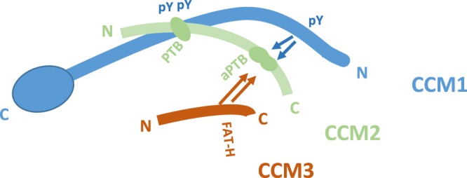
Schematic representation of binding interaction among CCM proteins within CSC complex. Our current data suggests that CCM1 utilizes 2nd and 3rd NPXY motifs (pY) in its center portion to bind to CCM2 classic PTB domain (PTB). The remaining 1st NPXY motif competes with the CCM3 (FAT-H) to bind to the newly defined atypical PTB domain (aPTB) present at the C – terminus of the CCM2, suggesting CCM2 plays a central role in the CSC. CCM1: blue color; CCM2: light green color; CCM3: red color.
In summary, our data suggest that CCM2 can play pleiotropic cellular roles by enhancing the variation of its coding sequences and domains through alternative splicing processes in various cells and tissues. We illustrated that alternatively spliced CCM2 isoforms have isoform-specific cellular expression and subcellular localization patterns among different cells and tissues. Our data validate our previous belief that CSC can mediate signaling activities of multiple pathways through its multiple molecular forms. In conclusion, we proposed that the differential cellular expression and subcellular localization patterns are partially due to the difference of transcriptional regulation among different isoforms. Various isoforms also diversify its cellular localization and dynamics, creating multiple layers of representation of this single protein. Combination of these cellular events will certainly increase the complexity of CSC mediated events in angiogenesis. This project provides new insights into CSC-mediated signaling pathways and angiogenesis, which may revolutionize the current concepts of vascular malformations and molecular mechanism of angiogenesis, leading to new therapeutic strategies.
Materials and Methods
Isolation of novel exons, open reading frame, isoforms, and conserved domains in CCM2 gene
Multi-tissue reference RNAs pool from four different suppliers (Super Array, BiotaQ, Biochain, NTomics) were used to amplify CCM2 cDNA fragments with SuperScript® III reverse transcriptase (Invitrogen). Rapid amplification of cDNA end (RACE) was applied for using either human brain Marathon-Ready cDNA or SMART RACE kit reagents (BD Clontech) to define full-length cDNA. A second round of amplification was performed to enhance the specificity and yield using nested primers (Suppl. Table 1).
The RACE and RT-PCR resulting products were sequenced and further analyzed with Vector NTI (Invitrogen) and CLC genomic workbench (Qiagen) to define the potential novel fragments and open reading frames (ORFs) as described before43. After the start-codon exons of isoforms were defined, all full–length CCM2 isoforms were amplified from the cDNA pool with the proper primers and Platinum Pfx50 DNA Polymerase (Invitrogen) (Suppl. Table 1). Potential conserved domains (CD) on the protein were searched on both CDD and Pfam databases in NCBI CD search site (http://www.ncbi.nlm.nih.gov/Structure/cdd/wrpsb.cgi).
Real time quantitative PCR analysis (qPCR)
Allele-specific real-time quantitative PCR (qPCR) assays were designed using boundary-spanning primer sets (Suppl. Table 2) and applied to quantify the RNA levels of the endogenously expressed CCM2 isoforms using Power SYBR Green Master Mix with ViiA 7 Real-Time PCR System (Applied Biosystems). TissueScan™ Real-Time PCR panels (HMRT100, 103, CSRT502) with Human ß-actin control primer set (Origene, MD) were used to determine the endogenous expression levels of various CCM2 isoforms in different tissues. qPCR plates with human brain tissues and cell lines were prepared using an epMotion 5075 automated liquid handling systems (Eppendorf) and were performed in triplicates (n = 3). V5-tag and Neomycin allele-specific qPCR primers (Suppl. Table 2) were used to perform allele-specific qPCR to quantify the ectopic expression levels of each isoform-pair. The qPCR data were analyzed as described before20.
Plasmid construction, subcellular localization, western blots, and cellular dynamics
CCM2 isoforms were inserted into corresponding gene-expression vectors as described previously7,16,20,43. The efficient expression for each CCM2 isoform in both transcriptional and translational levels was first confirmed through both qPCR and Western blots and subsequently enabling further experiments, such as transient gene expression associated with immune-fluorescence (IF) analysis, quantitative flow cytometry (qFCM), and RNA decays assays. Anti-CCM2 (Sigma), β-Actin, and α-Actinin (Santa Cruz) antibodies with anti-GST (Promega) and HIS-tag (Invitrogen) beads were used in Western Blots.
Multiple primary endothelial (HUVEC, HMVEC, hCMEC/D3) cell lines were cultured in six well plates and transfected with different CCM2 isoform-V5 constructs to determine their subcellular localization and expression level as described2,7,8,14,54,55. Transfected cells were then fixed, and subcellular localization of each isoform was determined with IF analysis using NIKON TiE imaging system. Cyclin B, known to accumulate in the nucleus after leptomycin treatment, served as a positive control during IF screening.
For qFCM assay, several isoform-pairwise constructs (100/200, 101/201, 102/210, 107/212, and 600/206) with nearly identical coding sequence differing only in the start-codon exon, exon 1 (isoform A group) or exon1A (isoform B group) at 5′ terminus, were used to study the protein expression pattern of these two major groups of CCM2 isoforms. The ectopically expressed proteins from each isoform pairwise construct were measured separately in the same batch of 293 T cells with triplicate repeats, with standard transfection regime. Twenty-four hours after transfection, cells were detached with 0.05% Trypsin-EDTA solution (Sigma-Aldrich) and counted in a hemocytometer (American Optical). The cells were fixed in 4% PFA for 20 min at room temperature and then permeabilized by 90% cold methanol on ice for 30 min. The cell concentration was adjusted to 1 × 10⁶cells/mL by blocking buffer and was incubated at room temperature for 1 hour. The cells were further divided in two parts: one was stained with 2 μg/mL sample of anti-V5-FITC mouse monoclonal IgG2a (Invitrogen) and the other one with anti-neomycin phosphotransferase II Rabbit polyclonal IgG (Millipore) and Alexa Fluor 647 Goat Anti-Rabbit IgG (H + L) (Invitrogen). Normal Rabbit IgG (Millipore) followed by Alexa Fluor 647 Goat Anti-Rabbit IgG (H + L) was used as the background control. The expression level of neomycin phosphotransferase II was used as an internal control for transfection efficiency. The cells were washed 3 times with ice cold PBS at 500 g/5 min after each step in cell staining. The cells were then resuspended in 0.5 mL PBS and measured by GalliosTM flow cytometer (Beckman Coulter). The flow cytometry data were analyzed by Kaluza® Analysis Software (Beckman Coulter).
For RNA decay assay, 293 T cells (1 × 10⁶cells/mL) were cultured and transiently transfected with isoform-pairwise CCM2-100/200 constructs as previously described. After 24 hours, 5 μM Actinomycin D (Life Technologies) was added and cell concentration was adjusted to 2 × 10⁶cells/mL by cell counting with a hemocytometer. Treated cells were collected at five different time points (0 h, 2 h, 6 h, 10 h, 24 h) for RNA extraction and qPCR analysis.
Yeast two-hybrid analysis, protein preparation, and co-immunoprecipitation
Yeast two-hybrid analysis was performed as previously described2,7,20. In initial Co-IP experiments, potential target proteins were expressed with TNT® Quick Coupled Transcription/Translation and E. coli S30 Extract Systems (Promega) with S35-labeled methionine (PerkinElmer). The expressions were confirmed by Western blots. In vitro pull-down co-immunoprecipitation was performed as previously described2,7,20.
Measurement of molecular interactions and modeling of tertiary structures
Recombinantly expressed proteins were purified with chromatography: GST-tagged fragments from PTB domains were purified with GSTrap HP column (GE), while HIS-tagged NPXY motifs were purified using HiTrap TALON column (GE), followed by size-exclusion column, S100-HR (GE). The binding interaction between fragments from PTB domains and NPXY motifs were quantified by Bio-Layer Interferometry (BLI) and Isothermal Titration Calorimetry (ITC). Subsequent data acquisition and analysis were performed as described before20.
The molecular modeling of the tertiary structure of atypical PTB (aPTB) domain was established by Iterative Threading ASSEmbly Refinement (I-TASSER), with RasMol (version 2.7.5) for the structure visualization, as described before20.
Statistical analysis
One-way analysis of variance (ANOVA) was used to detect the differences in the mean values among the treatment groups. All pairwise multiple comparison procedures were analyzed using Tukey t-test to test the difference between each treatment. Plots and charts were constructed and produced by SigmaPlot 12.0 (Systat Software, Inc.).
Ethical approval
This article does not contain any studies with human participants performed by any of the authors, nor studies with animals performed by any of the authors.
Supplementary information
Acknowledgements
We wish to thank Junli Zhang, Amna Siddiqui, Saafan Malik, Adam Banda, Lillian Dominguez, Joshua Kallman, Carly Levin, Jasmine Cazares and Edna Lopez, Pallavi Dubey, Aileen Zhang, Zhuoran Yao, and Elias Gonzalez at Texas Tech University Health Science Center El Paso (TTUHSC) for their technical help during the experiments; Drs. Couraud at INSERM in France and Ulrich Bickel and Xiaoming Gong at TTUHSC for their technical help on hCMEC/D3 and several cancer cell lines. This study was funded by Coldwell Foundation.
Author contributions
J.Z. designed and performed experiments, analyzed data, prepared the manuscript and Figures. J.Z., A.P., J.A.F. and A.B. contributed in writing the main manuscript text. J.Z., X.J., A.P., Y.Q. and S.S. contributed in cloning and genomic studies, J.Z., Y.Q. and S.S. contributed in cellular localization, immune-fluorescence (IF) analysis, quantitative flow cytometry (qFCM), and RNA decays assays, and J.Z., X.J., A.P., J.A.F. and A.B. contributed in yeast-two hybrid and co-immunoprecipitation. All authors reviewed the manuscript.
Data availability
The datasets generated and/or analyzed during the current study are available from the corresponding author on reasonable request. Some data generated or analyzed during this study have been included in this published article (and its Supplementary Information files).
Competing interests
The authors declare no competing interests.
Footnotes
Publisher’s note Springer Nature remains neutral with regard to jurisdictional claims in published maps and institutional affiliations.
Supplementary information
is available for this paper at 10.1038/s41598-019-52386-0.
References
- 1.Del Curling O, Jr., Kelly DL, Jr., Elster AD, Craven TE. An analysis of the natural history of cavernous angiomas. J Neurosurg. 1991;75:702–708. doi: 10.3171/jns.1991.75.5.0702. [DOI] [PubMed] [Google Scholar]
- 2.Zhang J, Clatterbuck RE, Rigamonti D, Chang DD, Dietz HC. Interaction between krit1 and icap1alpha infers perturbation of integrin beta1-mediated angiogenesis in the pathogenesis of cerebral cavernous malformation. Hum Mol Genet. 2001;10:2953–2960. doi: 10.1093/hmg/10.25.2953. [DOI] [PubMed] [Google Scholar]
- 3.Zawistowski JS, Serebriiskii IG, Lee MF, Golemis EA, Marchuk DA. KRIT1 association with the integrin-binding protein ICAP-1: a new direction in the elucidation of cerebral cavernous malformations (CCM1) pathogenesis. Hum. Mol. Genet. 2002;11:389–396. doi: 10.1093/hmg/11.4.389. [DOI] [PubMed] [Google Scholar]
- 4.Uhlik MT, et al. Rac-MEKK3-MKK3 scaffolding for p38 MAPK activation during hyperosmotic shock. Nat Cell Biol. 2003;5:1104–1110. doi: 10.1038/ncb1071. [DOI] [PubMed] [Google Scholar]
- 5.Zhang J, Carr C, Badr A. The cardiovascular triad of dysfunctional angiogenesis. Translational stroke research. 2011;2:339–345. doi: 10.1007/s12975-011-0065-5. [DOI] [PMC free article] [PubMed] [Google Scholar]
- 6.Padarti A, Zhang J. Recent advances in cerebral cavernous malformation research. Vessel Plus. 2018;2:1–23. doi: 10.20517/2574-1209.2018.34. [DOI] [PMC free article] [PubMed] [Google Scholar]
- 7.Zhang, J., Rigamonti, D., Dietz, H. C. & Clatterbuck, R. E. Interaction between krit1 and malcavernin: implications for the pathogenesis of cerebral cavernous malformations. Neurosurgery, 60, 353–359, discussion 359 (2007). [DOI] [PubMed]
- 8.Zhang, J., Basu, S., Rigamonti, D., Dietz, H. C. & Clatterbuck, R. E. krit1 modulates beta1-integrin-mediated endothelial cell proliferation. Neurosurgery, 63, 571–578, discussion 578 (2008). [DOI] [PubMed]
- 9.Mably JD, et al. santa and valentine pattern concentric growth of cardiac myocardium in the zebrafish. Development. 2006;133:3139–3146. doi: 10.1242/dev.02469. [DOI] [PubMed] [Google Scholar]
- 10.Hogan BM, Bussmann J, Wolburg H, Schulte-Merker S. ccm1 cell autonomously regulates endothelial cellular morphogenesis and vascular tubulogenesis in zebrafish. Hum. Mol. Genet. 2008;17:2424–2432. doi: 10.1093/hmg/ddn142. [DOI] [PubMed] [Google Scholar]
- 11.Jin S-W, et al. A transgene-assisted genetic screen identifies essential regulators of vascular development in vertebrate embryos. Developmental Biology. 2007;307:29–42. doi: 10.1016/j.ydbio.2007.03.526. [DOI] [PMC free article] [PubMed] [Google Scholar]
- 12.Liu H, Rigamonti D, Badr A, Zhang J. Ccm1 assures microvascular integrity during angiogenesis. Transl. Stroke Res. 2010;1:146–153. doi: 10.1007/s12975-010-0010-z. [DOI] [PMC free article] [PubMed] [Google Scholar]
- 13.Liu H, Rigamonti D, Badr A, Zhang J. Ccm1 regulates microvascular morphogenesis during angiogenesis. JOURNAL OF VASCULAR RESEARCH. 2011;48:130–140. doi: 10.1159/000316851. [DOI] [PMC free article] [PubMed] [Google Scholar]
- 14.Zhang, J., Basu, S., Clatterbuck, R. E., Rigamonti, D. & Dietz, H. C. Pathogenesis of cerebral cavernous malformation: Depletion of Krit1 leads to perturbation of 1 integrin-mediated endothelial cell mobility and survival. Am J Hum Genet suppl, S222 (2004).
- 15.Fisher OS, Boggon TJ. Signaling pathways and the cerebral cavernous malformations proteins: lessons from structural biology. Cell Mol Life Sci. 2014;71:1881–1892. doi: 10.1007/s00018-013-1532-9. [DOI] [PMC free article] [PubMed] [Google Scholar]
- 16.Zhang J, Clatterbuck RE, Rigamonti D, Chang DD, Dietz HC. Novel insights regarding the pathogenesis of cerebral cavernous malformation (CCM) Am J Hum Genet. 2001;69:178–178. doi: 10.1093/hmg/10.25.2953. [DOI] [PubMed] [Google Scholar]
- 17.Draheim KM, et al. CCM2-CCM3 interaction stabilizes their protein expression and permits endothelial network formation. J Cell Biol. 2015;208:987–1001. doi: 10.1083/jcb.201407129. [DOI] [PMC free article] [PubMed] [Google Scholar]
- 18.Tumbarello DA, Brown MC, Turner CE. The paxillin LD motifs. FEBS Lett. 2002;513:114–118. doi: 10.1016/S0014-5793(01)03244-6. [DOI] [PubMed] [Google Scholar]
- 19.Vanarotti MS, Miller DJ, Guibao CD, Nourse A, Zheng JJ. Structural and mechanistic insights into the interaction between Pyk2 and paxillin LD motifs. J Mol Biol. 2014;426:3985–4001. doi: 10.1016/j.jmb.2014.08.014. [DOI] [PMC free article] [PubMed] [Google Scholar]
- 20.Zhang J, et al. Novel functions of CCM1 delimit the relationship of PTB/PH domains. Biochim Biophys Acta. 2017;1865:1274–1286. doi: 10.1016/j.bbapap.2017.07.002. [DOI] [PMC free article] [PubMed] [Google Scholar]
- 21.Briknarova K, et al. The serine-rich domain from Crk-associated substrate (p130cas) is a four-helix bundle. J Biol Chem. 2005;280:21908–21914. doi: 10.1074/jbc.M501258200. [DOI] [PubMed] [Google Scholar]
- 22.Barrett A, Pellet-Many C, Zachary IC, Evans IM, Frankel P. p130Cas: a key signalling node in health and disease. Cell Signal. 2013;25:766–777. doi: 10.1016/j.cellsig.2012.12.019. [DOI] [PubMed] [Google Scholar]
- 23.Craig HD, et al. Multilocus linkage identifies two new loci for a mendelian form of stroke, cerebral cavernous malformation, at 7p15-13 and 3q25.2-27. Hum Mol Genet. 1998;7:1851–1858. doi: 10.1093/hmg/7.12.1851. [DOI] [PubMed] [Google Scholar]
- 24.Liquori CL, et al. Different spectra of genomic deletions within the CCM genes between Italian and American CCM patient cohorts. Neurogenetics. 2008;9:25–31. doi: 10.1007/s10048-007-0109-x. [DOI] [PubMed] [Google Scholar]
- 25.Liquori CL, et al. Low frequency of PDCD10 mutations in a panel of CCM3 probands: potential for a fourth CCM locus. Hum Mutat. 2006;27:118. doi: 10.1002/humu.9389. [DOI] [PubMed] [Google Scholar]
- 26.Sahoo T, et al. Mutations in the gene encoding KRIT1, a Krev-1/rap1a binding protein, cause cerebral cavernous malformations (CCM1) Hum Mol Genet. 1999;8:2325–2333. doi: 10.1093/hmg/8.12.2325. [DOI] [PubMed] [Google Scholar]
- 27.Zhang, J., Clatterbuck, R. E., Rigamonti, D. & Dietz, H. C. Mutations in KRIT1 in familial cerebral cavernous malformations. Neurosurgery, 46, 1272–1277; discussion 1277-1279 (2000). [DOI] [PubMed]
- 28.Eerola I, et al. KRIT1 is mutated in hyperkeratotic cutaneous capillary-venous malformation associated with cerebral capillary malformation. Hum Mol Genet. 2000;9:1351–1355. doi: 10.1093/hmg/9.9.1351. [DOI] [PubMed] [Google Scholar]
- 29.Huang WQ, et al. A Novel CCM2 Gene Mutation Associated with Familial Cerebral Cavernous Malformation. Front Aging Neurosci. 2016;8:220. doi: 10.3389/fnagi.2016.00220. [DOI] [PMC free article] [PubMed] [Google Scholar]
- 30.Verlaan DJ, Siegel AM, Rouleau GA. Krit1 missense mutations lead to splicing errors in cerebral cavernous malformation. Am J Hum Genet. 2002;70:1564–1567. doi: 10.1086/340604. [DOI] [PMC free article] [PubMed] [Google Scholar]
- 31.Yang, C. et al. Identification of a Novel Deletion Mutation (c.1780delG) and a Novel Splice-Site Mutation (c.1412-1G>A) in the CCM1/KRIT1 Gene Associated with Familial Cerebral Cavernous Malformation in the Chinese Population. J Mol Neurosci (2016). [DOI] [PubMed]
- 32.Nilsen TW, Graveley BR. Expansion of the eukaryotic proteome by alternative splicing. Nature. 2010;463:457–463. doi: 10.1038/nature08909. [DOI] [PMC free article] [PubMed] [Google Scholar]
- 33.Munaut C, Colige AC, Lambert CA. Alternative splicing: a promising target for pharmaceutical inhibition of pathological angiogenesis? Curr Pharm Des. 2010;16:3864–3876. doi: 10.2174/138161210794455012. [DOI] [PubMed] [Google Scholar]
- 34.Ladomery MR, Harper SJ, Bates DO. Alternative splicing in angiogenesis: the vascular endothelial growth factor paradigm. Cancer Lett. 2007;249:133–142. doi: 10.1016/j.canlet.2006.08.015. [DOI] [PubMed] [Google Scholar]
- 35.Li H, et al. Possible participation of pICln in the regulation of angiogenesis through alternative splicing of vascular endothelial growth factor receptor mRNAs. Endothelium. 2004;11:293–300. doi: 10.1080/10623320490904250. [DOI] [PubMed] [Google Scholar]
- 36.Ventura E, et al. Alternative splicing of the angiogenesis associated extra-domain B of fibronectin regulates the accessibility of the B-C loop of the type III repeat 8. PLoS One. 2010;5:e9145. doi: 10.1371/journal.pone.0009145. [DOI] [PMC free article] [PubMed] [Google Scholar]
- 37.Giacca M. Non-redundant functions of the protein isoforms arising from alternative splicing of the VEGF-A pre-mRNA. Transcription. 2010;1:149–153. doi: 10.4161/trns.1.3.13229. [DOI] [PMC free article] [PubMed] [Google Scholar]
- 38.Harper SJ, Bates DO. VEGF-A splicing: the key to anti-angiogenic therapeutics? Nat Rev Cancer. 2008;8:880–887. doi: 10.1038/nrc2505. [DOI] [PMC free article] [PubMed] [Google Scholar]
- 39.Nowak DG, et al. Expression of pro- and anti-angiogenic isoforms of VEGF is differentially regulated by splicing and growth factors. J Cell Sci. 2008;121:3487–3495. doi: 10.1242/jcs.016410. [DOI] [PMC free article] [PubMed] [Google Scholar]
- 40.Retta SF, et al. Identification of Krit1B: a novel alternative splicing isoform of cerebral cavernous malformation gene-1. Gene. 2004;325:63–78. doi: 10.1016/j.gene.2003.09.046. [DOI] [PubMed] [Google Scholar]
- 41.Spiegler S, et al. High mutation detection rates in cerebral cavernous malformation upon stringent inclusion criteria: one-third of probands are minors. Mol Genet Genomic Med. 2014;2:176–185. doi: 10.1002/mgg3.60. [DOI] [PMC free article] [PubMed] [Google Scholar]
- 42.Francalanci F, et al. Structural and functional differences between KRIT1A and KRIT1B isoforms: a framework for understanding CCM pathogenesis. Exp Cell Res. 2009;315:285–303. doi: 10.1016/j.yexcr.2008.10.006. [DOI] [PubMed] [Google Scholar]
- 43.Zhang J, Clatterbuck RE, Rigamonti D, Dietz HC. Cloning of the murine Krit1 cDNA reveals novel mammalian 5′ coding exons. Genomics. 2000;70:392–395. doi: 10.1006/geno.2000.6410. [DOI] [PubMed] [Google Scholar]
- 44.Rosen JN, Sogah VM, Ye LY, Mably J. D. ccm2-like is required for cardiovascular development as a novel component of the Heg-CCM pathway. Dev Biol. 2013;376:74–85. doi: 10.1016/j.ydbio.2013.01.006. [DOI] [PMC free article] [PubMed] [Google Scholar]
- 45.Cullere X, Plovie E, Bennett PM, MacRae CA, Mayadas TN. The cerebral cavernous malformation proteins CCM2L and CCM2 prevent the activation of the MAP kinase MEKK3. Proc Natl Acad Sci USA. 2015;112:14284–14289. doi: 10.1073/pnas.1510495112. [DOI] [PMC free article] [PubMed] [Google Scholar]
- 46.Zhang T, Haws P, Wu Q. Multiple variable first exons: a mechanism for cell- and tissue-specific gene regulation. Genome Res. 2004;14:79–89. doi: 10.1101/gr.1225204. [DOI] [PMC free article] [PubMed] [Google Scholar]
- 47.Xin D, Hu L, Kong X. Alternative promoters influence alternative splicing at the genomic level. PLoS One. 2008;3:e2377. doi: 10.1371/journal.pone.0002377. [DOI] [PMC free article] [PubMed] [Google Scholar]
- 48.Turner JD, Schote AB, Macedo JA, Pelascini LP, Muller CP. Tissue specific glucocorticoid receptor expression, a role for alternative first exon usage? Biochem Pharmacol. 2006;72:1529–1537. doi: 10.1016/j.bcp.2006.07.005. [DOI] [PubMed] [Google Scholar]
- 49.Slobodin B, Agami R. Transcription initiation determines its end. Mol Cell. 2015;57:205–206. doi: 10.1016/j.molcel.2015.01.006. [DOI] [PubMed] [Google Scholar]
- 50.Hattori Y, Ishii H, Morita A, Sakuma Y, Ozawa H. Characterization of the fundamental properties of the N-terminal truncation (Delta exon 1) variant of estrogen receptor alpha in the rat. Gene. 2015;571:117–125. doi: 10.1016/j.gene.2015.06.086. [DOI] [PubMed] [Google Scholar]
- 51.Curran JA, Weiss B. What Is the Impact of mRNA 5′ TL Heterogeneity on Translational Start Site Selection and the Mammalian Cellular Phenotype? Front Genet. 2016;7:156. doi: 10.3389/fgene.2016.00156. [DOI] [PMC free article] [PubMed] [Google Scholar]
- 52.Ghersi E, Noviello C, D’Adamio L. Amyloid-beta protein precursor (AbetaPP) intracellular domain-associated protein-1 proteins bind to AbetaPP and modulate its processing in an isoform-specific manner. J Biol Chem. 2004;279:49105–49112. doi: 10.1074/jbc.M405329200. [DOI] [PubMed] [Google Scholar]
- 53.Ghersi E, Vito P, Lopez P, Abdallah M, D’Adamio L. The intracellular localization of amyloid beta protein precursor (AbetaPP) intracellular domain associated protein-1 (AIDA-1) is regulated by AbetaPP and alternative splicing. J Alzheimers Dis. 2004;6:67–78. doi: 10.3233/JAD-2004-6108. [DOI] [PubMed] [Google Scholar]
- 54.Zhang J, Basu S, Rigamonti D, Dietz HC, Clatterbuck RE. Depletion of KRIT1 leads to perturbation of beta 1 integrin-mediated endothelial cell angiogenesis in the pathogenesis of cerebral cavernous malformation. Stroke. 2005;36:425–425. [Google Scholar]
- 55.Weksler BB, et al. Blood-brain barrier-specific properties of a human adult brain endothelial cell line. FASEB J. 2005;19:1872–1874. doi: 10.1096/fj.04-3458fje. [DOI] [PubMed] [Google Scholar]
Associated Data
This section collects any data citations, data availability statements, or supplementary materials included in this article.
Supplementary Materials
Data Availability Statement
The datasets generated and/or analyzed during the current study are available from the corresponding author on reasonable request. Some data generated or analyzed during this study have been included in this published article (and its Supplementary Information files).


