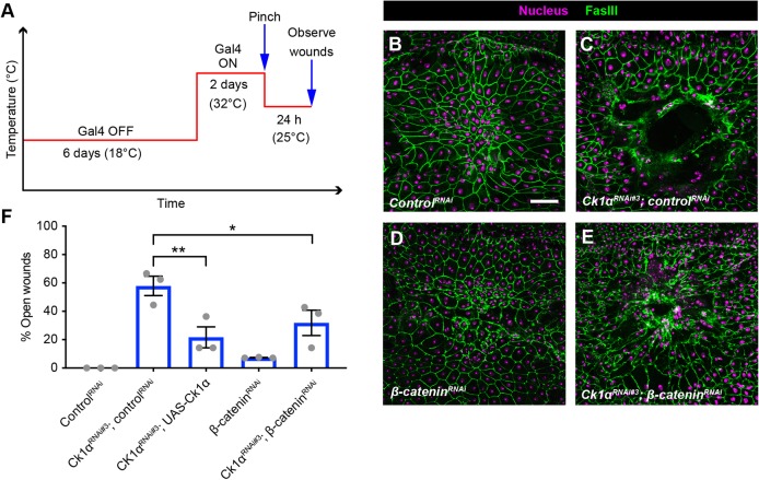
Fig. 3.
Silencing β-catenin partially rescues the Ck1αRNAi-induced wound closure defect. (A) Schematic of the experimental design/temperature shift regimen for using Gal80ts to inducibly express UAS-dependent transgenes in the larval epidermis. (B-E) Dissected epidermal whole mounts of pinch-wounded third instar larvae expressing Gal80ts transgene driven by a tubulin promoter, UAS-DsRed2Nuc (nuclei, magenta) via the e22c-Gal4 driver, and the indicated transgenes. Cell boundaries were immunostained using anti-Fasciclin III antibodies (green). (B) ControlRNAi, (C) UAS-Ck1αRNAi#3 and controlRNAi, (D) UAS-β-cateninRNAi, (E) Ck1αRNAi#3 and UAS-β-cateninRNAi. Scale bar: 100 μm. (F) Quantitation of the percentage of open wounds in larvae expressing indicated transgenes via the e22c-Gal4 driver. Each dot represents one set of n≥8 for each genotype. Data are mean±s.e.m. *P<0.05, **P<0.01 (one-way ANOVA).

