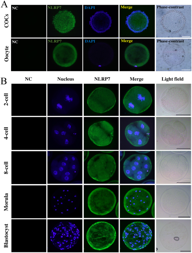
Figure 2.

Expression pattern of NLRP7 protein in ovine COC, oocyte and parthenogenetic embryos. (A) Immunofluorescence with NLRP7 antibody (N3C2, Gene Tex) in COCs (cumulus oocyte complexes) and denuded oocyte.(B) Immunofluorescence of NLRP7 protein in parthenogenetic embryos. Scale bar = 50 µm. Omission of NLRP7 primary antibody shows no signal as negative control (NC). Each sample was counterstained with DAPI to visualize nucleus (blue).

 This work is licensed under a