Abstract
The low solubility of the plant-extracted agent like D-limonene in cancer therapy is a critical problem. In this study, we prepared D-limonene-loaded niosomes (D-limonene/Nio) for cancer therapy through in vitro cytotoxicity assay of HepG2, MCF-7, and A549 cell lines. The niosomal formulation was prepared by film hydration technique with Span® 40: Tween® 40: cholesterol (35:35:30 molar ratio) and characterized for vesicle distribution size, morphology, entrapment efficiency (EE%), and in vitro release behaviour. The obtained niosomes showed a nanometric size and spherical morphology with EE% about 87 ± 1.8%. Remarkably prolonged release of D-limonene from niosomes compared to free D-limonene observed. The loaded formulation showed significantly enhanced cytotoxic activity with all three cancer cell lines (HepG2, Macf-7 and A549) at the concentration of 20 μM. These results indicated that niosome loaded with phytochemicals can be a promising nano-carrier for cancer therapy applications
Keywords: Cancer therapy, D-limonene, Niosome, Solubility
INTRODUCTION
Cancer in the less developed countries is the primary cause of death. It is expected that the prevalence of cancer in the world can be increased because of population increasing and aging. Moreover, smoking, inappropriate diet, and physical inactivity increase the risk of cancer (1). Liver, breast, and lung cancers are among the most common ones. Liver cancer, which is more common in men than women, is the second leading cause of death in the men (2). Conventional treatment methods for liver cancer are surgery, radiation, and chemotherapy. Breast cancer is one of the main causes of female mortality and is one of the most recognizable types of cancer. Hormonal and reproductive factors, treatment for menopause with hormone, and obesity increase the risk of breast cancer, while accouchement and breastfeeding can reduce this risk. Lung cancer is one of the most common cancers among women and men, mainly due to exposure to occupational and environmental carcinogens such as asbestos, polycyclic aromatic hydrocarbons, and others (3). Although chemotherapy prevents the proliferation of liver cancer cells, they have some adverse side effects. Therefore, the critical need to find alternative medicines for treating this type of malignancy is necessary (4,5). In recent years, the trends towards the herbal medicines and use of phytochemicals in the prevention and treatment of diseases have increased (6-8). Medicinal plants are herbs with one or more organs containing an active substance which has medicinal properties that affect living organisms (9,10).
D-limonene is a monocyclic monoterpenes and one of the most commonly used terpenes in nature, found in a variety of citrus fruits. Previous studies identified that it has anti-inflammatory and antioxidant activity and has been developed as an effective ingredient in the prevention and treatment of various types of cancer (11). D-limonene by inhibiting free radicals and lipids peroxidation prevents damage to cell membranes, reduces physiological and psychological stress, and also reduces blood pressure and cardiovascular response to stress (12).
D-Limonene stimulates the carcinogen- metabolizing enzymes (cytochrome P-450), which convert chemical carcinogens into less toxic compounds and prevent their reactions with DNA (11). Yu et al. found that D-limonene inhibits the proliferation of lung cancer cells and through induction of autophagy increases apoptosis of lung cancer cells (13). Previously, Lu et al. confirmed the effect of D-limonene on induction of apoptosis in BGC-823 gastric cancer cells (14). D-Limonene is also effective in primary chemotherapy for hepatocellular carcinoma (15).
The compounds extracted from medicinal plants such as D-limonene, despite many interesting properties and therapeutic applications, have some limitations and challenges. These limitations are due to low solubility, reduced permeability, low bioavailability, and instability in the body's biological fluids. Since herbal extracts have much lower toxicity than chemical drugs (16), researches in this field are critical to increase the efficacy of these compounds. Using herbal ingredients like D-limonene as a free entity would diminish their effectiveness and efficiencies (17). Thus, in order to enhance solubility and bioavailability of D-limonene, using efficient nanocarrier systems such as noisome are highly warranted.
Nanocarriers improve pharmacokinetics and the bioavailability of therapeutic agents and, consequently, minimize their toxicity by their specific accumulation at the target tissues (18,19). Currently, the use of nanocarriers for the delivery of phytochemicals has been highly considered (20). Nanocarrier systems offer many benefits which include modification of pharmacokinetic and pharmacodynamic profiles of the entrapped compounds, reduction of drug adverse effects, control of release rate of the compound, and thus increasing the life quality of the patients (21).
Amongst various nanocarriers, niosomes have attracted a lot of attention. Niosomes are bilayers vesicles that consist of non-ionic surfactants and cholesterol. Because of the nonionic nature of surfactants, niosomes have very low toxicity. In drug delivery approaches, biocompatibility and controlled release of the constituents play a great role in the treatment outcomes (22,23). Though, niosomes have many advantages, little researches have been conducted for the delivery of phytochemicals using niosome nanocarriers (24).
In this study, we aimed to evaluate the anti-cancer effect of D-limonene-loaded noisome. At first, the purity of D-limonene was investigated by gas chromatography-mass spectrometry (GC-MS) analysis. Secondly, D-limonene was loaded into niosomes using thin film hydration method. The encapsulation efficiency and release rate of D-limonene from niosome formulations assayed using dialysis bag and analuzed spectrophotometrically. Finally, anticancer effect of D-limonene on human hepatocellular carcinoma (HepG2), human breast cancer (MCF-7) and human lung cancer (A549) cell lines was evaluated using MTT assay.
MATERIALS AND METHODS
Materials
D-limonene (1-methyl-4-(1-methylethenyl)- cyc lohexane), cholesterol, Span® 40 (sorbitan monopalmitate) and Tween® 40 (polyoxyethylene sorbitan monopalmitate) were purchased from Sigma-Aldrich (St. Louis, MO, USA). MTT reagent (3-(4,5-dimethylthiazol-2yl)-2,5- diphenyltetrazolium bromide) was purchased from MELFORD Biolaboretories Ltd, UK. Chloroform was obtained from Merck (Germany). HepG2, MCF-7, and A549 cell lines were supplied by the Pasteur Institute of Iran, Tehran, I.R. Iran. Dialysis membrane (MWCO: 12,400) were purchased from Sigma Aldrich, Inc. (Milwaukee, USA). All other chemicals and components for buffer solutions were of analytical grade preparations.
Gas chromatography-mass spectrometry
D-limonene dissolved in chloroform was analyzed using a Varian gas chromatograph (CP 3800, Palo Alto, CA, USA) equipped with a mass detector (Varian, 2200, Mass/Mass). A fused silica capillary CP-Sil 8 CB (30 cm long, 0.25 mm ID, 0.25 μm film thickness) column was used. Helium as the carrier gas was set at the constant flow of 1 mL/min. D-limonene (1 μL) was injected at spit ratio of 50:1. The temperature of the column started at 110°C and increased to 200 °C at temperature rate 3°C/min. Total time of analysis was 30 min. MSD ChemStation software (Agilent, USA) was used to acquire mass spectrometric data. MS was operated in electron ionization mode (EI, 70 eV) and 60 to 650 m/z were scanned. The temperature of the transfer line was 270 °C. The mass spectra of each compound were compared with the NIST library of instrument software.
Preparation of D-limonene-loaded niosome
Film hydration technique was utilized to fabricate niosomes as already reported (25,26). Briefly, cholesterol, Span® 40, and Tween® 40 at molar ratio 30:35:35 in 10 mL of chloroform were dissolved. Then, a certain volume of the prepared solution and 10.88 μL of D-limonene (1, 10, and 20 μM) was transfered into a 50-mL round bottom flask. Chloroform was removed by a rotary evaporator (Heidolph, LABOROTA 4003-control-WB, Germany) at 60 °C for 30 min. The resulted lipid film further heated with a hair dryer for 1 min to remove chloroform completely. After solvent removal, 3 mL of phosphate buffered saline (PBS, pH 7.4) added to lipid layer and rotated for 30 min at 60 °C to obtain niosomes. For niosome purification, the obtained niosome formulations were centrifuged at 15,000 rpm for 15 min (MPW, MED INSTRUMENTS-150R, Germany). At the end, formulations were kept at 4 °C for further studies.
Characterization of niosomes
To observe the shape of vesicles, a drop of niosome was placed on a microscope slide and seen using an optical microscope (ICC50 W, Germany).
To evaluate the size of niosomes, the dynamic light scattering (DLS) technique was used (Malvern Instruments Ltd., Worcestershire, UK). The analyses were performed with He-Ne laser (633 nm) at the fixed scattering angle of 90°. Samples (20 μL) with 2 mL ultrapure water were diluted and filtered. Evaluations of size were conducted in triplicate for each sample.
The topography of the niosomes was studied using scanning electron microscope (SEM) (SBC-12, KYKY, China). A drop of niosome put on a silver tape on an aluminium base. The bases were kept under vacuum overnight and then sputter coated using gold.
Entrapment efficiency of D-limonene-loaded niosomes
D-limonene-loaded niosomes were dissolved in 1 mL of ethanol and then centrifuged at 15000 rpm for 30 min (model 5415D, Eppendorf, Germany) (27). D-limonene absorbance in the supernatant was measured spectrophotometrically at 255 nm (Agilent Technologies, Cary 60, USA). Loading efficiency of formulations was calculated as the difference between the total amount of D-limonene added to niosome and the D-limonene content measured in the supernatant by means of below equation:

In vitro release study
The dialysis method was used to investigate D-limonene release rate from loaded formulation and also free D-limonene. In this method, 1 mL of each sample (free D-limonene and D-limonene- loaded niosome) at same concentrations (20 μM) was placed in the dialysis bag, and the two ends of the bag were tighten (molecular weight cut off 12,000 Da). The bag was placed in a beaker containing 30% (v/v) ethanol in 50 mL of PBS 7.4 and stirred by a magnetic stirrer at 150 rpm and 37 °C (Heidolph, MR3002G, Germany). Then, at predestinated period, up to 12 h, 1 mL of the PBS solution inside the beaker was withdrawn and, instantly, 1 mL of fresh PBS 7.4 was added to the beaker (n = 3). The obtained samples were directly read by a spectrophotometer at 255 nm (Agilent Technologies, Cary 60 UV-Vis, USA) (28).
Cell culture
HepG2, MCF-7, and A549 cell lines were cultured in RPMI-1640 medium (Gibco, USA) augmented with 10% fetal bovine serum (FBS), 100 IU/mL penicillin antibiotic and 100 μg/mL streptomycin inside the incubator at 37 °C, sufficient humidity and 5% carbon dioxide (13).
Cell viability assay
To investigate the effect of D-limonene and niosomal containing D-limonene on growth and proliferation of cancer cells, MTT assay was used. In this method, 104 cells were cultured at 96-well plates at 100 μL/well, and then incubated overnight. Subsequently, different concentrations of empty niosomes (0.5, 1.5, 5 μM), various concentrations of free D-limonene (1-400 μM), D-limonene- loaded niosomes at concentrations of1, 10, and 20 μM (based on loaded D-limonene) were added to the cells. After 24 h of incubation, 10 mL of MTT solution was added to each well, and the plate was placed in a 37 °C incubator at 5% carbon dioxide for 4 h. After the required time, the medium containing MTT were carefully discarded and 100 mL of DMSO added to each well, then, the absorption of every well was read by ELISA readers at 570 nm (BIO-TEK INSTRUMENT, USA) (29).

Statistical analysis
For statistical analysis of different examinations, one-way ANOVA was exploited. To examine the ANOVA test, Dunnett's test was performed. A P value < 0.05 was considered significant. All values are reported as the mean ± standard deviation.
RESULTS
Gas chromatography-mass spectrometry analysis
As shown in Fig. 1, there is a sharp and intense peak at the retention time of 9.8 min that relates to D-limonene. The purity of D-limonene was found to be about 71%.
Fig. 1.
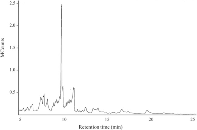
Gas chromatography-mass spectrometry chromatogram of D-limonene
Characterization of formulations
As shown in Fig. 2, most of the niosomes appeared circular or slightly off circular shape. Also, the microscopic appearances of the formulation after loading with D-limonene indicated spherical multilamellar vesicles with a normal distribution, as seen in Fig. 2C and 2D.
Fig. 2.
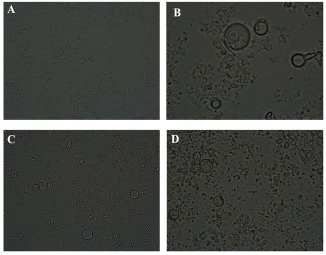
Images of (A and B) free and (C and D) D-limonene-loaded niosomes in different magnifications (40× and 100×) by optical microscopy.
The size distribution and polydispersity index (PDI) of formulations was determined using DLS. Figure 3 shows the mean vesicle size of free and D-limonene-loaded niosomes. The size distribution of niosomes before D-limonene loading had a narrow size distribution peak. The vesicle size of blank niosomes was 90-102 nm and increased to 110-1100 nm after loading with D-limonene. PDI of free niosome and loaded niosome was 0.21 and 0.37, respectively that means both niosome formulations demonstrated appropriate size homogeneity with reasonable PDI value. Stability properties of formulations depend on the zeta potential. A zeta potential near ±30 mV could guarantee a long term stable formulation (30). Zeta potentials for empty niosome and D-limonene-loaded niosome were -21 mV and -45 mV, respectively (Fig. 4A and 4B). Loading of D-limonene into niosome resulted in more negative zeta values.
Fig. 3.
Mean vesicle size of (A) empty and (B) D-limonene-loaded niosomes.
Fig. 4.
Zeta potential of (A) empty niosomes and (B) D-limonene-loaded niosomes by dynamic light scattering analysis.
The morphology of the free and loaded formulations was shown in Fig. 5. All the niosomes showed a spherical unilamellar vesicular structure with size under 200 nm. It can be found that the D-limonene-loaded niosomes distributed independently with the similar spherical shape. Both niosome formulations were stable and free of coagulation. Based on SEM analysis, niosome containing D-limonene and free niosomes had similar particle size. EE of D-limonene was about 87 ± 1.8%, which was satisfactory.
Fig. 5.
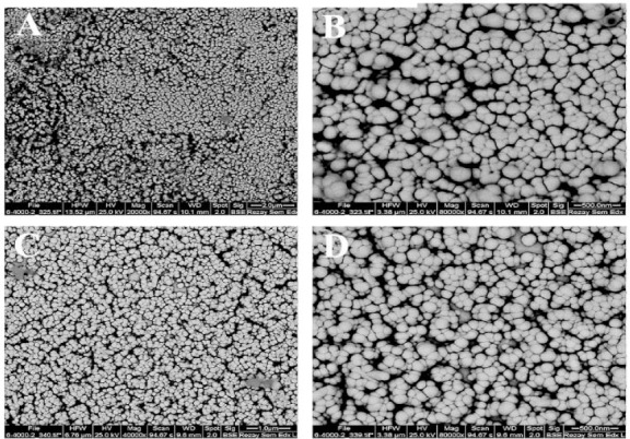
Scanning electron microscope images of (A and B) empty and (C and D) D-limonene-loaded niosomes at different resolutions.
In vitro release of D-limonene
Percentage of D-limonene release was obtained by calculating the ratio of the contents of released D-limonene in release medium and the amount of total D-limonene initially loaded in the niosomes. The cumulative release profile of formulations is shown in Fig. 6. The experiment was conducted in triplicate. As observed in Fig. 6, D-limonene was released from niosomes slowly, and after 12 h ~54% D-limonene was released from the formulation which demonstrated a sustained release manner. The release content of D-limonene increased in a controlled fashion as a function of time without a critical burst release.
Fig. 6.
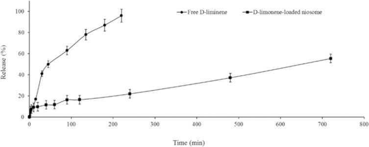
Drug release profiles of free D-limonene and D-limonene-loaded niosomes. Bars represent ± SD.
Almost all free D-limonene released from dialysis bag after 4 h.
In vitro cytotoxicity
To identify the potential of prepared niosomes as pharmaceutical carriers for D-limonene against cancer cell lines, the human hepatocellular carcinoma (HepG2) cell line was initially incubated with empty niosomes at different concentrations (0.5, 1.5, and 5 μM) and free D-limonene at various concentrations (1-400 μM) for 24 h using MTT assay. As shown in Fig. 7, by increasing the concentrations of empty niosomes the viability of cells decreased.
Fig. 7.
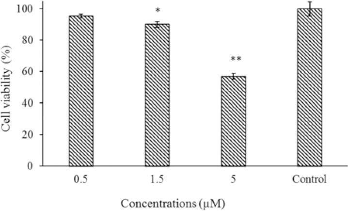
The effect of different concentrations of empty niosomes on viability of HepG2 cells. Data are presented as mean ± SD. * P < 0.05 and ** P < 0.01 indicate significant differences compared to the control group.
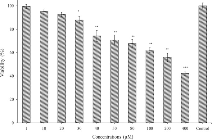
At concentration of 1.5 μM, the viability was 90%, and thus this concentration of empty niosome used for next experiments. The low toxicity of empty niosomes provides good safety for healthy cells. In addition, the cell toxicity of different concentrations of D-limonene is shown in Fig. 8. As presented in this figure, at higher concentrations of D-limonene, the viability of tumor cells decreased.
Fig. 8.
The effect of different concentrations of D-limonene on viability of HepG2 cells. Data are presented as mean ± SD. * P < 0.05, ** P < 0.01, and *** P < 0.001 indicates significant differences compared to the control group.
For anticancer assay of D-limonene loaded D-niosomes, we used concentrations of 1, 10 and 20 μM and Their anticancer effect was evaluated on 3 different cell lines of HepG2, A549, and MCF-7. As shown in Fig. 8 free D-limonene at concentration of 1-20 μM did not exhibited significant cytotoxicity as expected based on the results illustrated in Fig.7, but according to the Fig. 9 which shows cell viability of D-limonene loaded niosomes on 3 tested cell lines, D-limonene-loaded niosomes at these concentrations showed a noticeable anticancer effect after 24 h of incubation. Also, this result proved that high concentration of D-limonene in niosomes can enhance anti-tumor activity of D-limonene.
Fig. 9.
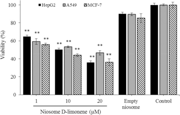
Viability percentage of HepG2, A549, and MCF-7 after treatment with various concentrations of D-limonene-loaded niosome (1, 10, 20 μM) and empty niosome at 1.5 μM. The cytotoxicity of niosomes was evaluated by MTT assay and data represent the mean ± SD. ** Indicates significant differences compared to the corresponding control group, P< 0.01.
DISCUSSION
Many researchers have demonstrated that D-limonene possess anticancer activity (14,31). However, low solubility of D-limonene in biological fluids is a crucial problem and has limited its application in cancer treatment. To address this issue, nano-niosomes containing D-limonene fabricated and characterized as a potential carrier for D-limonene.
GC-MS spectrum of chloroform-solvated D-limonene obtained to verify its purity (Fig. 1). The obtained chromatogram by GC-MS analysis used to compare the relative percentage of each component. High purity of D-limonene confirmed by GC-MS is necessary for in vitro assessments.
When the niosome formulations were examined by optical microscopy, different shapes with various diameters were observed. Hemati et al. investigated the synthesis of niosomes containing quercetin, doxorubicin, and siRNA. The observation of the optical microscope has revealed multi-layered spheres, whose formation and morphology are similar to our findings (32). One of the most experimental approaches used to determine the formation of drug delivery carriers is size distribution measurement. DLS analysis showed that loading of D-limonene into niosomes, increased the size of vesicles. It has been suggested that the carrier sizes of approximately several hundred nm were desirable for cellular uptake (33). In general, PDI value lower than 0.4 means a good size distribution for particles but for higher PDI value we can see aggregate of nanoparticles.
PDI of free niosomes and loaded niosomes was 0.21 and 0.37, respectively meaning both niosome formulations demonstrated suitable size homogeneity with reasonable PDI values. Also, zeta potential can represent the stability of the formulation. The zeta potential of prepared formulations is in the range that guarantees high stability of emulsions by electrostatic repulsion. The loading process of D-limonene can increase the size and PDI value of formulations. The difference in size measurements between SEM and DLS method might be due to the drying process during the SEM imaging. EE% of D-limonene in niosome formulation was about 87%. High EE% may be attributed to a good interaction between surfactant, cholesterol, and D-limonene which makes walls of the vesicles rigid and prevents leakage of D-limonene from niosomes (34). This is also could be explained by the presence of amphiphilic moieties, which form a rigid membrane having characteristics of a barrier against drug leakage. These factors result in higher entrapment efficiency (35). Thakkar et al. manufactured niosomes containing primaquine and curcumin as an anti-malarial compound with a particle size of 220 nm and EE% of 82.12 % (36).
Generally, an applicable nanocarrier should protect bioactive core substance under harsh surroundings environment, which means that the wall material might be resistant against this condition and release its loaded drug in a sustained manner (34). The loaded niosome formulation had a controlled release rate and released the content within 12 h. Hemati et al. reported, under the normal temperature and pH of the body, minimum release rates for doxorubicin, 27.4% and 45.3% for quercetin, after 72 h (32). Ismail et al. improved the bioavailability and release profile of acyclovir by loading the drug into nisomes to the extent that about 85% of the content released within 6 hm while in our study, the release rate was about 30% within 6 h (37). In another study, Abbasi et al. stated that the release rate of curcumin from niosome was about 66% after 24 h (38). The results of the release studies in our work are in line with previous reports as mentioned above.
To illustrate the potential of these niosomes as a pharmaceutical carrier for D-limonene against hepatocellular carcinoma, HepG2 cells were incubated with empty niosomes, free D-limonene, and loaded niosome at different concentrations for 24 h and their cytotoxicity were assessed using MTT assay. Results showed that D-limonene loaded in niosomes was more effective than free D-limonene. The enhanced cytotoxicity of D-limonene loaded in niosomes was due to a synergistic activity of better solubility and consequently better internalization to the cell and a controlled release of D-limonene. The viability results obtained from three cancer cell lines suggested loading of D-limonene into niosome can effectively enhance the antitumour activity of D-limonene. In the study of Kanaani et al., which examined the effect of cisplatin and its niosomal form on breast cancer cells considerable cytotoxic effects of cisplatin observed and concluded that niosomes could be a good carrier system for targeted delivery of the cisplatin in the treatment of breast cancer (39). Tabrizi et al. found that IC50 of the formulation of PEGylated nano niosomal gingerol is less than the free drug which is in line with our findings (40).
CONCLUSION
Phytochemicals have a potential anticancer ability, but their low solubility always is a problem. In this study, we investigated the potentials of D-limonene-loaded niosomes as a promising anticancer delivery system on three cancer cell lines, namely HepG2, A549, and MCF-7. The loaded niosome showed an improved anticancer effect against different cancer cell lines compared to free phytochemicals. This report showed that cancer treatment ability of phytochemicals could be increased by loading them into niosomes.
ACKNOWLEDGEMENT
The authors are grateful to Graduate University of Advanced Technology, Kerman, I.R. Iran and Rafsanjan University of Medical Sciences, Rafsanjan, I.R. Iran for their excellent technical and financial supports (Grant No. 96076).
REFERENCES
- 1.Arnold M, Soerjomataram I, Ferlay J, Forman D. Global incidence of oesophageal cancer by histological subtype in 2012. Gut. 2015;64(3):381–387. doi: 10.1136/gutjnl-2014-308124. [DOI] [PubMed] [Google Scholar]
- 2.Thun M, Linet MS, Cerhan JR, Haiman ChA, Schottenfeld D, editors. Cancer epidemiology and prevention. 3rd edition. United Kingdom: Oxford University Press; 2006. pp. 763–786. [Google Scholar]
- 3.Torre LA, Bray F, Siegel RL, Ferlay J, Lortet-Tieulent J, Jemal A. Global cancer statistics, 2012. CA Cancer J Clin. 2015;65(2):87–108. doi: 10.3322/caac.21262. [DOI] [PubMed] [Google Scholar]
- 4.Mugada V, Ramineni H, Padala D. 5-Fluorouracil induced severe febrile neutropenia and death. J Young Pharm. 2017;9(1):133–134. [Google Scholar]
- 5.Li WJ, Lian YW, Guan QS, Li N, Liang WJ, Liu WX, et al. Liver-targeted delivery of liposome-encapsulated curcumol using galactosylated-stearate. Exp Ther Med. 2018;16(2):925–930. doi: 10.3892/etm.2018.6210. [DOI] [PMC free article] [PubMed] [Google Scholar]
- 6.Lazutka JR, Mierauskiene J, Slapsyte G, Dedonyte V. Genotoxicity of dill (Anethum graveolens L.), peppermint (Mentha piperita L.) and pine (Pinus sylvestris L.) essential oils in human lymphocytes and Drosophila melanogaster. Food Chem Toxicol. 2001;39(5):485–492. doi: 10.1016/s0278-6915(00)00157-5. [DOI] [PubMed] [Google Scholar]
- 7.Rattanamaneerusmee A, Thirapanmethee K, Nakamura Y, Bongcheewin B, Chomnawang MT. Chemopreventive and biological activities of Helicteres isora L. fruit extracts. Res Pharm Sci. 2018;13(1):484–492. doi: 10.4103/1735-5362.245960. [DOI] [PMC free article] [PubMed] [Google Scholar]
- 8.Eghbali-Feriz S, Taleghani A, Al-Najjar H, Emami SA, Rahimi H, Asili J, et al. Anti-melanogenesis and anti-tyrosinase properties of Pistacia atlantica subsp. mutica extracts on B16F10 murine melanoma cells. Res Pharm Sci. 2018;13(1):533–545. doi: 10.4103/1735-5362.245965. [DOI] [PMC free article] [PubMed] [Google Scholar]
- 9.Liu R. H. Health benefits of fruit and vegetables are from additive and synergistic combinations of phytochemicals. Am J Clin Nutr. 2003;78(3 Suppl):517S–520S. doi: 10.1093/ajcn/78.3.517S. [DOI] [PubMed] [Google Scholar]
- 10.Nili-Ahmadabadi A, Alibolandi P, Ranjbar A, Mousavi L, Nili-Ahmadabadi H, Larki-Harchegani A, et al. Thymoquinone attenuates hepatotoxicity and oxidative damage caused by diazinon: an in vivo study. Res Pharm Sci. 2018;13(1):500–508. doi: 10.4103/1735-5362.245962. [DOI] [PMC free article] [PubMed] [Google Scholar]
- 11.11 Sun J.D-Limonene: safety and clinical applications. Altern Med Rev. Sun J D-Limonene: safety and clinical applications Altern Med Rev 2007;12(3):259-264. [PubMed] [Google Scholar]
- 12.Jing L, Zhang Y, Fan S, Gu M, Guan Y, Lu X, et al. Preventive and ameliorating effects of citrus D-limonene on dyslipidemia and hyperglycemia in mice with high-fat diet-induced obesity. Eur J Pharmacol. 2013;715(1-3):46–55. doi: 10.1016/j.ejphar.2013.06.022. [DOI] [PubMed] [Google Scholar]
- 13.Yu X, Lin H, Wang Y, Lv W, Zhang S, Qian Y, et al. D-limonene exhibits antitumor activity by inducing autophagy and apoptosis in lung cancer. OncoTargets Ther. 2018;11:1833–1847. doi: 10.2147/OTT.S155716. [DOI] [PMC free article] [PubMed] [Google Scholar]
- 14.Lu XG, Feng BA, Zhan LB, Yu ZH. D-limonene induces apoptosis of gastric cancer cells. Zhonghua Zhong Liu Za Zhi. 2003;25(4):325–327. [PubMed] [Google Scholar]
- 15.Guyton KZ, Kensler TW. Prevention of liver cancer. Curr Oncol Rep. 2002;4(1):464–470. doi: 10.1007/s11912-002-0057-4. [DOI] [PubMed] [Google Scholar]
- 16.Murali R, Karthikeyan A, Saravanan R. Protective effects of D-limonene on lipid peroxidation and antioxidant enzymes in streptozotocin-induced diabetic rats. Basic Clin Pharmacol Toxicol. 2013;112(3):175–181. doi: 10.1111/bcpt.12010. [DOI] [PubMed] [Google Scholar]
- 17.Perchyonok VT, Souza J, Zhang Sh, Moodley D, Grobler S. Bio-active designer materials and dentures: from design to application. Int J Med Nano Res. 2015;2(1):2–12. [Google Scholar]
- 18.Alexis F, Rhee JW, Richie JP, Radovic-Moreno AF, Langer R, Farokhzad OC. New frontiers in nanotechnology for cancer treatment. Urol Oncol. 2008;26(1):74–85. doi: 10.1016/j.urolonc.2007.03.017. [DOI] [PubMed] [Google Scholar]
- 19.Khajeh Ebrahimi A, Barani M, Sheikhshoaie I. Fabrication of a new superparamagnetic metal- organic framework with core-shell nanocomposite structures: Characterization, biocompatibility, and drug release study. Mater Sci Eng C. 2018;92:349–355. doi: 10.1016/j.msec.2018.07.010. [DOI] [PubMed] [Google Scholar]
- 20.Uchegbu IF, Vyas SP. Non-ionic surfactant based vesicles (niosomes) in drug delivery. Int J Pharm. 1998;172(1-2):33–70. [Google Scholar]
- 21.Farokhzad OC, Langer R. Impact of nanotechnology on drug delivery. ACS Nano. 2009;3(1):16–20. doi: 10.1021/nn900002m. [DOI] [PubMed] [Google Scholar]
- 22.Barani M, Mirzaei M, Torkzadeh-Mahani M, Nematollahi MH. Lawsone-loaded niosome and its antitumor activity in MCF-7 breast cancer cell line: a Nano-herbal treatment for Cancer. DARU J Pharm Sci. 2018;26(1):11–17. doi: 10.1007/s40199-018-0207-3. [DOI] [PMC free article] [PubMed] [Google Scholar]
- 23.Davarpanah F, Khalili Yazdi A, Barani M, Mirzaei M, Torkzadeh-Mahani M. Magnetic delivery of antitumor carboplatin by using PEGylated-Niosomes. DARU J Pharm Sci. 2018 doi: 10.1007/s40199-018-0215-3. DOI: 10.1007/s40199-018-0215-3. [DOI] [PMC free article] [PubMed] [Google Scholar]
- 24.Moghassemi S, Hadjizadeh A. Nano-niosomes as nanoscale drug delivery systems: an illustrated review. J Control Release. 2014;185:22–36. doi: 10.1016/j.jconrel.2014.04.015. [DOI] [PubMed] [Google Scholar]
- 25.Cui F, Shi K, Zhang L, Tao A, Kawashima Y. Biodegradable nanoparticles loaded with insulin-phospholipid complex for oral delivery: preparation, in vitro characterization and in vivo evaluation. J Control Release. 2006;114(2):242–250. doi: 10.1016/j.jconrel.2006.05.013. [DOI] [PubMed] [Google Scholar]
- 26.Stockwell AF, Davis SS, Walker SE. In vitro evaluation of alginate gel systems as sustained release drug delivery systems. J Control Release. 1986;3(1-4):167–175. [Google Scholar]
- 27.Rahdar A, Taboada P, Hajinezhad MR, Barani M, Beyzaei H. Effect of tocopherol on the properties of Pluronic F127 microemulsions: Physico-chemical characterization and in vivo toxicity. J Mol Liq. 2019;277:624–630. [Google Scholar]
- 28.Rajera R, Nagpal K, Singh SK, Mishra DN. Niosomes: a controlled and novel drug delivery system. Biol Pharm Bull. 2011;34(2):945–953. doi: 10.1248/bpb.34.945. [DOI] [PubMed] [Google Scholar]
- 29.Thangapazham RL, Puri A, Tele S, Blumenthal R, Maheshwari RK. Evaluation of a nanotechnology- based carrier for delivery of curcumin in prostate cancer cells. Int J Oncol. 2008;32(5):1119–1123. [PMC free article] [PubMed] [Google Scholar]
- 30.Hans ML, Lowman AM. Biodegradable nanoparticles for drug delivery and targeting. Curr Opin Solid St M. 2002;6(4):319–327. [Google Scholar]
- 31.Vandresen F, Falzirolli H, Almeida Batista SA, da Silva-Giardini AP, de Oliveira DN, Catharino RR, et al. Novel R-(+)-limonene-based thiosemicarbazones and their antitumor activity against human tumor cell lines. Eur J Med Chem. 2014;79:110–116. doi: 10.1016/j.ejmech.2014.03.086. [DOI] [PubMed] [Google Scholar]
- 32.Hemati M, Haghiralsadat F, Yazdian F, Jafari F, Moradi A, Malekpour-Dehkordi Z. Development and characterization of a novel cationic PEGylated niosome-encapsulated forms of doxorubicin, quercetin and siRNA for the treatment of cancer by using combination therapy. Artif Cells Nanomed Biotechnol. 2018;(1):47, 1295–1311. doi: 10.1080/21691401.2018.1489271. [DOI] [PubMed] [Google Scholar]
- 33.Khan M, Ang CY, Wiradharma N, Yong LK, Liu S, Liu L, et al. Diaminododecane-based cationic bolaamphiphile as a non-viral gene delivery carrier. Biomaterials. 2012;33(18):4673–4680. doi: 10.1016/j.biomaterials.2012.02.067. [DOI] [PubMed] [Google Scholar]
- 34.Sarwa KK, Suresh PK, Debnath M, Ahmad MZ. Tamoxifen citrate loaded ethosomes for transdermal drug delivery system: preparation and characterization. Curr Drug Deliv. 2013;10(4):466–476. doi: 10.2174/1567201811310040011. [DOI] [PubMed] [Google Scholar]
- 35.Hariharan S, Bhardwaj V, Bala I, Sitterberg J, Bakowsky U, Ravi Kumar MN. Design of estradiol loaded PLGA nanoparticulate formulations: a potential oral delivery system for hormone therapy. Pharm Res. 2006;23(1):184–195. doi: 10.1007/s11095-005-8418-y. [DOI] [PubMed] [Google Scholar]
- 36.Thakkar M, Brijesh S. Brijesh, Physicochemical investigation and in vivo activity of anti-malarial drugs co-loaded in Tween 80 niosomes. J Liposome Res. 2017;(4):28, 315–321. doi: 10.1080/08982104.2017.1376684. [DOI] [PubMed] [Google Scholar]
- 37.Attia IA, El-Gizawy SA, Fouda MA, Donia AM. Influence of a niosomal formulation on the oral bioavailability of acyclovir in rabbits. AAPS PharmSciTech. 2007;8(4):206–212. doi: 10.1208/pt0804106. [DOI] [PMC free article] [PubMed] [Google Scholar]
- 38.Abbasi S, Kajimoto K, Harashima H. Critical parameters dictating efficiency of membrane-mediated drug transfer using nanoparticles. Int J Pharm. 2018;553(1-2):398–407. doi: 10.1016/j.ijpharm.2018.10.042. [DOI] [PubMed] [Google Scholar]
- 39.Kanaani L. Effects of cisplatin-loaded niosomal nanoparticleson BT-20 human breast carcinoma cells. Asian Pac J Cancer Prev. 2017;18(2):365–368. doi: 10.22034/APJCP.2017.18.2.365. [DOI] [PMC free article] [PubMed] [Google Scholar]
- 40.Behroozeh A, Tabrizi MM, Kazemi SM, Choupani E, Kabiri N, Ilbeigi D, et al. Evaluation the anti-cancer effect of pegylated nano-niosomal gingerol, on breast cancer cell lines (T47D), in-vitro. Asian Pac J Cancer Prev. 2018;19(3):645–648. doi: 10.22034/APJCP.2018.19.3.645. [DOI] [PMC free article] [PubMed] [Google Scholar]


