Abstract
An HIV-positive female on antiretroviral therapy (ART) presented with an annular eruption diagnosed as a drug reaction based on histology of a lichenoid dermatitis. She responded to oral steroid therapy and discontinuation, but progressed to develop features in keeping with cutaneous lupus. Although the antinuclear factor remained negative, her low serum complement levels, histology, and clinical features pointed to a diagnosis of subacute lupus in the setting of HIV infection. She responded well to antimalarial therapy and recommenced ART.
Keywords: Subacute lupus, HIV infection, Antiretroviral therapy
Introduction
Lupus is uncommon in HIV infection, but both conditions present with multisystem disease and a range of dermatological manifestations that evolve over time and correlate with systemic activity. There is some overlap in the cutaneous conditions seen in systemic lupus erythematosus (SLE) with those of HIV infection, and in addition, there may be a false-positive antinuclear factor (ANF) in HIV infection. Therapy for lupus with immunosuppressants could be a challenge in HIV-positive patients. The case below illustrates some of these challenges.
Case Report
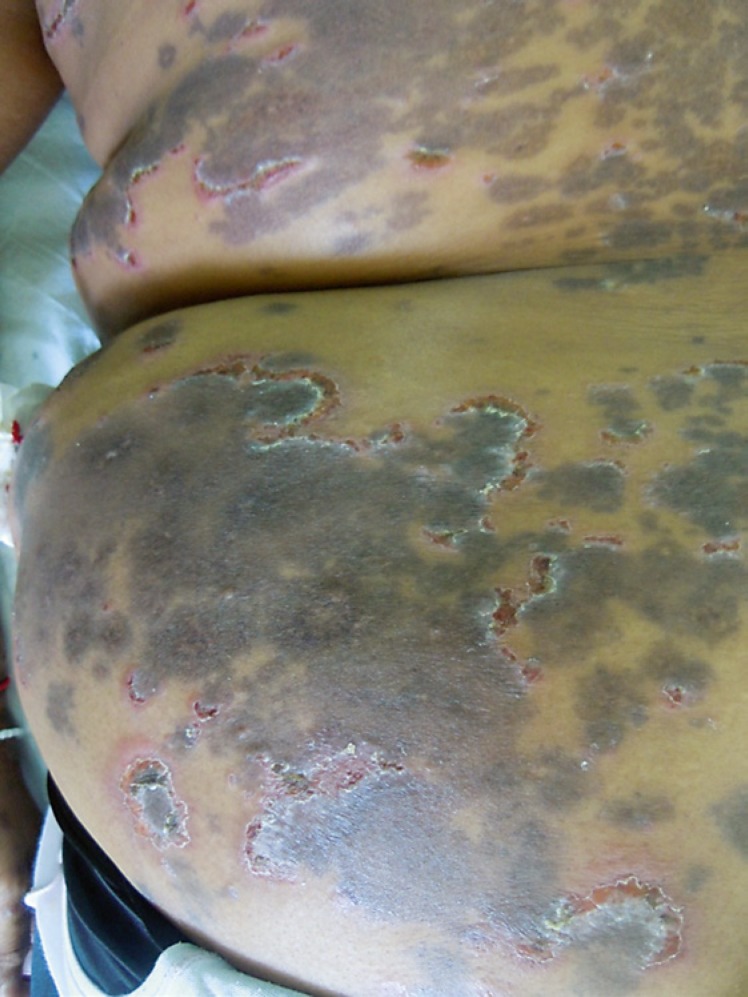
A 40-year-old, HIV-positive female presented with a 1-month history of an annular truncal eruption (Fig. 1). She had been on highly active antiretroviral therapy (ART) for 1 year (efavirenz, lamivudine, and tenofovir) without trimethoprim-sulfamethoxazole prophylaxis and had a CD4 count of 316 cells/mm3. In view of the annularity of the lesions, differentials of secondary syphilis, a drug eruption, and subacute lupus were entertained. Her rapid plasma reagin and Treponema pallidum hemagglutination assay were negative, ANF was negative, and histology showed a lichenoid eruption with basal cell vacuolization and colloid bodies (Fig. 2). A diagnosis of ART-induced lichenoid interface dermatitis was made, and the patient was commenced on oral steroids at 0.5 mg/kg and her ART was discontinued for a 2-week period. During this time her rash improved markedly and the prednisone was slowly tapered, but the rash recurred despite discontinuation of her ART.
Fig. 1.
Annular eruption healing with postinflammatory pigmentation.
Fig. 2.
Lichenoid eruption with basal cell vacuolation and colloid bodies.
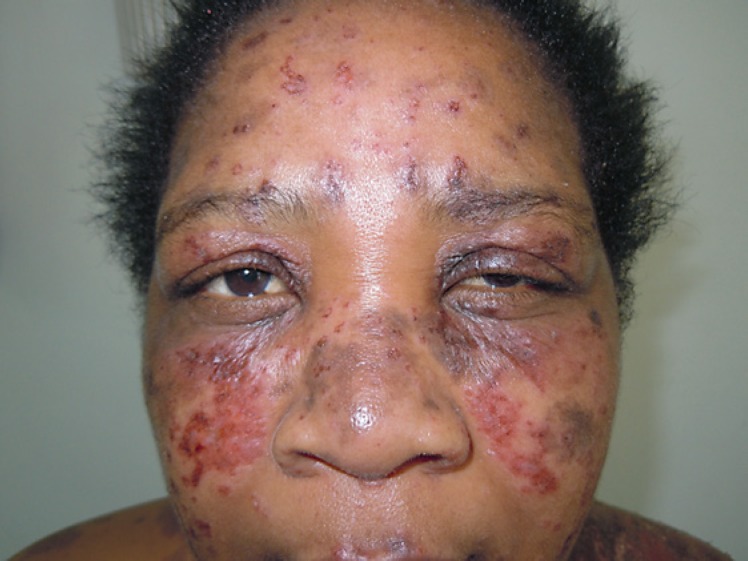
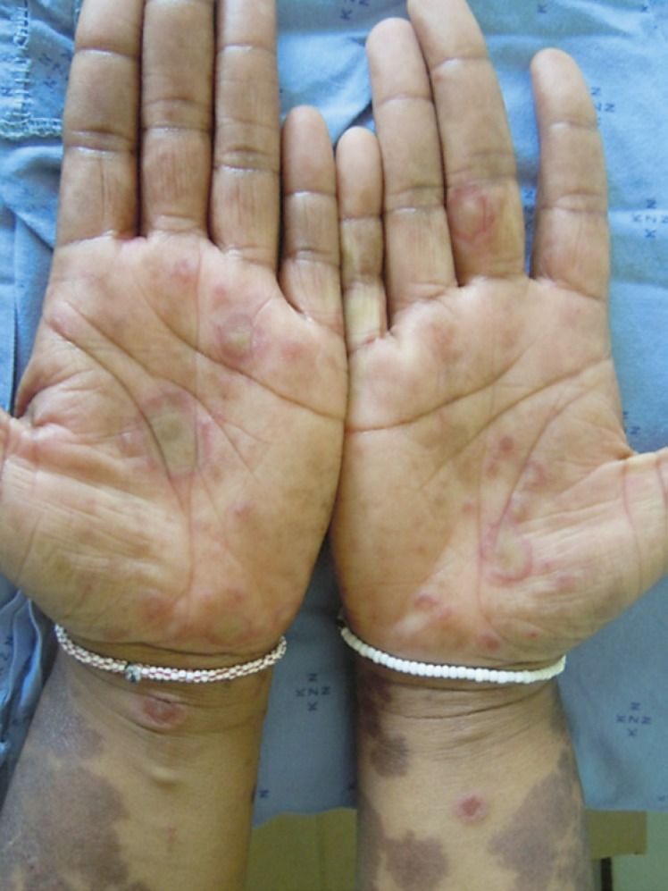
In addition to the presenting rash, she developed an intercurrent herpes zoster infection and was treated with antivirals. Subsequently she began developing an erythematous rash involving the malar area (Fig. 3), annular erythematous plaques on the trunk, specifically on the “V” of the neck (Fig. 4), and on the upper limbs with targetoid lesions on the palms (Fig. 5) and vasculitic lesions on the limbs. Her repeat ANF remained negative with a low serum complement level C3 (0.7 g/L) and C4 (0.14 g/L) and a positive anti-Ro SSA antibody. The patient was then diagnosed as having subacute cutaneous lupus with some features of cutaneous lupus in the setting of HIV infection.
Fig. 3.
Erythematous plaques in a malar distribution.
Fig. 4.
Annular erythematous plaques involving the trunk.
Fig. 5.
Annular erythematous plaques involving the hands.
Her ART was restarted with chloroquine at 200 mg daily and she tolerated both well, with a good clinical response (Fig. 6).
Fig. 6.
Resolution of lesions with postinflammatory hyperpigmentation.
Discussion
SLE is uncommon in HIV infection, with the first case documented in 1988 [1]. Cutaneous lupus is rarer, with only a few cases of discoid lupus and subacute lupus reported in HIV infection [2, 3, 4]. Subacute lupus has been described as a side effect of isoniazid therapy in patients with tuberculosis [5]. In addition, a few cases of cutaneous lupus have been reported as a manifestation of the immune reconstitution syndrome [6, 7].
There may be underreporting, but it is more likely explained by immunologic exclusivity. Autoantibody formation is central to the presentation of lupus whilst immunosuppression is the key event in HIV infection and is likely to decrease the risk of developing HIV infection [8]. There is certainly overlap of cutaneous features in both SLE and HIV infection, e.g., the malar rash of lupus could be confused with seborrheic eczema in HIV infection, and oral ulcers are common in both conditions, as are vasculitis and the predisposition to opportunistic infections. Furthermore, there is a 20% chance of false positivity of ANF in the setting of HIV infection, and up to 10% may have titers of >1:160 [9], adding to the dilemma. However, the discerning clinician aided by the pathologist would be able to come to an accurate diagnosis. This is crucial, as therapy with immunosuppressants and steroids could cause HIV viral replication; however, chloroquine is safe in HIV infection.
In sub-Saharan Africa with its high HIV prevalence, clinicians' knee-jerk response is to diagnose drug reactions, especially with the multitude of drugs patients are prescribed. This is borne out in this case, and even the therapeutic trial of steroids would have caused resolution of both conditions, and hence there was a delay in making the diagnosis. It was only on reappearance of the rash whilst the ART was discontinued and the progression of the rash involving the malar area, annular plaques of the trunk, and erythema multiforme-like lesions of the hands that a diagnosis of lupus was considered. Although the ANF was negative, it was the hypocomplementemia and positive anti-Ro SSA that supported the diagnosis. The patient did not fulfil all criteria for Rowell's syndrome, and hence a diagnosis of subacute lupus with some overlap features of SLE was made. The response to chloroquine was remarkable and further supports the diagnosis.
Conclusion
This case highlights a few important lessons. Clinicians need to follow up patients and take note of the evolution of a rash. Lupus is a great mimicker even if a serological test is negative; clinicians need to be aware of the subtle histological variants of lupus, and dermatologists in South Africa need to consider other causes of eruptions in HIV-positive patients on ART. Clinicopathological correlation is essential in making an appropriate diagnosis and prescribing the correct therapy.
Statement of Ethics
The patient's consent was obtained for use of her photographs.
Disclosure Statement
The authors have no conflicts of interest to declare. No funding was received.
Acknowledgments
The authors thank the Department of Anatomical Pathology, National Health Laboratory Service, Inkosi Albert Luthuli Central Hospital for the histopathology images.
References
- 1.Palacios R, Santos J. Human immunodeficiency virus infection and systemic lupus erythematosus. Int J STD AIDS. 2004 Apr;15((4)):277–8. doi: 10.1258/095646204773557857. [DOI] [PubMed] [Google Scholar]
- 2.Calza L, Manfredi R, Colangeli V, D'Antuono A, Passarini B, Chiodo F. Systemic and discoid lupus erythematosus in HIV-infected patients treated with highly active antiretroviral therapy. Int J STD AIDS. 2003 May;14((5)):356–9. doi: 10.1258/095646203321605585. [DOI] [PubMed] [Google Scholar]
- 3.Two A, So JK, Paravar T. Discoid Lupus and Human Immunodeficiency Virus: A Retrospective Chart Review to Determine the Prevalence and Progression of Co-occurrence of these Conditions at a Single Academic Centre. Indian J Dermatol. 2017 Mar-Apr;2((2)):226. doi: 10.4103/0019-5154.201750. doi: . [DOI] [PMC free article] [PubMed] [Google Scholar]
- 4.Luwanda LB, Gamell A. Isoniazid-induced subacute cutaneous lupus erythematosus in an HIV-positive woman: a rare side effect to be aware of with the current expansion of isoniazid preventive therapy. Pan Afr Med Jnl 2018 Apr; 6;29:200. d. eCollection. 2018 doi: 10.11604/pamj.2018.29.200.12081. [DOI] [PMC free article] [PubMed] [Google Scholar]
- 5.Hazarika I, Chakravarty BP, Dutta S, Mahanta N. Emergence of manifestations of HIV infection in a case of systemic lupus erythematosus following treatment with IV cyclophosphamide. Clin Rheumatol. 2006 Feb;25((1)):98–100. doi: 10.1007/s10067-005-1133-6. [DOI] [PubMed] [Google Scholar]
- 6.Chamberlain AJ, Hollowood K, Turner RJ, Byren I. Tumid lupus erythematosus occurring following highly active antiretroviral therapy for HIV infection: a manifestation of immune restoration. J Am Acad Dermatol. 2004 Nov;51((5Suppl)):S161–5. doi: 10.1016/j.jaad.2004.04.020. [DOI] [PubMed] [Google Scholar]
- 7.Soria A, Canestri A, Bournerias I, Le Pelletier F, Bricaire F, Caumes E. [Cutaneous chronic lupus, a new cutaneous manifestation of the immune reconstitution in human immunodeficiency virus infection] Presse Med. 2009 Oct;38((10)):1541–3. doi: 10.1016/j.lpm.2009.01.018. [DOI] [PubMed] [Google Scholar]
- 8.Daikh BE, Holyst MM. Lupus-specific autoantibodies in concomitant human immunodeficiency virus and systemic lupus erythematosus: case report and literature review. Semin Arthritis Rheum. 2001 Jun;30((6)):418–25. doi: 10.1053/sarh.2001.23149. [DOI] [PubMed] [Google Scholar]
- 9.Zandman-Goddard G, Shoenfeld Y. HIV and autoimmunity. Autoimmun Rev. 2002 Dec;1((6)):329–37. doi: 10.1016/s1568-9972(02)00086-1. [DOI] [PubMed] [Google Scholar]


