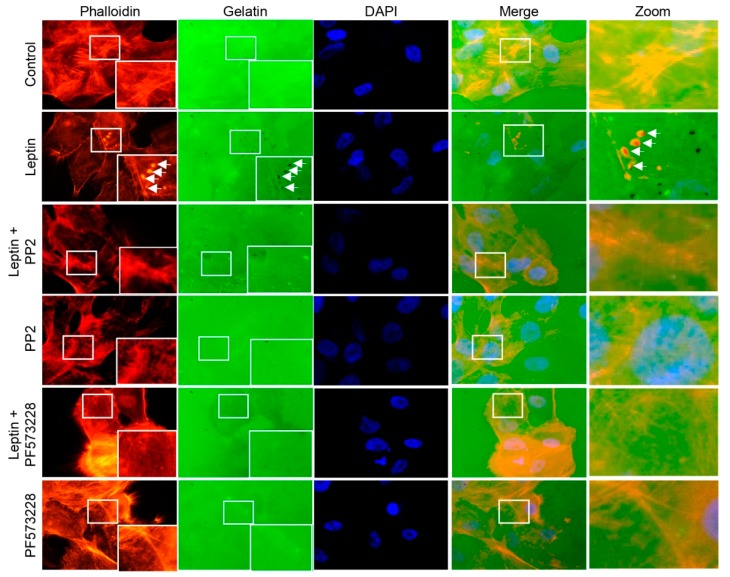
Figure 6.
Src and FAK regulate the invasion capacity of MCF10A cells stimulated with leptin. Representative images of MCF10A cells pre-treated or not with PP2 or PF-573228 (10 µM) for 30 min and subsequently incubated in the presence or absence of leptin (400 ng/mL) for 24 h. Actin puncta was detected with phalloidin (red) and Alexa 488-labeled gelatin (green) was used as a specific substrate for the membrane-bound MMP-14 located at the edge of invadopodia. Arrows indicate areas of gelatin degradation. Images were acquired using a 100× magnification.

