Abstract
The term “microbiome” defines the collective genome of all commensal, symbiotic, and pathogenic microbes living in the human body. The composition of microbiota in the gut and skin is influenced by many factors such as the stage of life, nutrition, lifestyle, and gender. In the past few years, several scientific papers have demonstrated an implication of microbiota in many immune-mediated diseases, for example, diabetes, ulcerative colitis, and multiple sclerosis. The alterations in the proportion of gut microbiota have emerged as potential immunomodulators with the capacity to induce physiologic as well as pathologic immune responses against the human body, causing inflammation and destruction of tissues or organs. The microbiota influences the differentiation of adaptive immune cells not only in the gut but also in the skin. Alopecia areata (AA) is a dermatologic disorder which causes hair loss in most cases resistant to treatment. There are some clinical and experimental evidences indicating that AA is the demonstration of autoimmune attack against hair follicles. The factors that may implicate such an autoimmunity in AA still remain unknown. Despite more and more evidences demonstrate that human microbiome plays a key role in human health and diseases, to the best of our knowledge, no study has been conducted to analyze an implication of microbiome in the pathogenesis of AA. Undoubtedly, there is a need to performing a study which might explain the involvement of gut and skin microbiota in the unclear pathogenesis of AA and lead to alternative treatment options for numerous patients suffering from current treatment limitations.
Key words: Alopecia areata, dermatology, gut microbiome, pathophysiology, skin microbiome
INTRODUCTION
The term “microbiome” defines the collective genome of all commensal, symbiotic, and pathogenic microbes living in the human body. The healthy intestinal and skin microbiota is an ecological community of trillions of microorganisms containing viruses, bacteria, archaea, and fungi that share human body space.[1,2] Among them are not only commensal and symbiotic organisms occurring on the skin, mouth, gastrointestinal tract, and in the respiratory and urinary tracts but also those that cause pathological conditions. The composition of microbiota in the gut and skin is influenced by many factors such as the stage of life, nutrition, lifestyle, and gender.[3]
GUT MICROBIOTA AND IMMUNE SYSTEM
In the past few years, several scientific papers have demonstrated an implication of microbiota in many immune-mediated diseases, for example, diabetes, Crohn's disease, ulcerative colitis, and multiple sclerosis. The interpretation and exploration of such findings occurring in many studies published recently are a subject of much debate now. The prevalence of autoimmune diseases is growing, especially in Western countries, affecting majorly women.[4,5] It has been considered that modern era lifestyle can influence the normal flora composition effecting in deregulation of the immune system. The alterations in the proportion of gut microbiota have emerged as potential immunomodulators with the capacity to induce physiologic as well as pathologic immune responses against the human body, causing inflammation and destruction of tissues or organs. Imbalances in the gut microbiota, described as dysbiosis, may trigger several disorders through the manipulation of activity of T-cells that are both near to and distant from the site of their induction.[6] Particular bacteria inhabiting defined niches transmit distinct signals and may affect functions of innate and adaptive immune system. Thus microbiome may effect in distal to the site of colonization systemic process. Culturing and characterization of human commensal bacteria gave the possibility to assess their influence on the host's immune system as well as provide new tools for defining which cell types and signaling tracks are relevant for inducing of the distinct immune responses. A valuable advance was the identification of immunoglobulin A (IgA)-coated gut bacteria in humans, which provides an idea of the bacteria that can be sensed by the cells of the adaptive immune system. This investigation enables a comparison of species of bacteria that elicit T-cell-dependent and T-cell-independent IgA-mediated responses in the host's immune system.[7,8,9] Mucosal IgA is secreted across the epithelium which binds to the immunoglobulin receptor. IgA can bind to microbes, several components of diet or antigens in the lumen of the intestine. As the result of this process is a formation of IgA-coated elements which prevent direct interaction with host's immune system. Thus, it controls the genes' expression by intestinal microbes as well as it can provide a physical barrier. Notably, gut microbiome affects the accumulation of cells that may express IgA and both the level and the diversity of IgA in the lumen.[3]
SKIN MICROBIOTA AND IMMUNE SYSTEM
The microbiota influences the differentiation of adaptive immune cells not only in the gut but also in the skin. Although immunological communication occurs between mucosal tissues such as the intestine and the lung or the nasopharynx, it seems to be specific for compartment immunological regulation in the skin.[8,9,10] A study suggested that Th17 cells in the skin are affected by the skin microbiota independently of gut microbiota. The production of interleukin (IL)-17A by T-cells in the skin requires the expression of IL1R but not IL23R, in contrast to the requirements of Th17 cells in the gut and in line with compartment-specific mechanism for T-cell regulation. This condition might be caused by different types of challenges that skin and mucosal sites are faced with, which effect in the requirement of distinct pathways to control their local immune responses.[11,12,13]
GUT–SKIN AXIS?
On the other hand, some studies suggest that the intestinal microbiome's influence extends beyond the gut and thus contributes to the function and dysfunction of distant organ system.[12] In this ever-expanding field, scientists are exploring how the local microbiome influences immunity at distal sites such as skin, clarifying the existence of “gut–skin axis.”[14,15] It is still undiscovered whether the altered composition of gut microbiota may precede the development of the skin disease, and thereby shift the immune system, disrupting the gut epithelial barrier. On the other hand, it has been considered whether skin and gut microbiome are autonomous and independent from each other. Skin as a physical barrier constantly interacting with external factors such as solar irradiation, grades of humidity or dryness, and microorganisms might influence integrity, homeostasis, and the composition of skin microbiota.[13] These factors may lead to the skin dysbiosis and thus contribute to the disruption of immune homeostasis in the skin and promote the development of diverse skin diseases regardless of gut microbiome.
COMPLEX AND UNEXPLORED PATHOPHYSIOLOGY OF ALOPECIA AREATA AND NEW PERSPECTIVES
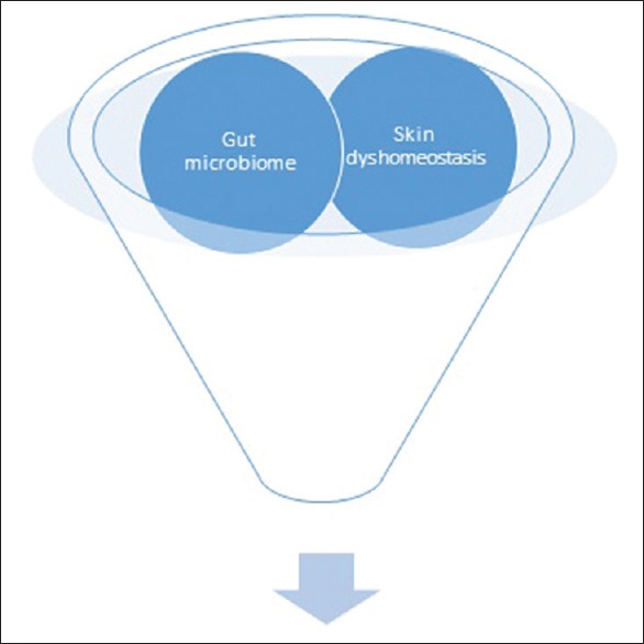
Alopecia areata (AA) is a dermatologic disorder occurring in the population at a frequency of 0.1%–0.2%, which causes hair loss in most cases resistant to treatment. The disease negatively influences the shaping of the body's image and self-esteem, which makes an important role in the process of socialization, causing in many cases inefficient functioning in the social environment and decreased quality of life.[16] The etiology and precise pathomechanism of AA is speculative [Table 1]. There are some clinical and experimental evidences indicating that AA is the demonstration of autoimmune attack against hair follicles which causes an inflammatory condition of the hair follicle.[17,18] The autoimmunity is the defect in immune tolerance, resulting in the production and proliferation of autoreactive T-cells and antibodies. The factors that may implicate such an autoimmunity in AA, however, remain still unknown.[19,20] Despite more and more evidences demonstrate that human microbiome plays a key role in human health and diseases, to the best of our knowledge, no study has been conducted to analyze an implication of microbiome in the pathogenesis of AA. There is only one case report describing hair growth in two patients suffering from AA after fecal microbiota transplant, which may suggest the potential role of gut microbiota in the disease mechanism.[21] Undoubtedly, there is a need to performing a study which might explain the involvement of gut and skin microbiota in the unclear pathogenesis of AA, which leads to alternative treatment options for numerous patients suffering from current treatment limitations [Figure 1]. Simultaneous analysis of both gut and skin microbiome in patients with AA will provide an exceptional insight if observed autoimmunity in AA is caused from the “inside” (gut–skin axis) or from the “outside” (skin).
Table 1.
Recently explored etiopathology of alopecia areata
| Category | Factors | References |
|---|---|---|
| Loss of immune privilege and immune dysregulation | Increased secretion of TNFα, IL 15 that activate target immune cells via the Janus kinase-signal transducer and activator of transcription (JAK-STAT) signaling pathway Supressed function of regulatory T cells and promoted proliferation of natural killer cells and T cells |
16, 17, 18, 22, 23 |
| Genetic predisposition (selected alopecia areata associated genes) |
HLA-DQB1*03 ACOXL/BCL2L11 – proteins regulating apoptosis GARP (LRRC32) – connected with activated Tregs TNF (inflammatory cytokine) IL13 (Th2 cytokine) TNFSF4 (OX40 ligand regulating T cell activation) THADA (thyroid adenoma associated protein) FASLG (FAS ligand controlling apoptosis) |
18, 22, 24, 25, 26 |
| Vitamines and hormones | Retinoic acid Vitamin D PTH Stress hormones |
18, 22, 27 |
| Emotional stress | Childhood trauma Total lifetime traumatic events |
22, 28 |
| Other | Infections Drugs Vaccinations Diet |
18, 22, 29 |
Figure 1.

What's new in etiopathology of alopecia areata – possible role of skin and gut microbiome
CONCLUSION
The proper understanding of the role of skin and gut microbiome might be undoubtedly a novel insight at the still unexplored pathophysiology of AA. Explanation of mechanisms that distinguish between physiologic and pathogenic microbiome–host interactions might identify therapeutic targets for preventing or modulating that disease. Therapeutic microbiome manipulation might represent an innovative treatment option for AA and undoubtedly has to be an important subject for future research regarding the important role of these dermatologic diseaseS in the abnormal process of socialization, decreasing the quality of life.
Financial support and sponsorship
Nil.
Conflicts of interest
There are no conflicts of interest.
REFERENCES
- 1.Human Microbiome Project Consortium. A framework for human microbiome research. Nature. 2012;486:215–21. doi: 10.1038/nature11209. [DOI] [PMC free article] [PubMed] [Google Scholar]
- 2.Sommer F, Bäckhed F. The gut microbiota – Masters of host development and physiology. Nat Rev Microbiol. 2013;11:227–38. doi: 10.1038/nrmicro2974. [DOI] [PubMed] [Google Scholar]
- 3.Honda K, Littman DR. The microbiota in adaptive immune homeostasis and disease. Nature. 2016;535:75–84. doi: 10.1038/nature18848. [DOI] [PubMed] [Google Scholar]
- 4.Bengmark S. Gut microbiota, immune development and function. Pharmacol Res. 2013;69:87–113. doi: 10.1016/j.phrs.2012.09.002. [DOI] [PubMed] [Google Scholar]
- 5.Wu HJ, Wu E. The role of gut microbiota in immune homeostasis and autoimmunity. Gut Microbes. 2012;3:4–14. doi: 10.4161/gmic.19320. [DOI] [PMC free article] [PubMed] [Google Scholar]
- 6.Peterson CT, Sharma V, Elmén L, Peterson SN. Immune homeostasis, dysbiosis and therapeutic modulation of the gut microbiota. Clin Exp Immunol. 2015;179:363–77. doi: 10.1111/cei.12474. [DOI] [PMC free article] [PubMed] [Google Scholar]
- 7.Palm NW, de Zoete MR, Cullen TW, Barry NA, Stefanowski J, Hao L, et al. Immunoglobulin A coating identifies colitogenic bacteria in inflammatory bowel disease. Cell. 2014;158:1000–10. doi: 10.1016/j.cell.2014.08.006. [DOI] [PMC free article] [PubMed] [Google Scholar]
- 8.Bunker JJ, Flynn TM, Koval JC, Shaw DG, Meisel M, McDonald BD, et al. Innate and adaptive humoral responses coat distinct commensal bacteria with immunoglobulin A. Immunity. 2015;43:541–53. doi: 10.1016/j.immuni.2015.08.007. [DOI] [PMC free article] [PubMed] [Google Scholar]
- 9.Öhman L, Törnblom H, Simrén M. Crosstalk at the mucosal border: Importance of the gut microenvironment in IBS. Nat Rev Gastroenterol Hepatol. 2015;12:36–49. doi: 10.1038/nrgastro.2014.200. [DOI] [PubMed] [Google Scholar]
- 10.Berbers RM, Nierkens S, van Laar JM, Bogaert D, Leavis HL. Microbial dysbiosis in common variable immune deficiencies: Evidence, causes, and consequences. Trends Immunol. 2017;38:206–16. doi: 10.1016/j.it.2016.11.008. [DOI] [PubMed] [Google Scholar]
- 11.Nagy G, Huszthy PC, Fossum E, Konttinen Y, Nakken B, Szodoray P. Selected aspects in the pathogenesis of autoimmune diseases. Mediators Inflamm. 2015;2015:351732. doi: 10.1155/2015/351732. [DOI] [PMC free article] [PubMed] [Google Scholar]
- 12.Salem I, Ramser A, Isham N, Ghannoum MA. The gut microbiome as a major regulator of the gut-skin axis. Front Microbiol. 2018;9:1459. doi: 10.3389/fmicb.2018.01459. [DOI] [PMC free article] [PubMed] [Google Scholar]
- 13.Lee SY, Lee E, Park YM, Hong SJ. Microbiome in the gut-skin axis in atopic dermatitis. Allergy Asthma Immunol Res. 2018;10:354–62. doi: 10.4168/aair.2018.10.4.354. [DOI] [PMC free article] [PubMed] [Google Scholar]
- 14.Naik S, Bouladoux N, Wilhelm C, Molloy MJ, Salcedo R, Kastenmuller W, et al. Compartmentalized control of skin immunity by resident commensals. Science. 2012;337:1115–9. doi: 10.1126/science.1225152. [DOI] [PMC free article] [PubMed] [Google Scholar]
- 15.Borde A, Šstrand A. Alopecia areata and the gut-the link opens up for novel therapeutic interventions. Expert Opin Ther Targets. 2018;22:503–11. doi: 10.1080/14728222.2018.1481504. [DOI] [PubMed] [Google Scholar]
- 16.Wang ECE, Christiano AM. The changing landscape of alopecia areata: The translational landscape. Adv Ther. 2017;34:1586–93. doi: 10.1007/s12325-017-0540-9. [DOI] [PMC free article] [PubMed] [Google Scholar]
- 17.Pratt CH, King LE, Jr, Messenger AG, Christiano AM, Sundberg JP. Alopecia areata. Nat Rev Dis Primers. 2017;3:17011. doi: 10.1038/nrdp.2017.11. [DOI] [PMC free article] [PubMed] [Google Scholar]
- 18.Dainichi T, Kabashima K. Alopecia areata: What's new in epidemiology, pathogenesis, diagnosis, and therapeutic options? J Dermatol Sci. 2017;86:3–12. doi: 10.1016/j.jdermsci.2016.10.004. [DOI] [PubMed] [Google Scholar]
- 19.Juárez-Rendón KJ, Rivera Sánchez G, Reyes-López MÁ, García-Ortiz JE, Bocanegra-García V, Guardiola-Avila I, et al. Alopecia areata. Current situation and perspectives. Arch Argent Pediatr. 2017;115:e404–e411. doi: 10.5546/aap.2017.eng.e404. [DOI] [PubMed] [Google Scholar]
- 20.Ito T, Tokura Y. The role of cytokines and chemokines in the T-cell-mediated autoimmune process in alopecia areata. Exp Dermatol. 2014;23:787–91. doi: 10.1111/exd.12489. [DOI] [PubMed] [Google Scholar]
- 21.Rebello D, Wang E, Yen E, Lio PA, Kelly CR. Hair growth in two alopecia patients after fecal microbiota transplant. ACG Case Rep J. 2017;4:e107. doi: 10.14309/crj.2017.107. [DOI] [PMC free article] [PubMed] [Google Scholar]
- 22.Strazzulla LC, Wang EHC, Avila L, Lo Sicco K, Brinster N, Christiano AM, et al. Alopecia areata: Disease characteristics, clinical evaluation, and new perspectives on pathogenesis. J Am Acad Dermatol. 2018;78:1–2. doi: 10.1016/j.jaad.2017.04.1141. [DOI] [PubMed] [Google Scholar]
- 23.Xing L, Dai Z, Jabbari A, Cerise JE, Higgins CA, Gong W, et al. Alopecia areata is driven by cytotoxic T lymphocytes and is reversed by JAK inhibition. Nat Med. 2014;20:1043–9. doi: 10.1038/nm.3645. [DOI] [PMC free article] [PubMed] [Google Scholar]
- 24.Betz RC, Petukhova L, Ripke S, Huang H, Menelaou A, Redler S, et al. Genome-wide meta-analysis in alopecia areata resolves HLA associations and reveals two new susceptibility loci. Nat Commun. 2015;6:5966. doi: 10.1038/ncomms6966. [DOI] [PMC free article] [PubMed] [Google Scholar]
- 25.Redler S, Angisch M, Heilmann S, Wolf S, Barth S, Basmanav BF, et al. Immunochip-based analysis: High-density genotyping of immune-related loci sheds further light on the autoimmune genetic architecture of alopecia areata. J Invest Dermatol. 2015;135:919–21. doi: 10.1038/jid.2014.459. [DOI] [PubMed] [Google Scholar]
- 26.Jagielska D, Redler S, Brockschmidt FF, Herold C, Pasternack SM, Garcia Bartels N, et al. Follow-up study of the first genome-wide association scan in alopecia areata: IL13 and KIAA0350 as susceptibility loci supported with genome-wide significance. J Invest Dermatol. 2012;132:2192–7. doi: 10.1038/jid.2012.129. [DOI] [PubMed] [Google Scholar]
- 27.Zhang X, Yu M, Yu W, Weinberg J, Shapiro J, McElwee KJ. Development of alopecia areata is associated with higher central and peripheral hypothalamic-pituitary-adrenal tone in the skin graft induced C3H/HeJ mouse model. J Invest Dermatol. 2009;129:1527–38. doi: 10.1038/jid.2008.371. [DOI] [PMC free article] [PubMed] [Google Scholar]
- 28.Willemsen R, Vanderlinden J, Roseeuw D, Haentjens P. Increased history of childhood and lifetime traumatic events among adults with alopecia areata. J Am Acad Dermatol. 2009;60:388–93. doi: 10.1016/j.jaad.2008.09.049. [DOI] [PubMed] [Google Scholar]
- 29.McElwee KJ, Niiyama S, Freyschmidt-Paul P, Wenzel E, Kissling S, Sundberg JP, et al. Dietary soy oil content and soy-derived phytoestrogen genistein increase resistance to alopecia areata onset in C3H/HeJ mice. Exp Dermatol. 2003;12:30–6. doi: 10.1034/j.1600-0625.2003.120104.x. [DOI] [PubMed] [Google Scholar]


