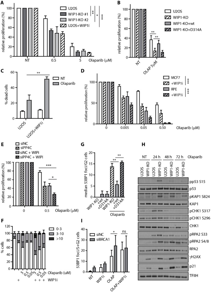
Figure 5.
WIP1 deficient cells are more sensitive to PARP inhibition. (A) Cell survival of parental U2OS, two independent U2OS-WIP1-KO cell lines with or without combined treatment with WIP1i was evaluated 7 days after treatment with indicated doses of olaparib using resazurin viability assay. Plotted is mean +/− SD, n ≥ 3. Statistical significance evaluated by two-way ANOVA. (B) Cell survival of parental U2OS, U2OS-WIP1-KO cells and cell lines complemented with wild-type or phosphatase-dead (D314A) mutant of WIP1 in response to 5 μM olaparib as in A. Statistical significance evaluated by two-tailed t-test (n ≥ 3). (C) Percentage of dead cells was evaluated by Hoechst 33258 staining and FACS analysis 7 days after treatment with 5 μM olaparib in U2OS cell line with or without combined treatment with WIP1i. Plotted is mean +/− SD. (D) Cell survival of RPE and MCF7 cell lines with or without combined treatment with WIP1i was evaluated 7 days after treatment with indicated doses of olaparib using resazurin viability assay. Plotted is mean +/− SD. N ≥ 3. Statistical significance evaluated by two-way ANOVA. (E) Cells were transfected with control siRNA (siNC) or siRNA to PP4C (siPP4C). Cell survival was evaluated after 7 days of treatment with olaparib and DMSO or WIP inhibitor. Statistical significance evaluated by two-tailed t-test (n = 3). (F) Quantification of 53BP1 foci number 3 days after treatment with olaparib. U2OS cells were treated with indicated doses of olaparib together with or without WIP1i for 3 days, fixed, stained with 53BP1 antibody and percentage of cells having 0–3, 3–10 and >10 foci were quantified. Mean +/− SD is plotted, n ≥ 3. (G) Quantification of 53BP1 foci after treatment with olaparib. U2OS-WIP1-KO cells and cell lines complemented with wild-type or phosphatase-dead (D314A) mutant of WIP1 were treated with WIP1i and olaparib for 3 days, fixed after pre-extraction and stained with 53BP1 antibody. Number of 53BP1 foci in S/G2 cells was evaluated using DAPI content of >2 n to gate S-G2 cells. Mean of median foci number +/− SD is plotted, n ≥ 3. Statistical significance evaluated by two-tailed t-test. (H) Response of U2OS and U2OS-WIP1-KO cell lines to treatment with 5 μM olaparib for 24–72h was analyzed by Western blotting using indicated antibodies. I) Quantification of 53BP1 foci 3 days after treatment with olaparib. MCF7 cells were transfected with indicated siRNAs and treated after 2 days with WIP1i and olaparib alone or combined for further 3 days. Cells were fixed after pre-extraction and stained with 53BP1 antibody. Number of 53BP1 foci in S/G2 cells was evaluated using DAPI content of >2 n to gate S-G2 cells. Mean of median foci number +/− SD is plotted. Statistical significance evaluated by two-tailed t-test.

