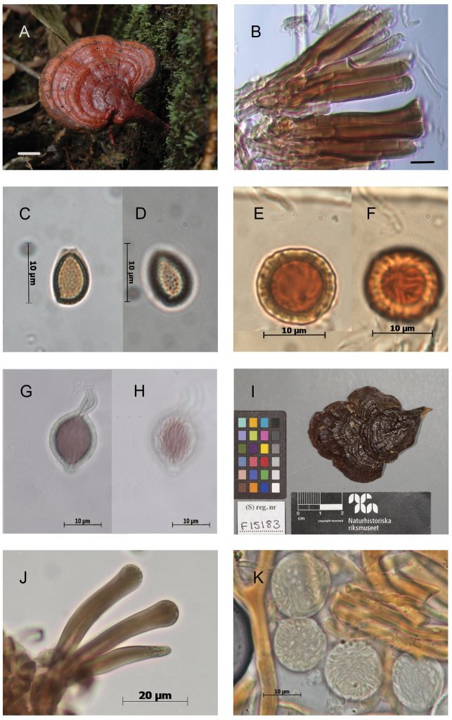
Figure 4.
Morphological features and microscopic structures of Ganoderma parvulumA–H MUCL 53123 A pilear surface B cuticular cells C–D basidiospores with free to subfree pillars E–H chlamydospores ornamented with free to partially anastomosed ridges E–F from context G–H from culture I–K E. Ule 2748 (as G. subamboinense, holotype) I upper surface of basidiomata, copyright: Naturhistoriska riksmuseet, Stockholm J cuticular cells K chlamydospores ornamented with partially anastomosed ridges, from context, in KOH. Scale bars: 1 cm (A); 5 μm (B).

