Genome sequence analysis has demonstrated that there is widespread lateral gene transfer among strains within the species C. trachomatis and with other closely related Chlamydia species in laboratory experiments. This is in contrast to the complete absence of foreign DNA in the genomes of sequenced clinical C. trachomatis strains. There is no understanding of any mechanisms of genetic transfer in this important group of pathogens. In this report, we demonstrate that interspecies genetic exchange can occur but that the nature of the fragments exchanged is different than those observed in intraspecies crosses. We also generated a large hybrid strain library that can be exploited to examine important aspects of chlamydial disease.
KEYWORDS: Chlamydia, lateral gene transfer, recombination, transposon
ABSTRACT
Lateral gene transfer (LGT) among Chlamydia trachomatis strains is common, in both isolates generated in the laboratory and those examined directly from patients. In contrast, there are very few examples of recent acquisition of DNA by any Chlamydia spp. from any other species. Interspecies LGT in this system was analyzed using crosses of tetracycline (Tc)-resistant C. trachomatis L2/434 and chloramphenicol (Cam)-resistant C. muridarum VR-123. Parental C. muridarum strains were created using a plasmid-based Himar transposition system, which led to integration of the Camr marker randomly across the chromosome. Fragments encompassing 79% of the C. muridarum chromosome were introduced into a C. trachomatis background, with the total coverage contained on 142 independent recombinant clones. Genome sequence analysis of progeny strains identified candidate recombination hot spots, a property not consistent with in vitro C. trachomatis × C. trachomatis (intraspecies) crosses. In both interspecies and intraspecies crosses, there were examples of duplications, mosaic recombination endpoints, and recombined sequences that were not linked to the selection marker. Quantitative analysis of the distribution and constitution of inserted sequences indicated that there are different constraints on interspecies LGT than on intraspecies crosses. These constraints may help explain why there is so little evidence of interspecies genetic exchange in this system, which is in contrast to very widespread intraspecies exchange in C. trachomatis.
IMPORTANCE Genome sequence analysis has demonstrated that there is widespread lateral gene transfer among strains within the species C. trachomatis and with other closely related Chlamydia species in laboratory experiments. This is in contrast to the complete absence of foreign DNA in the genomes of sequenced clinical C. trachomatis strains. There is no understanding of any mechanisms of genetic transfer in this important group of pathogens. In this report, we demonstrate that interspecies genetic exchange can occur but that the nature of the fragments exchanged is different than those observed in intraspecies crosses. We also generated a large hybrid strain library that can be exploited to examine important aspects of chlamydial disease.
INTRODUCTION
The chlamydiae are obligate intracellular bacteria that cause a wide spectrum of diseases in humans and animals of veterinary importance. The genus Chlamydia includes closely related pathogens of humans (Chlamydia trachomatis), pigs (C. suis), and mice (C. muridarum) and more distantly related species infecting a wide variety of hosts. Sequence analysis of the genomes of different Chlamydia spp. demonstrated a highly reduced physiological capacity encoded by genomes of approximately 1,000 kb. The mechanisms of pathogenicity and host tropism are issues remaining to be resolved. In some cases, modest differences in genome structure are correlated with significant tropism differences (1). An example of this resides in the host tropism difference between C. trachomatis, a primarily human pathogen that does not naturally infect mice, and C. muridarum, a mousetropic species that does not cause zoonotic disease. The genetic basis of these tropism differences is unclear. While the genomes of C. trachomatis and C. muridarum are highly syntenic and otherwise quite similar, there is a single region of high diversity, termed the plasticity zone (PZ), that varies in size and structure between species (2). It is hypothesized that genetic differences in the PZ might be a key factor in the ability of each species to adapt to its particular host.
Demars and colleagues showed that lateral gene transfer (LGT) was possible in C. trachomatis, using parental strains with different antibiotic resistance phenotypes (3). Subsequent genome sequence analysis confirmed these analyses and demonstrated that single transferred fragments can account for up to ∼40% of the chromosome (4, 5). Recent genomics data from a variety of laboratories have shown that genomic mosaicism in clinical isolates is very common, perhaps universal, within C. trachomatis (6, 7). This is in contrast to the near absence of any DNA incorporated from a different species either outside or inside the genus Chlamydia. The sole example of a foreign, laterally acquired sequence in recent evolutionary history is the C. suis tet(C) island, a set of genes acquired from very distantly related bacteria that is widespread in C. suis strains (8). Our group has demonstrated that the C. suis tet(C) island can be readily recombined into C. trachomatis and C. muridarum in the laboratory (9). Aside from this example, very little is known about how these organisms acquire exogenous DNA and integrate it into their genomes.
In this work, we describe a system to examine lateral gene transfer within the genus Chlamydia that has utility in the study of host tropism by the organisms. We used a recently generated random library of C. muridarum transposon mutants (10) as donor strains in mixed infections with C. trachomatis and examined the nature of interspecies lateral gene transfer among these chlamydiae. The collected progeny strains were then used to examine the possibilities and constraints of heterospecific lateral gene transfer within the genus Chlamydia.
RESULTS
Description of the system.
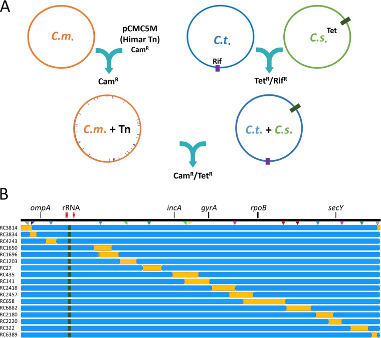
The overall strategy for developing a chimeric C. trachomatis × C. muridarum recombinant library is outlined in Fig. 1. The present study builds on the transposition system of Wang and colleagues (10), who describe the development of a library of independent transposon mutant C. muridarum strains that are marked with a Himar-generated transposon (chloramphenicol resistant [Camr]) at random positions around the chromosome. A similar system for C. trachomatis has been recently described by LaBrie et al. (11). The C. muridarum transposon library allows the production of a wide variety of recombinant C. trachomatis strains carrying syntenic C. muridarum chromosomal fragments. The tetracycline-resistant (Tcr) parent C. trachomatis strains are recombination products involving a rifampin-resistant C. trachomatis strain and a clinical Tcr C. suis strain isolated from an infected pig in a production facility (12). These C. suis strains readily recombine with C. trachomatis parents in vitro, leading to the transfer of a tet(C) allele into either C. trachomatis D/UW3, F/70, or L2/434 (9). The resulting Tcr C. trachomatis isolates each contain an ∼16-kb fragment of DNA from the C. suis parent, consisting of the tet(C) island and additional C. suis DNA, located between the paired rRNA operons. In all cases, the transposon parent is indicated with a “CM” designation, while recombinant progeny are indicated with an “RC” designation.
FIG 1.
Generating interspecies recombinants from crosses between C. muridarum and C. trachomatis parents. (A) Transposon mutagenesis, as described in reference 10, led to the creation of a variety of randomly mutated, chloramphenicol-resistant C. muridarum strains (C.m. + Tn). C. trachomatis parents were generated by transferring the C. suis tet(C) locus, plus adjacent sequences, to rifampin (Rif)-resistant C. trachomatis strains D/UW3, L2/434, and F/70, using previously described methods (8). Cocultures of individual C. muridarum and C. trachomatis parent strains were then incubated in the presence of both Cam and Tc to select for recombinant progeny. (B) Doubly antibiotic-resistant strains were cloned by limiting dilution, and their genomes were sequenced. A linear representation of the C. trachomatis chromosome is shown (black line). The locations of selected genes are included for reference purposes. The location of the tet(C) locus is indicated with green shading in the individual recombinants and is in this position in each of the progeny strains generated by LGT. Seventeen individual recombinant strains (orange bars), which collectively represent 79% of the C. muridarum genome, are indicated. The complete list of recombinant strains is given in Fig. S1 and Table S1.
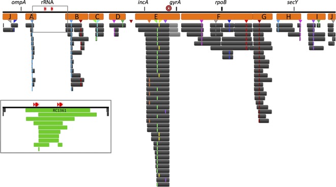
The focus of this work was the generation of C. trachomatis strains with different regions of C. muridarum recombined into the chromosome. However, progeny from every coculture event also included reciprocal crosses in which Tcr C. trachomatis fragments were transferred into a C. muridarum chromosomal background. The percentages of these reciprocal clones were highly variable between different parent combinations. A subset of the C. muridarum recipient strains were included in the genome sequencing, and each contained recombined inserts centered on the C. suis tet(C) island located between the rRNA operons (Fig. 2). The largest of these clones (RC1361) replaces approximately 8% of the genome with sequence from L2tet9 (Fig. 2; see Table S1 in the supplemental material). This is a region of the chromosome that is poorly represented in the C. trachomatis L2 backbone constructs because it is the location of the C. trachomatis parent Tcr marker.
FIG 2.
Recombinant progeny generated from mixed cultures of C. muridarum (Camr) and C. trachomatis (Tcr). Each black bar represents a segment of C. muridarum DNA transferred into an individual C. trachomatis parent. The three grey bars are clones generated without the Camr transposon insert. All the strains shown are cloned recombinant progeny from individual LGT experiments. The inset shows an expanded view of the chromosomal region containing the rRNA operons (red). The green sequences within the inset represent reciprocal clones, in which C. trachomatis L2tet9 DNA transferred into various C. muridarum backgrounds. The indicated reciprocal clone (RC1361) is described in Fig. S1 and Table S1. The orange bars show different sets of recombinants that are compared in Fig. 6. The transposon insertion sites of the individual parents used are shown as colored triangles and are indicated with tick marks in the relevant recombinant strains. Gene identities are included as in Fig. 1. The red target symbol near the center of the genome is the chromosomal ter sequence, a recombination hot spot. For scale, the rRNA operons and intervening sequence equal 43 kb.
Primary characterization of interspecies recombinants.
More than 500 C. trachomatis backbone interspecies recombinant strains were generated from a total of 33 C. trachomatis and C. muridarum parent combinations; 142 of them were cloned by limiting dilution, and the genomes were sequenced (Fig. 2; see Fig. S1 in the supplemental material). The strain library encompasses 79.3% of the C. muridarum chromosome introduced, in fragments, into C. trachomatis parent strains (see Fig. S1). The transferred fragments ranged in length from 558 to ∼124,000 bp (see Table S1). This range of sizes in the transferred genome fragments is in marked contrast to the intraspecies recombinants generated by our group (5) and in the work of DeMars et al. (4), in which fragments of 200 to 400 kb were routinely transferred in LGT events.
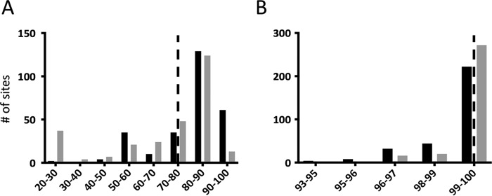
The location of the exchanged DNA was heavily influenced by the location of the selectable marker in both intraspecies and interspecies crosses. Genomic sequence identity differences led to limitations in the specific positions of recombination in the interspecies crosses, which were not apparent in the intraspecies crosses. While the overall chromosomal identity between C. trachomatis and C. muridarum is approximately 74%, the average sequence identity at individual recombination sites was over 92% when comparing either the 100 bp or the 1,000 bp immediately surrounding the crossover point (Fig. 3A). In contrast, genomic identify between the serovar L2, F, and J parents used in intraspecies recombination was above 99%, and virtually all the recombination targets shared greater than 97% identity (Fig. 3B). Therefore, the specific region involved in recombination was a function of the genetic identity between the parents, resulting in more limited regions of the chromosome being available for LGT in interspecies crosses.
FIG 3.
Recombination margins in Chlamydia intraspecies and interspecies lateral gene transfer events. The percentages of identity at the collected margins of recombination for chlamydial interspecies (A) and intraspecies (B) events are indicated for the collected primary recombination events. Intraspecies data were collected from the recombinant strains described by Jeffrey et al. (5). In each graph, the x axis indicates the percent identity between the parental strains of the recombinant progeny, and the y axis depicts the number of recombination margins falling in the indicated range. The gray bars indicate the percent identity within the 100 bp surrounding a crossover site (50 per side), and the black bars indicate the percent identity within the 1,000 bp surrounding a crossover site. The dashed line in each graph indicates the overall percent genomic identity for parents in the specific crosses.
Although the majority of crosses involved a single C. trachomatis parent (L2tet9), a small number of progeny strains were generated with different Tcr parents, C. trachomatis strains Dtet3 and Ftet11. Crosses between these C. trachomatis strains and C. muridarum CM007 led to C. trachomatis backbone progeny strains: RC103, RC106, and RC296, which resulted from crosses with Dtet3 (see Fig. S1 and Table S1) and a collection of uncharacterized serovar F progeny strains (not shown). These successful crosses indicated that our interspecies recombination system is not limited to a biotype LGV parent C. trachomatis strain.
The majority of strains were generated with single, discrete crossover events at each end of the introduced fragment. However, 32 of 142 primary recombinants (∼23%) had a mosaic sequence at one end, leading to secondary recombined fragments of C. muridarum DNA. These additional recombined sequences ranged in size from 7 to 8,005 bp, with a median length of 291 bp. The largest fragment introduced in a secondary recombination event was present in clone RC4102, in which an additional 8,005 bp of C. muridarum DNA sequence was included, in addition to the selected, primary recombined fragment (see Fig. S1). This is in contrast to C. trachomatis intraspecies LGT, in which virtually all the examined recombinants had extensive secondary recombination events, in some cases including fragments over 100 kb (5). A single recombinant (RC1420) contained an unselected, highly mosaic hybrid sequence within its chromosome that was not adjacent to the primary laterally transferred DNA. These ∼3,600 bp span open reading frames (ORFs) CTL0569 to CTL0574 (TC0591 to TC0596) and consist of approximately 58% C. muridarum DNA in a C. trachomatis L2tet9 background.
Wang and colleagues (10) describe the generation of the C. muridarum parents used in these LGT experiments. In that work, a subset of transposon mutants that contain two insertions in a single chromosome are described. Two of the mutants (CM007 and CM023) were used as C. muridarum parents in our crosses. A total of 31 cloned recombinant progeny strains were generated in crosses using these parents, and in each case, only one of the C. muridarum transposons was recombined into a progeny clone (see Table S1).
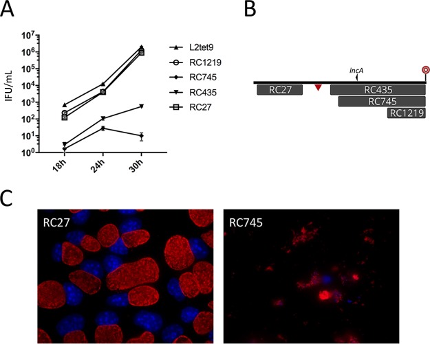
As C. muridarum grows much more quickly than C. trachomatis in vitro, one of our goals was to phenotype each recombinant for growth rate. For these experiments, the growth parameters were the production of infectious chlamydial particles, which was quantitatively assessed at three time points: 18, 24, and 30 h postinfection. These analyses did not identify any C. trachomatis backbone recombinants with a demonstrably higher growth rate (not shown). However, we did identify a set of clones in which a much smaller number of elementary bodies (EBs) were produced during growth (Fig. 4A). This set included all recombinants that contained C. muridarum DNA between clones RC27 and RC1219 (Fig. 4B; see Fig. S1), representing a large region of the genome between C. trachomatis ORFs CTL0320 and CTL0346 (C. muridarum TC0334 and TC0366). In each of these poorly growing recombinants, an early-lysis phenotype was demonstrated for both the inclusion and the host cell at approximately 20 to 26 h postinfection (Fig. 4C). All the other examined clones had growth parameters representative of the backbone strain.
FIG 4.
Examination of recombinant clones that express a growth defect. (A) Inclusion-forming units were determined at three time points following inoculation of McCoy cells with a set of four recombinants plus the C. trachomatis L2tet9 parent. The numbers on the vertical axis are inclusion-forming units per milliliter of lysed, infected cells. Monolayers were inoculated with approximately 30,000 IFU per ml. The error bars represent standard deviations. (B) Map of the recombinants tested in the growth experiments. This is a subset of the library of recombinants shown in Fig. 2 and includes recombinants from regions D (RC27) and E (RC435, RC745, and RC1219) in Fig. 2. The locations of incA and the 32-bp recombination hot spot can be identified here and in Fig. 2 for positioning. The triangle indicates the homologous position in C. muridarum that is targeted in transposon mutant CM013, for which no syntenic recombinants were generated. For scale, the C. muridarum insert shown for recombinant RC27 is 52 kb. (C) Immunofluorescence microscopic images of a methanol-fixed McCoy cell recombinant progeny strain with a wild-type inclusion morphology phenotype (RC27) and a recombinant expressing the early-lysis phenotype (RC745). The cells were fixed at 30 h and had identical multiplicities of infection. Red, MOMP; blue, DNA.
Recombination at nonhomologous target sites.
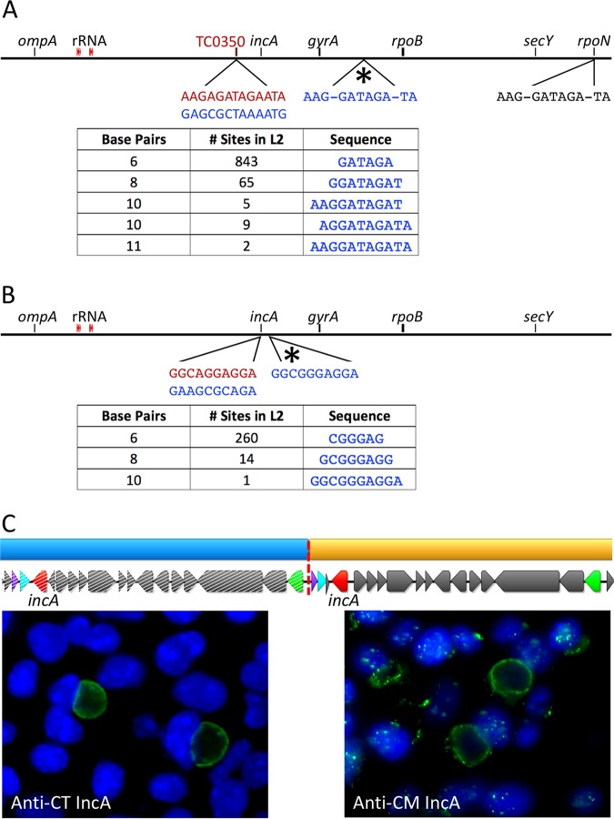
There were two examples of illegitimate recombination that led to progeny strains carrying C. muridarum DNA in nonhomologous regions of the chromosome. The first was a progeny strain carrying 16 kb of syntenic, similar DNA from each parent strain, which had a nonhomologous crossover event at the left end of the integrated fragment (RC1201) (see Table S1). The C. trachomatis target site used in this event shares 9 of 10 bases with the end of the inserted C. muridarum sequence, facilitating the recombination at the nonhomologous site. This recombination process led to a merodiploid status for most genes within this 16-kb region, including, for example, incA genes from both C. trachomatis and C. muridarum. Fluorescence microscopic analysis using species-specific anti-IncA monoclonal antibodies showed that IncA proteins from both species are produced and correctly localized to the inclusion membrane in strain RC1201 (Fig. 5C). Inclusions formed by recombinants encoding IncA from either or both parental species are fully fusion competent with inclusions formed by C. trachomatis or C. muridarum (see Fig. S2 in the supplemental material; data not shown).
FIG 5.
Examination of progeny strains using nonhomologous target sites in recombination. Strains RC936 (A) and RC1201 (B and C) have sequences that result from one (RC1201) or both (RC936) recombination endpoints occurring at nonhomologous target sites. (A and B) C. muridarum sequence is indicated in red, while C. trachomatis sequences are indicated in blue or black. In both cases, the predicted target of recombination shares no immediate identity with the C. muridarum donor DNA. The actual site of recombination, indicated with an asterisk above each linear chromosome, is either identical with the donor DNA sequence (RC1201) or very nearly identical (RC936). The tables show the numbers of occurrences in the recipient chromosome for increasingly long candidate target sites. In panel A, a second, identical target sequence is identified within rpoN. This target was not used in any identified recombinant. (C) The illegitimate recombination event at one end of the integrated DNA led to a duplication of ∼16 kb of chlamydial DNA. The recipient DNA (C. trachomatis; blue, with hatched ORFs) and the donor DNA (C. muridarum; tan, with solid ORFs) are largely homologous in this region. This clone carries homologous copies of incA (red) from each species, and the protein products of both genes are expressed and correctly localized to the inclusion membrane (green labeling in the micrographs). Host cell nuclei are labeled blue with DAPI (4′,6-diamidino-2-phenylindole) in each micrograph.
The second example of nonhomologous recombination resulted from crosses with the C. muridarum parent CM013, which contains a transposon insertion in TC0350 (Fig. 4B, triangle). Crosses with this parent yielded no C. trachomatis backbone clones that carried a homologous exchanged DNA sequence. Doubly resistant C. trachomatis backbone progeny were generated from crosses with CM013 as a parent, but each of them had the donated resistance allele in an incorrect position in the genome (Fig. 5).
Progeny strains that result from a crossover at a nonhomologous site in the chromosome allow measurement of a minimum target sequence that is required for successful recombination. This is most clearly demonstrated in progeny resulting from crosses with parent CM013. Six progeny strains from four independent crosses had identical insertion sites and sequences, represented by strain RC936 in Fig. S1 and Table S1. The transferred DNA contained the entire transposon sequence plus a total of 11 flanking base pairs, including the TA site used as a target for transposition of the parent (10). The predicted syntenic target site for integration of the transposon does not share any identity at these 11 bp (Fig. 5A), but there are two other positions in the recipient chromosome that do. This includes the actual position that was used as the recombination target in these strains—an intergenic site between ORFs CTL0506 and CTL0507—and an identical 11-bp site within rpoN. The latter position was not used in any of the LGT events.
The RC1201 clone resulted from an LGT event at a homologous position in the genome, with the right end terminating at the 32-bp recombination hot spot described above. However, the left end of this transferred DNA crossed over at a nonhomologous position, a 10-bp sequence that shares 9 bp with the donor DNA sequence and occurs only once in the chromosome (Fig. 5B). Similar to the CM013-based LGT, this 10-bp sequence shares no identity at the predicted syntenic site in the parental chromosomes. Therefore, in the two recombinant progeny strains that result from exchange at a nonhomologous position in the chromosome, a 10- or 11-bp nearly identical crossover point was used for recombination.
A bioinformatic approach was used to explore the minimum-size target that might be available for recombination in this system. This issue is most readily explored by examining recombination endpoints for RC936 and RC1201, the strains shown above to have undergone a nonhomologous-recombination event. In both cases, shorter identical candidate recombination targets are available in the recipient chromosome (Fig. 5A and B). This question is most readily addressed in the set of 6 progeny strains represented by RC936, each of which underwent recombination at the same, apparently random, 11-bp target sequence in the recipient chromosome. Shorter identical sequences are spread throughout the recipient genome, including a total of 14 candidate targets of 10 bp and over 900 candidate targets of 6 or 8 bp. Similarly, the left-end recombination endpoint for strain RC1201 involved a 10-bp nonsyntenic sequence, though there were many other, shorter potential crossover points in the recipient chromosome.
A statistical approach was used to examine the variation in length of different recombined fragments across the recipient chromosome. The collected primary LGT progeny strains were binned into 10 different groups, based on the position of their inserted DNA, regardless of their individual transposon parent (A to J in Fig. 2). All the regions represented by six or more independent recombinants were used to test the hypothesis that, in our LGT system, genetic exchange across the chromosome led to different-size C. muridarum inserts. A Weibull analysis of distribution was used to compare the average insert length for all recombinants (∼48,000 bp) to the average lengths of inserted DNA across these different regions of the chromosome. The analysis demonstrated that the average transferred lengths for most of the regions of the chromosome differed significantly from the mean (Fig. 6A), supporting a hypothesis that there are selective pressures within this interspecies LGT system that affect the length and nature of recombined fragments.
FIG 6.
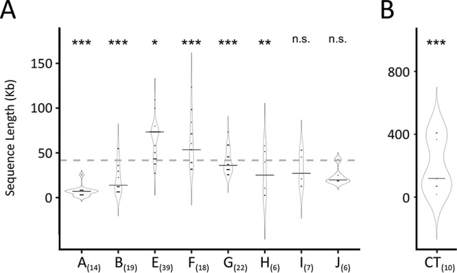
Lengths of primary recombined fragments generated in C. muridarum × C. trachomatis crosses. (A) Recombinant fragment lengths are indicated on the vertical axis, and the groups from Fig. 2 are listed on the horizontal axis. Each region shown in Fig. 2 that is represented by at least 6 individual clones was included in these analyses. The number of clones examined in each region is indicated in parentheses. The overall mean from all primary C. muridarum inserts into the C. trachomatis genome is indicated as a dashed line, while group medians are shown with solid lines within the violin plots. (B) Parallel data from recombinant progeny generated in intraspecies (C. trachomatis × C. trachomatis) recombinant strains described in reference 5. Note the different scales on the vertical axes in panels A and B. The difference between the mean length for each region and the overall mean for all recombined inserts was analyzed using a Weibull distribution. *, P value of ∼0.05; **, P < 0.05; ***, P < 0.001; n.s., not significant.
A similar approach was used to compare the sizes of primary recombined fragments in interspecies crosses to those in intraspecies crosses (Fig. 6B). The C. trachomatis intraspecies crosses described by Jeffrey et al. (5) had primary recombined fragments that ranged from ∼16,000 to 413,000 bp, with an average size of 181,000 bp. In contrast, the largest exchanged fragment in our interspecies crosses was ∼124,000 bp (strain RC658).
Recombination rate differences and results from multiparent crosses.
Coculture of C. trachomatis L2tet9 and every tested transposon parent yielded recombinant progeny. There were individual C. muridarum parents, however, that were uniquely efficient (CM020) or challenging (CM013) with respect to generation of recombinants. In direct comparisons, C. muridarum parent 020 was approximately 250-fold more prolific in crosses than C. muridarum parents 009 and 017 (not shown), and this efficiency led to a requirement for a much higher dilution of that parent in cocultures. Parent CM020 was the only tested parent that had a transposon in an intergenic region (between TC0080 and TC0081) (10). With these exceptions, every parent combination led to recombinant progeny with apparently similar efficiencies.
Cocultures of three different C. muridarum parents mixed with the single C. trachomatis L2tet9 were also conducted. Each C. muridarum parent used in these multistrain crosses was successful in the generation of progeny when mixed singly with C. trachomatis L2tet9, including the two strains (CM020 and CM013) described above that had unusual recombination rates. These multiple-parent crosses each generated recombinant L2 backbone strains, but the recombination and selection process led to biases favoring selected C. muridarum parents. In cross 1 (parents CM009, CM017, and CM020), the process led to a higher number of progeny derived from parent CM020, which was consistent with its high efficiency in the two-parent crosses. This multiparent cross also led to progeny containing CM009, but none originating from CM017. A second set of C. muridarum parents (CM012, CM013, and CM026) was used in a multiparent cross, and the experiment showed a strong bias toward CM012 and no progeny containing CM013 (see Table S2 in the supplemental material). These data demonstrate that a competitive hierarchy exists in LGT events when a mixture of parents is present and selected for in a cross.
Complete genome sequencing of 34 total multiple-parent-cross progeny identified no examples where more than one C. muridarum parent donated genomic DNA to a C. trachomatis progeny strain. This is consistent with mixed C. trachomatis × C. trachomatis crosses conducted in our laboratory (4). Therefore, in both interspecies and intraspecies crosses, all the progeny strains appear to result from interactions between single pairs of chlamydial developmental forms; alternatively, they could be the result of single interactions between a free DNA molecule and a recipient cell.
Lateral gene transfer surrounding the chlamydial PZ.
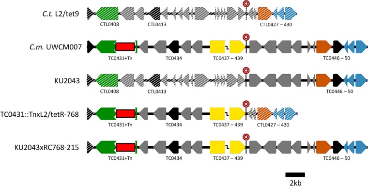
The PZ is a region of significant dissimilarity between strains and species against an overall genetically similar and syntenic set of genomes (2, 13, 14). The differences between the C. trachomatis L2/434 and the C. muridarum VR-123 plasticity zones are quite extensive (Fig. 7). Within C. muridarum, the region contains a set of ∼3-kb cytotoxicity-associated genes (TC0437 to TC0439; tox) whose functions are unclear (15). Additionally, the C. muridarum PZ has an apparent operon that encodes proteins predicted to function in purine metabolism (TC0444 to TC0441; add-gua). Both the tox and add-gua sequences are absent in C. trachomatis L2/434. In contrast, the much shorter C. trachomatis L2/434 plasticity zone encodes proteins involved in indole/tryptophan metabolism that are lacking in C. muridarum. With the goal of identifying recombinant strains carrying hybrid phenotypes in vitro or in vivo, we generated >50 independent recombinant strains in the PZ region, using four different C. muridarum parents (CM003, CM007, CM015, and CM023) in multiple experiments (Fig. 7; see Fig. S1). A variety of C. muridarum sequences were transferred in these crosses, ranging in length from ∼40,400 to 109,200 bp. Each PZ recombinant carried the entire set of three C. muridarum tox genes, although one of the tox genes carries an inactivating transposon in parent CM023, and this mutation was carried forward in several progeny strains (see Fig. S1 and Table S1).
FIG 7.
Open reading frame map of the plasticity zones for parental strains C. trachomatis L2tet9, C. muridarum transposon mutant CM007, and three PZ-based recombinants strains (KU2043, RC768, and RC215). C. muridarum ORFs are indicated with solid colors, while C. trachomatis ORFs are indicated with hatched colors. Homologous ORFs between strains are shown in black, green, orange, and blue. Plasticity zone genes unique to each species are shown in light gray, and the C. muridarum tox loci (truncated) are yellow. The transposed sequence within macP (TC0431) contributed by the CM007 parent is indicated in red. The 32-bp recombination target (ter) is indicated with a bullseye symbol. Homologous recombination at this target sequence forms a crossover point in each of the PZ-containing recombinant strains. Gene numbers are indicated by the species of origin.
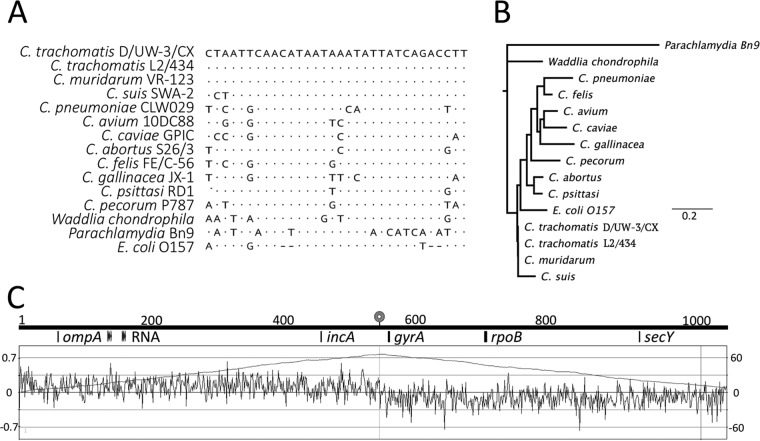
Surprisingly, each of these PZ-centered recombinant strains had an identical right margin: a 32-bp sequence of identity immediately to the right of the tox sequences (Fig. 8). These 32 bp represent a solitary island of identity between the PZs of the species, flanked by 3 to 10 kb of nonhomologous DNA on each side of the recombination target (Fig. 7). A sequence search of the chlamydial genome databases indicated that the 32-bp recombination target is strongly conserved within the chlamydiae, inside a genomic region that is among the most divergent in the genus.
FIG 8.
The 32-bp recombination target within the chlamydial PZ is conserved among strains and forms the chlamydial replication termination sequence. (A) BLAST analysis of the 32-bp recombination target among Chlamydia spp., Chlamydia-like organisms, and E. coli. The genomic sources of these data are indicated in Table S3 in the supplemental material. (B) Neighbor-joining phylogenetic tree built with the data from panel A. The scale bar represents the number of substitutions per site. (C) GCskew analysis of the C. trachomatis L2/434 chromosome showing an inflection point in the data at the position of the 32-bp recombination target (bullseye symbol), supporting the conclusion that this is the chlamydial replication terminator. Selected loci are indicated for reference purposes beneath a genome position scale (in kilobases).
BLAST-based analysis of other Gram-negative bacteria identified regions of the chromosomes that share identity with the 32-bp sequence, as shown for Escherichia coli O157A in Fig. 8. This motif has been described as the replication termination (ter) sequence, which is the point in a bacterial chromosome where replication forks meet (16). This is consistent in chlamydiae, as GCskew analysis in this work (Fig. 8) and by others (2) suggested the region surrounding the 32-bp sequence is a site of replication termination. There is evidence in other systems that this region of the chromosome is an active area of recombination (17).
In contrast to the highly selected hot spot on the right end of the PZ recombinants, crossover margins on the left end were more variable. Over 35 different PZ-based recombinants were genome sequenced, and C. muridarum inserts had 22 different left ends (Fig. 2; see Fig. S1 and Table S1). The left margin of the inserted C. muridarum sequence varied considerably among strains, with the largest inserted clone beginning adjacent to TC0366/CTL0346 (clone RC435) and the smallest beginning at TC0430 (clone RC768). Four independent PZ recombinants (RC49, RC104, RC259, and RC1560), generated with three different C. muridarum parents, had the same left and right margins and were therefore identical, except for a differently positioned transposon. Additionally, a progeny strain generated with the serovar D parent (clone RC103 [see Table S1]) also carried a recombined fragment identical to those seen in these strains. Therefore, 5 of 142 progeny strains generated from two different C. trachomatis parents and three different C. muridarum parents contained identical recombined fragments in the chromosome.
Our transposon-based LGT system yielded no progeny strains that contained sequence immediately to the right of the 32-bp recombination hot spot. Because the region contains gyrA, an alternate, quinolone-based selection strategy was used to generate recombinants covering this region of the PZ. Previous work in our laboratory led to the generation of ofloxacin-resistant (Oflr) C. muridarum strains (8), and they were used in the selection of Tcr-plus-Oflr chimeric progeny. Sequence analysis of the progeny strains showed that the transferred DNA began at the 32-bp recombination hot spot and extended right for ∼35 kb (strain KU2043) (Fig. 7; see Fig. S1).
At this point, our collection included PZ recombinants that extended to either the right or the left of the 32-bp recombination hot spot, but no strain contained the entire C. muridarum PZ in a C. trachomatis L2 background. To address this issue, clone KU2043 (Tcr plus Oflr) was crossed with either clone RC259 or clone RC768 (both Tcr plus Camr) to generate the Oflr-plus-Camr progeny strains RC8014 (C. trachomatis parent RC259) and RC215 (C. trachomatis parent RC768), strains that contained the entire C. muridarum PZ backbone in a C. trachomatis genomic context (Fig. 7).
DISCUSSION
The expansive list of fully genome-sequenced C. trachomatis strains has led to the conclusion that intraspecies LGT is nearly universal in the species. These sequencing efforts, in combination with laboratory-based LGT analyses, demonstrated that surprisingly large sections of a donor chromosome can be acquired by recipient strains. There is no evidence, however, of a clinical isolate carrying any DNA sequence from a non-C. trachomatis donor. Ecological constraints that restrict contact between recombination-competent C. trachomatis and other chlamydial species are most likely the primary factors limiting interspecies genetic exchange in this system. However, it is perhaps underappreciated that there are opportunities for closely related (18) and more distantly related (19) chlamydiae to occasionally occupy the same tissue as C. trachomatis. We undertook the above-described experiments to examine the constraints placed on interspecies recombination in vitro, with an overall goal of understanding the genetic limitations that may partially explain why clinical C. trachomatis strains carry no genomic evidence of DNA from a different bacterial species. The experiments also have created a recombinant library that we expect to be useful to individuals studying phenotypic differences between the two parent species.
Large-scale transfer of chromosomal DNA is not unique to the system. Genome sequencing of clinical Staphylococcus aureus isolates identified large chromosomal exchanges that are similar to what is documented for chlamydial intraspecies crosses (20). Distributive conjugal transfer, a type VII-dependent DNA exchange mechanism observed in Mycobacterium smegmatis, leads to progeny strains containing large fragments of homologous donor DNA within the recipient chromosome (21). Furthermore, there are many species for which acquisition of nonhomologous “genomic islands” from similar or dissimilar organisms is common. The chlamydial system is unique because it involves obligately intracellular bacteria, there do not yet appear to be any genomic determinants of transfer, and natural gene transfer is limited to intraspecies crosses.
The opportunity to examine global chromosomal transfer between chlamydial species evolved from in vitro transposition systems recently developed for C. trachomatis and C. muridarum, leading to random decoration of chlamydial chromosomes with antibiotic resistance markers (10). For our experiments, C. trachomatis parents were generated by recombining the C. suis tet(C) island (8) into C. trachomatis strain L2/434, DUW/3, or F/70, leading to C. trachomatis parents with a single anchored marker. These were crossed with C. muridarum strains carrying antibiotic resistance markers in a variety of positions on the chromosome. Reciprocal crosses were common but were redundant, as the C. trachomatis resistance marker was anchored at a single position in the chromosome (Fig. 2). In the present study, greater than 500 total progeny strains were generated, with 134 primary recombinants included in the analyses. Every single C. muridarum parent led to recombinant progeny, with both the C. muridarum and C. trachomatis parents serving as donor or recipient. In all but a single progeny strain, LGT led to classical homologous recombination at completely syntenic sites in recipient chromosomes. Two parents, CM013 and CM007, generated progeny clones that underwent illegitimate recombination, leading to nonhomologous-exchange events in the species. Our overall results are generally consistent with those observed in intraspecies C. trachomatis × C. trachomatis crosses, where no indels or other changes were detected following completely homologous sites of recombination.
While interspecies LGT was readily demonstrated with a large variety of different parent combinations, there were clear differences between interspecies and intraspecies gene transfer. In the interspecies crosses, the biggest fragment exchanged was 123,530 bp, and the majority of primary recombined fragments involved less than 12% of the genome. In contrast, C. trachomatis × C. trachomatis intraspecies LGT often led to exchanges of 30 to 40% of a donor parent’s chromosome. Differences were also observed in the genomic mosaicism that resulted from intraspecies versus interspecies LGT. In the intraspecies crosses previously described (n = 10), areas of high genomic mosaicism were observed in every recombinant, both in the regions surrounding the resistance alleles and in distal regions of the chromosome (5). These secondary recombination events were often over 200 kb from the fragment surrounding the transferred resistance allele. In the interspecies crosses, secondary recombination occurred in less than 25% of the crosses, and the lengths of these fragments were markedly less (median = 291 bp). Therefore, interspecies recombination led to both qualitative and quantitative differences in the genomes carried by progeny strains relative to intraspecies LGT.
Recombination at and around the chlamydial PZ was explored in detail. Eighteen different individual crosses were conducted, leading to 35 fully independent recombinant progeny. Remarkably, each of these progeny strains included a recombination event at the 32-bp recombination hot spot. This area is defined as the chlamydial ter, a sequence generally conserved among many bacteria. The replication terminator has been identified as a recombination hot spot in other species, as well (17). In chlamydiae, this highly conserved 32-bp sequence is located within a region of otherwise high dissimilarity and therefore is a logical recombination target in the PZ. In fact, this position in the chromosome has been used as a recombination endpoint in unlinked C. trachomatis strains taken from patients. Groups of clinical isolates characterized by Harris et al. (6) and represented by strains E/11023 and D(s)2923 (7) have a common recombination endpoint at or very near the 32-bp recombination hot spot. Other positions in the genome were used by multiple LGT events, including one spot adjacent to the PZ that is a crossover point in five independent strains (RC49, RC103, RC104, RC259, and RC1560). Therefore, hot spots are much more evident in vitro in cross-species LGT than was previously observed in intraspecies LGT.
Multiparent crosses were also conducted in this work. In these experiments, three different C. muridarum parents were combined with a single C. trachomatis parent. These crosses demonstrated that certain C. muridarum parents were much more successful in donating DNA than others in a mixed culture. However, sequence analysis of the progeny genomes demonstrated that no recipient strain received DNA from more than one C. muridarum donor parent. This is consistent with our intraspecies crosses, in which progeny strains generated in multiparent crosses carried DNA from only two of the parental strains.
One possible constraint to interspecies LGT is the genetic incompatibility of different regions of the parent chromosomes when mosaics are created via homologous recombination. Our recombination system identified one possible region in the chlamydial chromosome in which this appears to be the case. While doubly antibiotic-resistant C. trachomatis backbone recombinants were generated with C. muridarum parent CM013, 0 out of 6 clones were generated via classical homologous recombination. Each of the cloned progeny strains from these experiments, which were created in four fully independent mixed cultures, was the product of an identical illegitimate recombination event at a different position in the chromosome (Fig. 5). Progeny strains RC435 and RC745, which contain sequence adjacent to the homologous target for parent CM013, are the least fit strains in our collection, and this fitness defect manifested as an early-lysis phenotype (Fig. 4C). Additionally, recombinant RC1201 is the sole example of a strain containing a merodiploid sequence, and the region that is duplicated between the chromosomes is near the region targeted in CM013 parent crosses (Fig. 5). These collected data lead us to hypothesize that certain combinations of DNA sequences lead to phenotypically noncompetitive progeny in which the resulting proteome might contain, for example, incompatible members of a protein complex or biochemical pathway. The examination of such constraints awaits biological characterization of progeny strains, and experiments addressing these issues are under way in our laboratories.
MATERIALS AND METHODS
Creation of C. muridarum transposon mutants.
C. muridarum transposon mutants were generated and characterized by Wang et al. (10). Briefly, C. muridarum EBs (∼2 × 10⁷ inclusion-forming units [IFU]) were mixed with plasmid pCMC (∼10 μg) in 200 μl of CaCl2-Tris buffer for 20 to 30 min at room temperature (RT). The chlamydia-plasmid mixture was added to McCoy cells and incubated at RT for 15 min. Finally, the cell-chlamydia-plasmid mixture was aliquoted into a 6-well plate with 2 ml Dulbecco’s modified Eagle medium (DMEM) (Gibco) plus 10% fetal bovine serum (FBS) in each well. The plates were then incubated at 37°C in 5% CO2 for ∼2 h before being centrifuged at 700 × g for 30 to 60 min. After 6 h of incubation, the culture medium was replaced with DMEM supplemented with Cam (0.5 μg/ml) and cycloheximide (1 μg/ml). At 30 h postinfection, cultures from each well were harvested individually for further passages and selections in 0.5 μg/ml Cam. Fluorescence microscopy was used frequently to carefully check possible transposon mutants for green fluorescent protein (GFP) expression. GFP-positive Camr strains from successful transformation events were plaque cloned and passaged 3 or 4 times through McCoy cells. The resulting clones were expanded and then stored at –80°C until they were used.
Generation of recombinant libraries.
Crosses between tetracycline-resistant (Tcr) C. trachomatis strains and different Camr C. muridarum parent strains were performed using previously described methods (5, 9). Parental chlamydial stock titers were determined by culture and stored at −80°C until they were used. Briefly, multiple shell vials containing confluent monolayers of McCoy cells were inoculated sequentially with different C. muridarum transposon mutants and a single C. trachomatis parent. All but a very few crosses were conducted with the serovar L2 parent L2tet9. A small number of crosses resulted from crosses with different Tcr C. trachomatis parents: a serovar D strain (Dtet3) and a serovar F strain (Ftet11). These strains have a recombination-derived C. suis tet(C) island similar to that of L2tet9.
Unless otherwise indicated, the multiplicity of infection (MOI) was 1 for each pathogen in these mixed infections. All cells for mixed infections were centrifuged at 1,000 × g for 1 h prior to incubation in a cell culture incubator (37°C in 5% CO2). To accommodate growth differences between the species, cells were first infected with C. trachomatis and incubated for 6 h in culture medium (DMEM plus 1 mg/ml cycloheximide) prior to infection with C. muridarum. The medium was adjusted to a final concentration of 0.5 μg/ml Tc and 0.5 μg/ml Cam 60 min after the second inoculation, and the cultures were incubated for an additional 24 h. The infected cells were subjected to a freeze-thaw to release EBs, and aliquots were used as inocula onto fresh McCoy cells at an MOI of 1. The passage procedure was repeated until doubly resistant (Tcr-plus-Camr) recombinant strains were identified by fluorescence microscopy on duplicate wells with anti-chlamydia lipopolysaccharide (LPS) monoclonal antibody (EVI-H1) (22). An individual experiment was terminated if no resistant strains appeared after four passages. The isolated strains were then cloned by a 2-fold limiting-dilution method, and the antibiotic resistance phenotype was verified. One set of clones (KU2043, KU3106, and KU3220) was generated using Tc and Ofl in the selection process. All the strains were then evaluated via fluorescence microscopy for their major outer membrane protein (MOMP) phenotype using anti-MOMP monoclonal antibodies 33b (23) and L2-I45 (24). These antibodies allowed the identification of recipient strains as C. muridarum or C. trachomatis, respectively. All the progeny clones were expanded and frozen at −80°C. IncA proteins from both C. trachomatis and C. muridarum were characterized with species-specific monoclonal anti-IncA antibodies.
Multiparent interspecies recombination experiments were performed by inoculating single monolayers with C. trachomatis and three different C. muridarum transposon parents. These experiments were conducted as described above except that the MOI of each individual C. muridarum parent was reduced to 0.3. This reduction was required to avoid toxicity problems associated with infection by C. muridarum at high multiplicity (25).
Genome sequencing and GC skew analysis.
Purified EBs were centrifuged at 16,000 × g for 10 min, and the supernatant was discarded. The pellet was resuspended in water and treated with RQ1 DNase (Promega) at 37°C for 30 min. RQ1 stop solution was added to the solution for 10 min at 65°C. Dithiothreitol was added to a final concentration of 5 mM, and the lysates were incubated at 56°C for 1 h. Genomic DNA was then extracted using a DNeasy blood and tissue kit (Qiagen). Whole-genome-sequencing templates were prepared using a Nextera XT DNA library preparation (Illumina). Genome sequencing was conducted on an Illumina HiSeq 3000 at the Oregon State University Center for Genome Research and Biocomputing Core. The Illumina-generated reads were filtered and trimmed using Trimmomatic (26). The manicured read files were mapped to the parental strains using the programs Bowtie2 (27) and Trinity (28) and assembled using the bioinformatics software platform Geneious (29). The genome sequences were assembled using a reference-guided approach employing the genome sequences of C. muridarum NC_002620.2 and C. trachomatis L2tet9 (CP035484.1). Specific positions of recombination and transposon insertions were confirmed using traditional PCR and Sanger sequencing. For the purposes of this work, the left and right ends of a recombinant insert are determined based on its orientation in the linear representation of the chromosome in Fig. 1 and Fig. S1.
The chromosomal replication and termination sites were identified by assessing calculated GCskew plots (30). Data were generated using the program GCskew with a window size of 2,000 and a step size of 1,000.
Statistical analysis of recombination length differences.
The two-parameter Weibull distribution approach was used to compare each chromosomal region of recombination to the overall mean of the primary insertion length. The target chromosome was divided into regions A to J based primarily on convenient gaps in coverage of the chromosome. Only regions containing a minimum of 5 events in a region were considered for statistical analysis.
Supplementary Material
ACKNOWLEDGMENTS
We thank the Oregon State University Center for Genome Research and Biocomputing for sequencing and bioinformatics assistance. Savanna Avila-Crump is acknowledged for laboratory assistance. David Nelson (University of Indiana) is acknowledged for providing the monoclonal antibody to C. muridarum IncA. Steve Ramsey and Jeff Chang (Oregon State University) are acknowledged for critical review of the manuscript. Steve Ramsey is also acknowledged for his contributions to the statistical analysis of data in this study.
This work was supported by a grant from the National Institutes of Health, NIAID AI126785, to K. Hybiske and P. S. Hefty.
Footnotes
Supplemental material for this article may be found at https://doi.org/10.1128/JB.00365-19.
For a companion article on this topic, see https://doi.org/10.1128/JB.00366-19.
REFERENCES
- 1.Caldwell HD, Wood H, Crane D, Bailey R, Jones RB, Mabey D, Maclean I, Mohammed Z, Peeling R, Roshick C, Schachter J, Solomon AW, Stamm WE, Suchland RJ, Taylor L, West SK, Quinn TC, Belland RJ, McClarty G. 2003. Polymorphisms in Chlamydia trachomatis tryptophan synthase genes differentiate between genital and ocular isolates. J Clin Invest 111:1757–1769. doi: 10.1172/JCI17993. [DOI] [PMC free article] [PubMed] [Google Scholar]
- 2.Read TD, Brunham RC, Shen C, Gill SR, Heidelberg JF, White O, Hickey EK, Peterson J, Utterback T, Berry K, Bass S, Linher K, Weidman J, Khouri H, Craven B, Bowman C, Dodson R, Gwinn M, Nelson W, DeBoy R, Kolonay J, McClarty G, Salzberg SL, Eisen J, Fraser CM. 2000. Genome sequences of Chlamydia trachomatis MoPn and Chlamydia pneumoniae AR39. Nucleic Acids Res 28:1397–1406. doi: 10.1093/nar/28.6.1397. [DOI] [PMC free article] [PubMed] [Google Scholar]
- 3.Demars R, Weinfurter J, Guex E, Lin J, Potucek Y. 2007. Lateral gene transfer in vitro in the intracellular pathogen Chlamydia trachomatis. J Bacteriol 189:991–1003. doi: 10.1128/JB.00845-06. [DOI] [PMC free article] [PubMed] [Google Scholar]
- 4.DeMars R, Weinfurter J. 2008. Interstrain gene transfer in Chlamydia trachomatis in vitro: mechanism and significance. J Bacteriol 190:1605–1614. doi: 10.1128/JB.01592-07. [DOI] [PMC free article] [PubMed] [Google Scholar]
- 5.Jeffrey BM, Suchland RJ, Eriksen SG, Sandoz KM, Rockey DD. 2013. Genomic and phenotypic characterization of in vitro-generated Chlamydia trachomatis recombinants. BMC Microbiol 13:142. doi: 10.1186/1471-2180-13-142. [DOI] [PMC free article] [PubMed] [Google Scholar]
- 6.Harris SR, Clarke IN, Seth-Smith HM, Solomon AW, Cutcliffe LT, Marsh P, Skilton RJ, Holland MJ, Mabey D, Peeling RW, Lewis DA, Spratt BG, Unemo M, Persson K, Bjartling C, Brunham R, de Vries HJ, Morre SA, Speksnijder A, Bebear CM, Clerc M, de Barbeyrac B, Parkhill J, Thomson NR. 2012. Whole-genome analysis of diverse Chlamydia trachomatis strains identifies phylogenetic relationships masked by current clinical typing. Nat Genet 44:413–419. doi: 10.1038/ng.2214. [DOI] [PMC free article] [PubMed] [Google Scholar]
- 7.Jeffrey BM, Suchland RJ, Quinn KL, Davidson JR, Stamm WE, Rockey DD. 2010. Genome sequencing of recent clinical Chlamydia trachomatis strains identifies loci associated with tissue tropism and regions of apparent recombination. Infect Immun 78:2544–2553. doi: 10.1128/IAI.01324-09. [DOI] [PMC free article] [PubMed] [Google Scholar]
- 8.Dugan J, Rockey DD, Jones L, Andersen AA. 2004. Tetracycline resistance in Chlamydia suis mediated by genomic islands inserted into the chlamydial inv-like gene. Antimicrob Agents Chemother 48:3989–3995. doi: 10.1128/AAC.48.10.3989-3995.2004. [DOI] [PMC free article] [PubMed] [Google Scholar]
- 9.Suchland RJ, Sandoz KM, Jeffrey BM, Stamm WE, Rockey DD. 2009. Horizontal transfer of tetracycline resistance among Chlamydia spp. in vitro. Antimicrob Agents Chemother 53:4604–4611. doi: 10.1128/AAC.00477-09. [DOI] [PMC free article] [PubMed] [Google Scholar]
- 10.Wang Y, LaBrie SD, Carrell SJ, Suchland RJ, Dimond ZE, Kwong F, Rockey DD, Hefty PS, Hybiske K. 2019. Development of transposon mutagenesis for Chlamydia muridarum. J Bacteriol 201:e00366-19. doi: 10.1128/JB.00366-19. [DOI] [PMC free article] [PubMed] [Google Scholar]
- 11.LaBrie SD, Dimond ZE, Harrison K, Baid S, Wickstrum J, Suchland RJ, Hefty PS. 2019. Transposon mutagenesis in Chlamydia trachomatis identifies CT339 as a ComEC homolog important for DNA uptake and lateral gene transfer. mBio 10:e01343-19. doi: 10.1128/mBio.01343-19. [DOI] [PMC free article] [PubMed] [Google Scholar]
- 12.Lenart J, Andersen AA, Rockey DD. 2001. Growth and development of tetracycline-resistant Chlamydia suis. Antimicrob Agents Chemother 45:2198–2203. doi: 10.1128/AAC.45.8.2198-2203.2001. [DOI] [PMC free article] [PubMed] [Google Scholar]
- 13.Voigt A, Schofl G, Saluz HP. 2012. The Chlamydia psittaci genome: a comparative analysis of intracellular pathogens. PLoS One 7:e35097. doi: 10.1371/journal.pone.0035097. [DOI] [PMC free article] [PubMed] [Google Scholar]
- 14.Rajaram K, Giebel AM, Toh E, Hu S, Newman JH, Morrison SG, Kari L, Morrison RP, Nelson DE. 2015. Mutational analysis of the Chlamydia muridarum plasticity zone. Infect Immun 83:2870–2881. doi: 10.1128/IAI.00106-15. [DOI] [PMC free article] [PubMed] [Google Scholar]
- 15.Belland RJ, Scidmore MA, Crane DD, Hogan DM, Whitmire W, McClarty G, Caldwell HD. 2001. Chlamydia trachomatis cytotoxicity associated with complete and partial cytotoxin genes. Proc Natl Acad Sci U S A 98:13984–13989. doi: 10.1073/pnas.241377698. [DOI] [PMC free article] [PubMed] [Google Scholar]
- 16.Hill TM, Pelletier AJ, Tecklenburg ML, Kuempel PL. 1988. Identification of the DNA sequence from the E. coli terminus region that halts replication forks. Cell 55:459–466. doi: 10.1016/0092-8674(88)90032-3. [DOI] [PubMed] [Google Scholar]
- 17.Louarn JM, Louarn J, Francois V, Patte J. 1991. Analysis and possible role of hyperrecombination in the termination region of the Escherichia coli chromosome. J Bacteriol 173:5097–5104. doi: 10.1128/jb.173.16.5097-5104.1991. [DOI] [PMC free article] [PubMed] [Google Scholar]
- 18.De Puysseleyr K, De Puysseleyr L, Dhondt H, Geens T, Braeckman L, Morre SA, Cox E, Vanrompay D. 2014. Evaluation of the presence and zoonotic transmission of Chlamydia suis in a pig slaughterhouse. BMC Infect Dis 14:560. doi: 10.1186/s12879-014-0560-x. [DOI] [PMC free article] [PubMed] [Google Scholar]
- 19.Murao W, Wada K, Matsumoto A, Fujiwara M, Fukushi H, Kishimoto T, Monden K, Kariyama R, Kumon H. 2010. Epidemiology of Chlamydophila caviae-like Chlamydia isolated from urethra and uterine cervix. Acta Med Okayama 64:1–9. doi: 10.18926/AMO/32863. [DOI] [PubMed] [Google Scholar]
- 20.Robinson DA, Enright MC. 2004. Evolution of Staphylococcus aureus by large chromosomal replacements. J Bacteriol 186:1060–1064. doi: 10.1128/jb.186.4.1060-1064.2004. [DOI] [PMC free article] [PubMed] [Google Scholar]
- 21.Gray TA, Derbyshire KM. 2018. Blending genomes: distributive conjugal transfer in mycobacteria, a sexier form of HGT. Mol Microbiol 108:601–613. doi: 10.1111/mmi.13971. [DOI] [PMC free article] [PubMed] [Google Scholar]
- 22.Suchland RJ, Jeffrey BM, Xia M, Bhatia A, Chu HG, Rockey DD, Stamm WE. 2008. Identification of concomitant infection with Chlamydia trachomatis IncA-negative mutant and wild-type strains by genomic, transcriptional, and biological characterizations. Infect Immun 76:5438–5446. doi: 10.1128/IAI.00984-08. [DOI] [PMC free article] [PubMed] [Google Scholar]
- 23.Yang C, Whitmire WM, Sturdevant GL, Bock K, Moore I, Caldwell HD. 2017. Infection of hysterectomized mice with Chlamydia muridarum and Chlamydia trachomatis. Infect Immun 85:e00197-17. doi: 10.1128/IAI.00197-17. [DOI] [PMC free article] [PubMed] [Google Scholar]
- 24.Baehr W, Zhang YX, Joseph T, Su H, Nano FE, Everett KD, Caldwell HD. 1988. Mapping antigenic domains expressed by Chlamydia trachomatis major outer membrane protein genes. Proc Natl Acad Sci U S A 85:4000–4004. doi: 10.1073/pnas.85.11.4000. [DOI] [PMC free article] [PubMed] [Google Scholar]
- 25.Moulder JW, Hatch TP, Byrne GI, Kellogg KR. 1976. Immediate toxicity of high multiplicities of Chlamydia psittaci for mouse fibroblasts (L cells). Infect Immun 14:277–289. [DOI] [PMC free article] [PubMed] [Google Scholar]
- 26.Bolger AM, Lohse M, Usadel B. 2014. Trimmomatic: a flexible trimmer for Illumina sequence data. Bioinformatics 30:2114–2120. doi: 10.1093/bioinformatics/btu170. [DOI] [PMC free article] [PubMed] [Google Scholar]
- 27.Langmead B, Salzberg SL. 2012. Fast gapped-read alignment with Bowtie 2. Nat Methods 9:357–359. doi: 10.1038/nmeth.1923. [DOI] [PMC free article] [PubMed] [Google Scholar]
- 28.Grabherr MG, Haas BJ, Yassour M, Levin JZ, Thompson DA, Amit I, Adiconis X, Fan L, Raychowdhury R, Zeng Q, Chen Z, Mauceli E, Hacohen N, Gnirke A, Rhind N, di Palma F, Birren BW, Nusbaum C, Lindblad-Toh K, Friedman N, Regev A. 2011. Full-length transcriptome assembly from RNA-Seq data without a reference genome. Nat Biotechnol 29:644–652. doi: 10.1038/nbt.1883. [DOI] [PMC free article] [PubMed] [Google Scholar]
- 29.Kearse M, Moir R, Wilson A, Stones-Havas S, Cheung M, Sturrock S, Buxton S, Cooper A, Markowitz S, Duran C, Thierer T, Ashton B, Meintjes P, Drummond A. 2012. Geneious Basic: an integrated and extendable desktop software platform for the organization and analysis of sequence data. Bioinformatics 28:1647–1649. doi: 10.1093/bioinformatics/bts199. [DOI] [PMC free article] [PubMed] [Google Scholar]
- 30.Touchon M, Rocha EP. 2008. From GC skews to wavelets: a gentle guide to the analysis of compositional asymmetries in genomic data. Biochimie 90:648–659. doi: 10.1016/j.biochi.2007.09.015. [DOI] [PubMed] [Google Scholar]
Associated Data
This section collects any data citations, data availability statements, or supplementary materials included in this article.


