Abstract
Historically, honey is known for its anti-bacterial and anti-fungal activities and its use for treatment of wound infections. Although this practice has been in place for millennia, little information exists regarding which manuka honey components contribute to the protective nature of this product. Given that sugar accounts for over 80% of honey and up to 25% of this sugar is composed of oligosaccharides, we have investigated the anti-infective activity of manuka honey oligosaccharides against a range of pathogens. Initially, oligosaccharides were extracted from a commercially-available New Zealand manuka honey—MGO™ Manuka Honey (Manuka Health New Zealand Ltd.)—and characterized by High pH anion exchange chromatography coupled with pulsed amperiometric detection. The adhesion of specific pathogens to the human colonic adenocarcinoma cell line, HT-29, was then assessed in the presence and absence of these oligosaccharides. Manuka honey oligosaccharides significantly reduced the adhesion of Escherichia coli O157:H7 (by 40%), Staphylococcus aureus (by 30%), and Pseudomonas aeruginosa (by 52%) to HT-29 cells. This activity was then proven to be concentration dependent and independent of bacterial killing. This study identifies MGO™ Manuka Honey as a source of anti-infective oligosaccharides for applications in functional foods aimed at lowering the incidence of infectious diseases.
Keywords: Manuka honey, oligosaccharides, anti-adhesion, Escherichia coli, Pseudomonas aeruginosa, Staphylococcus aureus
1. Introduction
For millennia honey has been used for medicinal purposes. The ancient Egyptians, Chinese, Assyrians, Greeks, and Romans often consumed honey for treatment of pain and acute fever [1]. Indeed, historians have discovered many references to the application of honey in wound treatment and in oral health in ancient civilizations. Although the benefits of honey have been known for some time it was not until the 20th century that scientific studies reported on its anti-bacterial and anti-fungal activity and its value in treating infected surgical wounds, burns, and decubitus ulcers [2,3,4,5]. These discoveries have led to the development of various products, such as honey-containing wound gel and toothpaste, which assist conventional medicines in the fight against bacterial infection. Recently, honey has attracted increased interest due to the emergence of multi-drug resistant ‘superbugs’ and the need for alternative therapies to fight against infectious disease. Encouraging such interest is the fact that bactericidal components of honey have been shown to eliminate chronic and/or drug resistant infection in vivo [6,7,8,9,10,11,12]. Furthermore, honey can inhibit quorum-sensing networks used by pathogenic bacteria which could potentially reduce infection and disrupt virulence without the development of resistance [13,14]. Taking such studies into consideration, honey is now being recognized as a potential source of therapeutic agents capable of preventing chronic infections.
Honey is predominantly composed of water (17–20%) and sugar (~80%) but also contains proteins, enzymes, amino acids, organic acids, polyphenols, carotenoid-like substances, maillard reaction products, vitamins, and minerals [15,16]. Glucose (31%) and fructose (38%) are the most abundant sugars in honey, however, di-, tri-, and oligo- saccharides can also be found [17]. These more complex sugars are formed during the ripening stage in which enzymes and acids of honey are more productive [18]. Several excellent reviews have been dedicated to the characterization of honey composition and myriad of health benefits [19,20,21,22]. Often, the health promoting activity of honey is attributed to factors such as its low water activity, pH, and hydrogen peroxide and non-peroxide components [16]. However, research on honey has identified oligosaccharides as a potential bioactive ingredient. For example, Sanz et al. [23] demonstrated the prebiotic potential of honey oligosaccharides using an in vitro fermentation system. Similarly, Jan Mei et al. [24] have shown that two types of wild honey, Malaysian and Tualang, can support the growth of Bifidobacterium longum. These studies attributed this activity to the presence of fructooligosaccharides and the advantages of selectively stimulating the growth of these bacteria include the development of a more ‘balanced’ gut microbiota and increased resistance against pathogenic colonization [25,26,27]. This resistance occurs as commensal bacteria, such as bifidobacteria and lactobacilli, share mucosal carbohydrate binding specificities with enteric pathogens such as Campylobacter jejuni, Helicobacter pylori, Salmonella enterica and Escherichia coli. Therefore, occupancy of host cell surface receptors by commensal bacteria can lead to blocking invading pathogens thereby preventing the emergence of a diseased state. This anti-adhesive activity has also been demonstrated for other food sourced oligosaccharides as they mimic host cell surface receptors and block the initial attachment and/or compete with pre-existing attachments of microorganisms and toxins. To date, research in this area has mainly focused on the anti-adhesive activity of probiotics and bovine (BMO) and human milk oligosaccharides (HMO) [28,29,30,31]. In the current study, oligosaccharides from a commercially available New Zealand manuka honey, MGO™ Manuka Honey (Manuka Health New Zealand Ltd.), were isolated and the total oligosaccharide fraction was screened for anti-adhesive activity against Escherichia coli O157:H7, Listeria monocytogenes, Cronobacter sakazakii, Salmonella enterica serovar Typhimurium, and Pseudomonas aeruginosa. The main objective of this study was to determine the potential of using MGO™ Manuka Honey as a source of anti-infective oligosaccharides.
2. Materials and Methods
2.1. Extraction of Oligosaccharide From Manuka Honey
Methylglyoxal Manuka Honey produced by European honeybees (Apis mellifera) and made from the nectar of the native New Zealand manuka bush Leptospermum scoparium was supplied by Manuka New Zealand Health (grade MGO 100 or 100 mg/kg methylglycoxal, batch NO. 020710). This honey was tested and certified for MGO potency (by measuring the levels of the compound methylglyoxal), purity and quality. Chemical tests for 3-Phenyllactic acid, 2-Methoxyacetophenone, 2-Methoxybenzoic acid, and 4-Hydroxyphenyllactic acid are performed on all MGOTM honey. In addition, DNA testing is performed on MGOTM honey using a multiplex qPCR for the detection of Leptospermum scoparium DNA from pollen verifying its botanical origin. The isolation of oligosaccharides from manuka honey was carried out as per Sanz et al. [23]. Briefly, manuka honey (1 g) was dissolved in 40 mL of MillQ water and added to 250 mL of 10% (v/v) ethanol in water containing 6 g of activated charcoal Darco G-60, 100 mesh (Sigma-Aldrich®, Co. Wicklow, Ireland). This mixture was stirred for 30 min and then filtered under vacuum to remove the unbound monosaccharides. The oligosaccharides were recovered from the charcoal by mixing with 250 mL of 50% (v/v) ethanol. The mixture was stirred for 30 min and subsequently filtered under vacuum. Ethanol was then removed using a rotary evaporator and the sample was freeze dried.
2.2. Analysis of Honey Oligosaccharides
2.2.1. Oligosaccharide Standards
The oligosaccharide standards Kojibiose, Nigerose, Erlose, and D-Panose were purchased from Carbosynth Ltd. (Berkshire, UK). Maltose, maltriose, glucose and fructose were purchased from Sigma-Aldrich®. Of the eight sugars, two were mono-saccharides (glucose and fructose), three were disaccharides (kojibiose, nigerose and maltose) and three were tri-saccharides (erlose, panose, and maltriose). These specific standards were selected based on their abundance in honey [32].
2.2.2. High Performance Anion-Exchange Chromatography with Pulsed Amperometric Detection (HPAEC-PAD)
HPAEC-PAD was used to determine the oligosaccharide composition of the freeze-dried honey oligosaccharide powder. Analyses were performed on a Dionex ICS-3000 Series system (Dionex Corporation, Sunnyvale, CA, USA) equipped with an electrochemical detector. Carbohydrate separation was carried out by a CarboPac PA 100 (250 × 4 mm) connected to a CarboPac PA 100 guard column (Dionex Corporation, Sunnyvale, CA). The elution was carried out with the following gradient: 100 mM NaOH (Eluent A) and 100 mM NaOH, 500 mM NaAc (Eluent B) (t = 0–3 min 95% eluent A; t = 3–13 min 88% eluent A; t = 13–30 min 50% eluent A; t = 30–45 min equilibrated at 95% eluent A). Commercially available oligosaccharides (described above) were used as external standards.
2.3. Inhibition Studies
2.3.1. Bacterial Culture Conditions
The bacterial strains used in this study are listed in Table 1. All strains were grown under aerobic conditions overnight at 37 °C in their respective media (listed in Table 1). For inhibition studies the bacterial cells in the late exponential or early stationary phase were harvested from media, washed three times in phosphate buffer saline (PBS) and re-suspended in cell culture media to a concentration of 1 × 10⁸ CFU/mL.
Table 1.
List of bacterial strains.
| Pathogens | Growth Media | Strain information |
|---|---|---|
| Staphylococcus aureus ATCC 29213 | BHI ** | Human wound isolate |
| Escherichia coli DPC *P1432 | BHI ** | Non-toxigenic E. coli O157:H7 strain |
| Salmonella enterica serovar Typhimurium ATCC BAA-185 | BHI ** | Pig isolate |
| Cronobacter sakazakii NCTC 08155 | BHI ** | Infant formula isolate |
| Listeria monocytogenes NCTC 5348 | BHI ** | Isolated from mammal cerebrospinal fluid |
| Pseudomonas aeruginosa ATCC 33354 | LB ** | Serotype 6 |
* Dairy Products Research Centre, Teagasc, Moorepark, Fermoy, Co. Cork, Ireland. ** Brain heart infusion (BHI); Luria Broth (LB).
2.3.2. Cell Culture Conditions
The human colonic adenocarcinoma cell line, HT-29, was purchased from the American Type Culture Collection. HT-29 cells were routinely grown in McCoy’s 5A modified medium (Sigma-Aldrich®) supplemented with 10% fetal bovine serum (FBS). All cells were routinely maintained in 75 cm2 tissue culture flasks and incubated at 37 °C in 5% (v/v) CO2 in a humidified atmosphere. Cells were passaged when the confluency of the flask was approximately 90%. For inhibition studies, HT-29 cells were seeded into 12 well PVDF plates (Corning, Deeside, UK) at a density of 1 × 10⁵ cells/well. Cells were allowed to grow for 48 h and the media was changed to McCoy’s 5A modified medium supplemented with 2% FBS at least 24 h prior to inhibition studies.
2.3.3. Anti-Infective Assay
Bacteria harvested from broth after overnight growth at 37 °C were washed and diluted in McCoy’s 5A medium (2% FBS) (1 × 10⁸ CFU/mL) supplemented with 5 mg/mL extracted manuka honey oligosaccharides (MHO). This concentration was selected based on physiological concentrations of oligosaccharides present in human milk [33]. This mixture was incubated at 37 °C (5% CO2) for 1 h and then used to infect HT-29 cells. Infections were performed at different incubation times based on previously reported data [34]. Non-adherent bacteria were removed by washing the cells six times with PBS after 1 h (L. monocytogenes, C. sakazakii, S. aureus, and Pseudomonas aeruginosa) or 2 h (E. coli and S. enterica) incubation in 5% CO2 at 37 °C. The cell associated bacteria were recovered by lysing the HT-29 cells with Triton X-100 (0.1% v/v) in PBS at 37 °C which selectively disrupts host membranes but does not affect the viability of the bacterial cells [35]. Serial dilutions of the cell lysates were plated onto agar plates and incubated at 37 °C for 12 h after which bacterial CFU were counted. To determine the effect of oligosaccharide concentration on Pseudomonas aeruginosa, S. aureus, and E. coli infection of the HT-29 cells, the assay was repeated using 5, 2.5, 1.25, and 0.625 mg/mL of extracted honey oligosaccharides.
2.4. Effect of Honey Oligosaccharides on Bacterial Growth
To determine the growth of each bacterial strain in the presence and absence of the oligosaccharides, the bacteria were grown in optimal growth media and colonization media (McCoy’s 5A media supplemented with 2% FBS) supplemented with MHO (5 mg/mL). Briefly, bacteria harvested from an overnight culture were used to inoculate (1%) the test media. The growth of the bacteria was monitored by making serial dilutions of the inoculated media (0, 3, 5, and 24 h), plating onto BHI agar and incubating the plates for 18 h under aerobic conditions after which bacterial CFU were counted.
2.5. Statistical Analysis
All inhibition studies were carried out on at least three separate occasions in triplicate. Results are presented as mean ± standard deviations of replicate experiments. Graphs were drawn using Microsoft Excel and the unpaired student Test was used to determine statistically significant results. p ≤ 0.05 was considered significant.
3. Results
3.1. HPLC of Manuka Honey Oligosaccharides
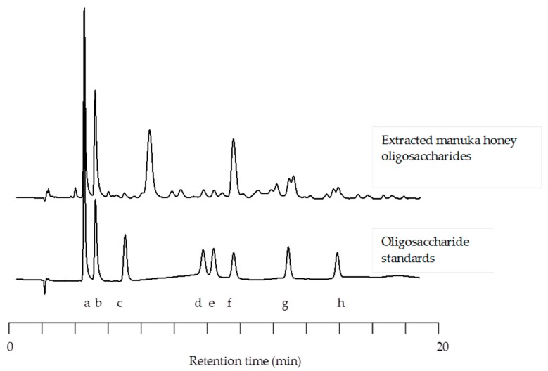
Extracted manuka honey oligosaccharides (MHO) were separated on a CarboPac PA100 column, detected using pulsed amperometric detection and quantified using external standards. HPAEC-PAD analysis revealed 28 peaks of interest. Due to the lack of commercially available standards many of these peaks could not be identified; however, those that could be identified and quantified included glucose, fructose, kojibiose, nigerose, maltose, erlose, D-panose and maltotriose. Although large amounts of glucose and fructose were removed during the extraction procedure; these simple sugars could be found in the final product (40 mg/g glucose and 48.5 mg/g fructose). The most abundant oligosaccharide structure quantified was erlose (179.5 mg/g) followed by panose (24.3 mg/g) and maltotriose (22.3 mg/g). Trace amounts of maltose (9.3 mg/g), nigerose (9.7 mg/g), and kojibiose (4.0 mg/g) were also identified (Figure 1).
Figure 1.
Comparative high performance anion-exchange chromatography with pulsed amperometric detection (HPAEC-PAD) chromatographs of manuka honey oligosaccharides with commercially available standards (Glucose (a), Fructose (b), Kojibiose (c), Nigerose (d), Maltose (e), Erlose (f), D-Panose (g), and Maltotriose (h)).
3.2. Anti-Adhesive Activity of Manuka Honey Oligosaccharides
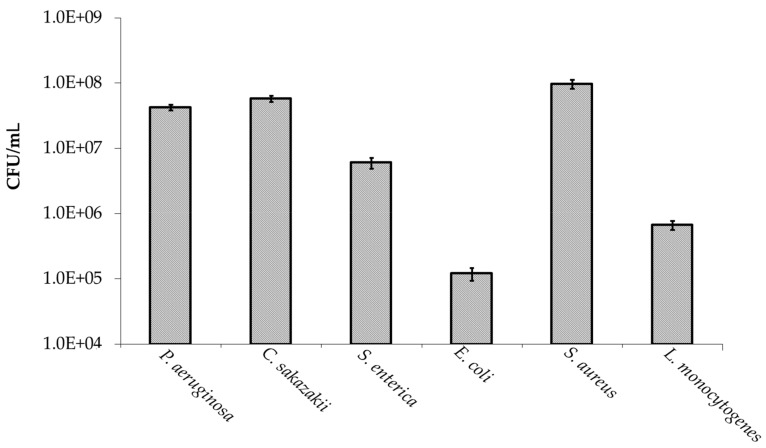
Oligosaccharides extracted from manuka honey were screened for anti-adhesive activity against a range of pathogenic bacteria including P. aeruginosa, C. sakazakii, S. enterica serovar Typhimurium, non-toxigenic E. coli O157:H7, S. aureus, and L. monocytogenes. These oligosaccharides were shown not to be cytotoxic to the bacterial cells and did not affect the viability of the human cells as confirmed by simple trypan blue staining and viability studies on an xCELLigence system (Roche©). As illustrated in Figure 2, all pathogens screened had the capacity to adhere to the human colonic adenocarcinoma cell line, HT-29. High levels of adhesion were observed for C. sakazakii (10% of the initial inoculum), P. aeruginosa (54%) and S. aureus (64%) and moderate to low levels of adhesion were observed for E. coli O157:H7 (0.19%), L. monocytogenes (1.1%), and S. enterica serovar Typhimurium (2.2%). The adhesion of these bacteria to HT-29 cells was then assessed after pre-incubation with 5 mg/mL MHO. This initial oligosaccharide concentration was used as it was representative of the daily consumption (5–10 g/L) of human milk oligosaccharides by infants, which has been linked with preventing pathogen colonization within the gastrointestinal tract [33].
Figure 2.
Adhesion of pathogenic bacteria to the human colonic adenocarcinoma cell line, HT-29.
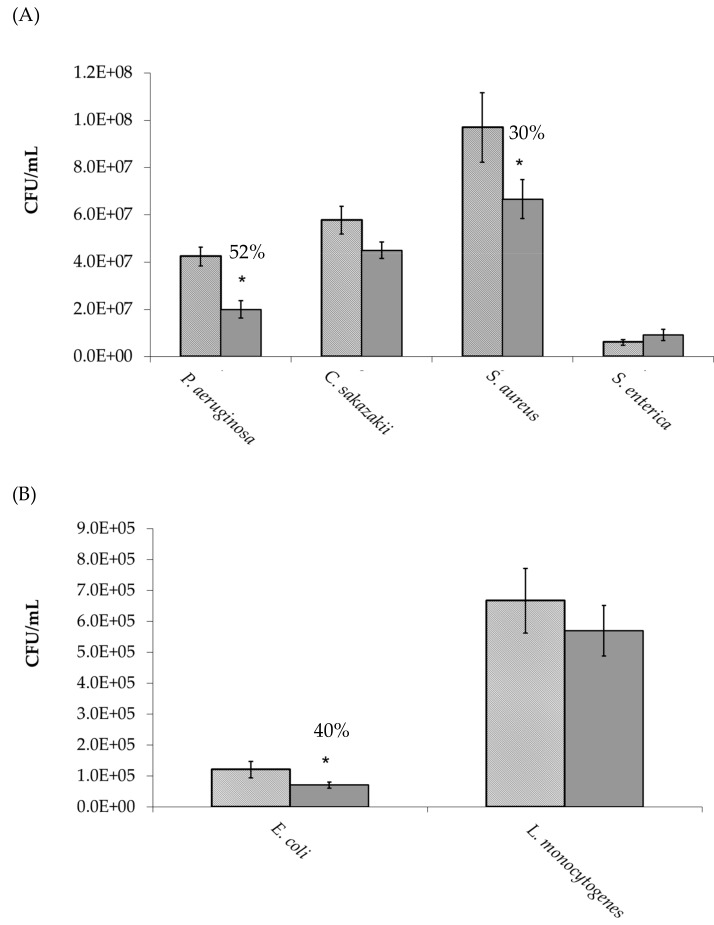
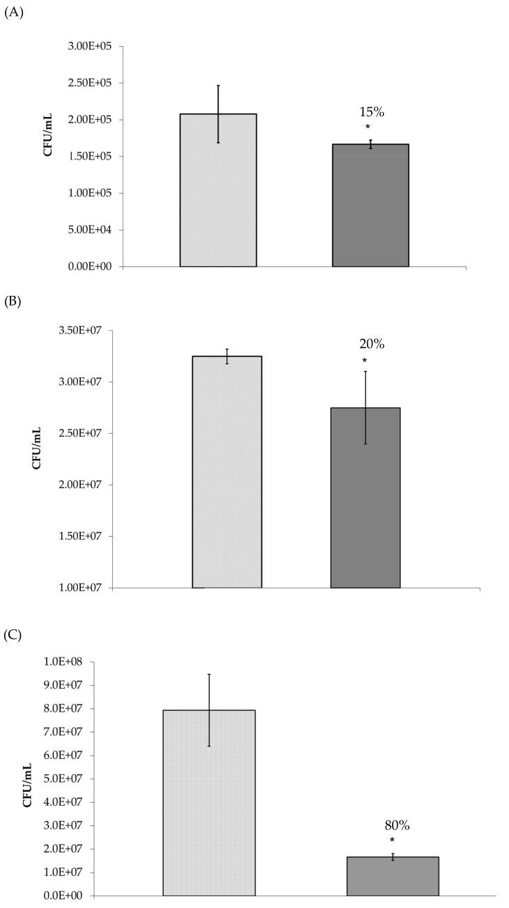
As observed in Figure 3A,B, P. aeruginosa, S. aureus and E. coli adherence to HT-29 cells was significantly (p ≤ 0.05) inhibited by 52%, 30%, and 40%, respectively, when compared to the control (no oligosaccharides). This anti-adhesive activity was then proven to be concentration dependent (Table 2). Indeed, P. aeruginosa adhesion to HT-29 cells was only reduced by MHO concentrations greater than 0.625 mg/mL and S. aureus and E. coli adhesion to HT-29 cells was only reduced by MHO concentrations greater than 1.25 mg/mL. As the pre-incubation of the bacteria with oligosaccharides prior to cell line infection may not be an accurate representation of an in-vivo situation; inhibition studies were also performed in the absence of this step (Figure 4). The oligosaccharides continued to demonstrate anti-adhesive activity under these conditions; however, the activity against P. aeruginosa and E. coli was significantly (p ≤ 0.05) reduced from 52% and 40% to 15% and 20%, respectively (Figure 4A,B). In contrast to this, the anti-adhesive activity of MHO against S. aureus dramatically increased to 80% under these conditions (Figure 4C). The MHO had no effect on the adhesion of the other pathogens examined (L. monocytogenes, C. sakazakii, and S. enterica serovar Typhimurium).
Figure 3.
Adhesion of bacteria to HT-29 cells in the  absence and
  presence of manuka honey oligosaccharides. * p ≤ 0.05 was considered significant.
Table 2.
Percentage Inhibition of adhesion.
| Concentration | ||||
|---|---|---|---|---|
| 5 mg/mL | 2.5 mg/mL | 1.25 mg/mL | 0.625 mg/mL | |
| Pseudomonas aeruginosa | 46 ± 5.7 | 42 ± 10 | 34 ± 15 | - |
| Staphylococcus aureus | 51 ± 8.2 | 31 ± 10 | - | - |
| Escherichia coli O157:H7 | 40 ± 13 | 20 ± 2.0 | 1 ± 9.0 | - |
Figure 4.
Adhesion of (A) Pseudomonas aeruginosa, (B) Escherichia coli, and (C) Staphylococcus aureus to HT-29 cells in the  absence and
  presence of manuka honey oligosaccharides where no pre-incubation step was performed. * p ≤ 0.05 was considered significant.
Growth analysis was also performed to ensure that the anti-adhesive activity observed for the extracted manuka honey oligosaccharide powder was not a consequence of bacterial cell death and to investigate the possibility that these oligosaccharides could increase the growth rate of the bacteria. When compared to the control (no oligosaccharides), no significant increase or decrease in bacterial cell numbers was observed for all bacterial strains tested at all time-points (3, 5, and 24 h) in the presence of the oligosaccharides (5 mg/mL).
4. Discussion
To date, less than 30 oligosaccharide structures have been reported in honey [36]. The most common oligosaccharides identified in honey include panose, sucrose, maltose, kojibiose, isomaltose, erlose, trehalose, raffinose, and turanose [37,38,39,40]. These oligosaccharides are often found at varying concentrations with dependence on the source of the honey. For example, blossom honey (polyfloral) can be discriminated from honeydew honey (forest) as the latter contains a higher concentration of melezitose and raffinose [41]. Honeydew honeys also have lower contents of monosaccharides than blossom honeys [32]. Honeydew honeys have also been characterized by significantly higher mean values of trehalose and isomaltose, and lower values of glucose, sucrose and turanose, than blossom honeys. However, no significant differences in the mean amounts of fructose [42], maltose [42] and sucrose [43] were found while the mean value of total sugars in blossom honey was higher than that in honeydew honeys [42]. The sum of glucose plus fructose has also been used to distinguish between blossom honey and honeydew honey. Blossom honey must have a fructose plus glucose content ≥ 60% (w/w) while honeydew honey and blends of honeydew honey with blossom honeys must have a fructose plus glucose content ≥ 45% (w/w) (EU Directive 110/20010).
HPAEC-PAD is widely used to profile the oligosaccharide content in honey, of which there are more than 300 different varieties. For example, Ouchemoukh et al. [39] exploited this technology to profile Algerian honey oligosaccharides and Cotte et al. [44] and Morales et al. [38] demonstrated the use of this technology to detect honey adulterations. In our study, HPAEC-PAD was used to profile the total oligosaccharide fraction isolated from a commercially available New Zealand manuka honey (MGO™ Manuka Honey). Twenty-eight peaks of interest were identified with the most abundant oligosaccharides being maltotriose, panose, and erlose. These findings correlated well with previously published works such as that of Weston and Brocklebank [40] who reported on the presence of 20 oligosaccharides, including isomaltose, kojibiose, turanose, nigerose, and maltose, in New Zealand manuka honeys. Swallow and Low [45] also separated 20 structurally similar carbohydrates using HPAEC-PAD in four honeys of known botanical origin. The authors noted that although relative oligosaccharide concentration varied from one honey to the next, the overall oligosaccharide pattern did not differ significantly and therefore these oligosaccharide patterns could be used as a “fingerprint” for honey authenticity. The supplier of the honey used in this study (MGO™ Manuka Honey) adheres to international standards established by the Codex Alimentarius Commission. Therefore, the purity and quality of the honey is examined and C4 sugar analysis is performed to confirm no adulteration has taken place. For this reason, the overall oligosaccharide pattern among samples of this manuka honey may not vary greatly.
Overall, we concluded that this fraction was significantly depleted in monosaccharides and contained a complex and diverse range of oligosaccharides with potential biological activity. This fraction was subsequently screened for anti-adhesive activity against a range of pathogens and a significant reduction in the adhesion of P. aeruginosa, E. coli O157:H7 and S. aureus to human colonic epithelial cells, HT-29 cells, was observed. The fact that this fructose and neutral oligosaccharide enriched fraction demonstrated anti-adhesive activity against P. aeruginosa may not be surprising given that this bacterium has been shown to bind to both fucosylated and sialylated epitopes during colonization. Indeed, a variety of studies have shown that other honeys can interfere with the binding capacity of this nosocomial pathogen. For example, Lerrer et al. [46] reported that four commercial honeys (‘wild flower’, ‘eucalyptus’, and ‘field flower’ honeys) provided excellent hemagglutination-like protection against PA-IIL-mediated P. aeruginosa adhesion and attributed this activity to the interaction of P. aeruginosa PA-IIL, a fucose > fructose/mannose binding lectin, with fructose and fructooligosaccharides. Together these studies highlight honey as a food source capable reducing or preventing chronic colonization of P. aeruginosa in the gastrointestinal tract (GIT). Although in-vivo studies are required to substantiate this hypothesis, these are significant findings given the increased antibiotic resistance of this bacterium and the need for alternative therapeutic treatments to prevent P. aeruginosa infection. Similarly, S. aureus infection has become notoriously difficult to treat due to its resistance to antibiotics, such as methicillin [47]. S. aureus is a highly versatile pathogen found in the human pharynx, perineum, axilla, and on the skin (hands, chest and abdomen) and is mainly associated with wound infections, in which the bacterium gains access to the blood stream causing septic shock, and gastroenteritis. To date, relatively little has been reported on S. aureus interactions with food sourced oligosaccharides. Previously, we reported on a direct interaction between S. aureus and a dominant human milk oligosaccharide, 2’-fucosyllatose [48] and here, we report that manuka honey oligosaccharides can prevent S. aureus adhesion to colonic epithelial cells. Although further work in needed to investigate these interactions; these results suggest that carbohydrate-based compounds may have potential in preventing S. aureus-associated gastroenteritis once consumed. As previously discussed, manuka honey is commonly used to prevent infections, such as S. aureus infection, in minor wounds and burns. This was thought to be mainly due to its anti-bacterial and anti-inflammatory activity however, our results suggest that manuka honey oligosaccharides could also play an important anti-adhesive role. Indeed, these compounds could be binding directly to the bacterium and/or epithelial cell surface receptors which could neutralize the threat of bacterial colonization.
E. coli O157:H7 is a highly virulent pathogen with an infectious dose as low as 5–50 cells and a major concern for the food industries such as the dairy industry [49]. The main source of this pathogen is bovine derived food products [50] and symptoms of infection include severe diarrhea, hemorrhagic colitis, and hemolytic-uremic syndrome. Various antibiotics and antibiotic combinations are often used to treat severe cases of E. coli O157:H7 infection and consequently, over the last 30 years, antibiotic resistant strains have emerged. Considering this, there is a need for alternative approaches to prevent and treat E. coli O157:H7 infections. Significantly, numerous E. coli strains have been shown to bind directly to food sourced glycans preventing their adhesion to target epithelial cell surface receptors. For example, Martin-Sosa et al. [51] reported on the ability of human milk oligosaccharides to prevent E. coli fimbriae-associated hemagglutination. Various groups have also demonstrated that bovine milk derived glycomacropeptide (GMP) reduces the adhesion of E. coli O157:H7 to human intestinal epithelial cells in-vitro [52,53]. Interestingly, this activity is mainly due to the presence of sialic acid at the terminal end of the O-linked glycans. Indeed, treatment of GMP with sialidase significantly reduced the binding of E. coli O157:H7 to GMP [53]. In our study, we demonstrate that manuka honey oligosaccharides can prevent the binding of E. coli O157:H7 to colonic intestinal epithelia in-vitro. Interestingly, honey does not contain sialylated oligosaccharides which would suggest that the activity of MHO is dissimilar to that of GMP. It should be noted that E. coli O157:H7 expresses multiple adhesins capable of binding both neutral and acidic oligosaccharides. Thus, neutralizing the threat of disease posed by this enteric pathogen through anti-adhesion therapy may require a complex mixture of oligosaccharides including neutral and acid oligosaccharides from various sources such as domestic animal milk and honey.
During our initial screening studies, pathogenic bacteria were pre-incubated with honey oligosaccharides prior to cell line infection. As previously discussed, this is not an accurate representation of the potential use of these compounds. Thus, we examined the anti-infective activity of the oligosaccharides in the absence of this pre-incubation step. Positively, this did not eliminate the activity of the oligosaccharides however we did observe a slight reduction in activity against Pseudomonas aeruginosa and E. coli O157:H7. The reason for this could be that these pathogens bind directly to food sourced oligosaccharides, which prevents adhesion to epithelial cells [51,53] and under these conditions the concentration of oligosaccharides available for binding is reduced as these biomolecules also bind directly to epithelial cell surface receptors. Therefore, the availability of oligosaccharides for binding to Pseudomonas aeruginosa and E. coli O157:H7 in-vivo is an important factor to consider. Interestingly, the anti-adhesive activity of the MHO against S. aureus increased in the absence of a pre-incubation step. This suggests that epithelial-oligosaccharide interactions, such as occupancy of bacterial binding sites and/or changes in epithelial cell surface expression, can significantly contribute to preventing S. aureus adhesion to human epithelial cells.
5. Conclusions
Overall, this study highlights MGO™ Manuka Honey as source of anti-infective oligosaccharides. Positively, these water-soluble biomolecules possess pH and thermal stability making them ideal for incorporation into dairy products, fruit juices, teas, snack bars, and other baked goods. Thus, the daily consumption of these compounds by individuals of all ages is possible and as demonstrated in this study could potentially reduce the risk of infection associated with multi-drug resistant bacteria such as Pseudomonas aeruginosa, Escherichia coli O157:H7, and Staphylococcus aureus. These biomolecules also exert a low selective pressure on invading pathogens as they are sourced from natural food products which would suggest that the risk of bacterial resistance is low. However, before an actual application can be considered, further questions must be addressed. The oligosaccharides are not 100% effective in preventing adhesion of the pathogens in this study. Would the concentration tested here in vitro be effective in vivo in humans? Also, would the cost of isolating oligosaccharides from manuka honey prohibit their use as food ingredients? The isolation method used in this study is not commercially viable and the amounts of honey required may not be available. A solution may be to identify the oligosaccharides in the honey responsible for bioactivity and synthesize these structures in a similar manner to human milk oligosaccharide manufacture (chemically, via fermentation or by enzymatic synthesis).
Author Contributions
Conceptualization, J.A.L. and R.M.H.; Formal analysis, H.S.; Investigation, J.A.L. and J.C.; Methodology, J.A.L.; Supervision, R.M.H.
Funding
This research received no external funding.
Conflicts of Interest
The authors declare no conflict of interest.
References
- 1.Zumla A., Lulat A. Honey-a remedy rediscovered. J. R. Soc. Med. 1989;82:384–385. doi: 10.1177/014107688908200704. [DOI] [PMC free article] [PubMed] [Google Scholar]
- 2.Bromfield R. Honey for decubitus ulcers. JAMA. 1973;224:905. doi: 10.1001/jama.1973.03220200061034. [DOI] [PubMed] [Google Scholar]
- 3.Bulman M.W. Honey as a surgical dressing. Middx. Hosp. J. 1955;55:188–189. [Google Scholar]
- 4.Cavanagh D., Beazley J., Ostapowicz F. Radical operation for carcinoma of the vulva. A new approach to wound healing. J. Obstet. Gynaecol. Br. Commonw. 1970;77:1037–1040. doi: 10.1111/j.1471-0528.1970.tb03455.x. [DOI] [PubMed] [Google Scholar]
- 5.Jeddar A., Kharsany A., Ramsaroop U.G., Bhamjee A., Haffejee I.E., Moosa A. The antibacterial action of honey. An in vitro study. S. Afr. Med. J. Suid-Afrikaanse tydskrif vir geneeskunde. 1985;67:257–258. [PubMed] [Google Scholar]
- 6.Al-Waili N.S., Lootah S.A., Shaheen W. Mixture of crude honey and olive oil in natural wax to treat chronic skin disorders. FASEB J. 1999;13:A846. [Google Scholar]
- 7.Al-Waili N.S., Lootah S.A., Shaheen W. Honey preparation with olive oil and natural wax to treat skin fungal infections. FASEB J. 1999;13:A848. [Google Scholar]
- 8.Al-Waili N.S., Saloom K.Y. Crude honey to treat seborrheic dermatitis of the scalp. FASEB J. 1999;13:A848. [Google Scholar]
- 9.Brudzynski K., Sjaarda C. Honey glycoproteins containing antimicrobial peptides, jelleins of the major royal jelly protein 1, are responsible for the cell wall lytic and bactericidal activities of honey. PLoS ONE. 2015;10:e0120238. doi: 10.1371/journal.pone.0120238. [DOI] [PMC free article] [PubMed] [Google Scholar]
- 10.Brudzynski K., Sjaarda C., Lannigan R. Mrjp1-containing glycoproteins isolated from honey, a novel antibacterial drug candidate with broad spectrum activity against multi-drug resistant clinical isolates. Front. Microbiol. 2015;6:711. doi: 10.3389/fmicb.2015.00711. [DOI] [PMC free article] [PubMed] [Google Scholar]
- 11.Robson V., Cooper R. Using leptospermum honey to manage wounds impaired by radiotherapy: A case series. Ostomy/Wound Manag. 2009;55:38–47. [PubMed] [Google Scholar]
- 12.Robson V., Dodd S., Thomas S. Standardized antibacterial honey (medihoney) with standard therapy in wound care: Randomized clinical trial. J. Adv. Nurs. 2009;65:565–575. doi: 10.1111/j.1365-2648.2008.04923.x. [DOI] [PubMed] [Google Scholar]
- 13.Hentzer M., Eberl L., Nielsen J., Givskov M. Quorum sensing—A novel target for the treatment of biofilm infections. Biodrugs. 2003;17:241–250. doi: 10.2165/00063030-200317040-00003. [DOI] [PubMed] [Google Scholar]
- 14.Wang R., Starkey M., Hazan R., Rahme L.G. Honey’s ability to counter bacterial infections arises from both bactericidal compounds and QS inhibition. Front. Microbiol. 2012;3:144. doi: 10.3389/fmicb.2012.00144. [DOI] [PMC free article] [PubMed] [Google Scholar]
- 15.Gheldof N., Wang X.H., Engeseth N.J. Identification and quantification of antioxidant components of honeys from various floral sources. J. Agric. Food Chem. 2002;50:5870–5877. doi: 10.1021/jf0256135. [DOI] [PubMed] [Google Scholar]
- 16.Manyi-Loh C.E., Ndip R.N., Clarke A.M. Volatile compounds in honey: A review on their involvement in aroma, botanical origin determination and potential biomedical activities. Int. J. Mol. Sci. 2011;12:9514–9532. doi: 10.3390/ijms12129514. [DOI] [PMC free article] [PubMed] [Google Scholar]
- 17.Wang J., Li Q.X. Chemical composition, characterization, and differentiation of honey botanical and geographical origins. Adv. Food Nutr. Res. 2011;62:89–137. doi: 10.1016/B978-0-12-385989-1.00003-X. [DOI] [PubMed] [Google Scholar]
- 18.Ball D.W. The chemical composition of honey. J. Chem. Educ. 2007;84:1643–1646. doi: 10.1021/ed084p1643. [DOI] [Google Scholar]
- 19.Alvarez-Suarez J.M., Gasparrini M., Forbes-Hernandez T.Y., Mazzoni L., Giampieri F. The composition and biological activity of honey: A focus on manuka honey. Foods. 2014;3:420–432. doi: 10.3390/foods3030420. [DOI] [PMC free article] [PubMed] [Google Scholar]
- 20.Alvarez-Suarez J.M., Giampieri F., Battino M. Honey as a source of dietary antioxidants: Structures, bioavailability and evidence of protective effects against human chronic diseases. Curr. Med. Chem. 2013;20:621–638. doi: 10.2174/092986713804999358. [DOI] [PubMed] [Google Scholar]
- 21.Erejuwa O.O., Sulaiman S.A., Wahab M.S.A. Effects of honey and its mechanisms of action on the development and progression of cancer. Molecules. 2014;19:2497–2522. doi: 10.3390/molecules19022497. [DOI] [PMC free article] [PubMed] [Google Scholar]
- 22.Nguyen H.T.L., Panyoyai N., Kasapis S., Pang E., Mantri N. Honey and its role in relieving multiple facets of atherosclerosis. Nutrients. 2019;11:167. doi: 10.3390/nu11010167. [DOI] [PMC free article] [PubMed] [Google Scholar]
- 23.Sanz M.L., Polemis N., Morales V., Corzo N., Drakoularakou A., Gibson G.R., Rastall R.A. In vitro investigation into the potential prebiotic activity of honey oligosaccharides. J. Agric. Food Chem. 2005;53:2914–2921. doi: 10.1021/jf0500684. [DOI] [PubMed] [Google Scholar]
- 24.Jan Mei S., Mohd Nordin M.S., Norrakiah A.S. Fructooligosaccharides in honey and effects of honey on growth of Bifidobacterium longum Bb 536 . Int. Food Res. J. 2010;17:557–561. [Google Scholar]
- 25.Gluck U., Gebbers J.O. Ingested probiotics reduce nasal colonization with pathogenic bacteria (Staphylococcus aureus, Streptococcus pneumoniae, and beta-hemolytic streptococci) Am. J. Clin. Nutr. 2003;77:517–520. doi: 10.1093/ajcn/77.2.517. [DOI] [PubMed] [Google Scholar]
- 26.Johnson-Henry K.C., Mitchell D.J., Avitzur Y., Galindo-Mata E., Jones N.L., Sherman P.M. Probiotics reduce bacterial colonization and gastric inflammation in H. pylori-infected mice. Dig. Dis. Sci. 2004;49:1095–1102. doi: 10.1023/B:DDAS.0000037794.02040.c2. [DOI] [PubMed] [Google Scholar]
- 27.Lin H.C., Su B.H., Chen A.C., Lin T.W., Tsai C.H., Yeh T.F., Oh W. Oral probiotics reduce the incidence and severity of necrotizing enterocolitis in very low birth weight infants. Pediatrics. 2005;115:1–4. doi: 10.1542/peds.2004-1463. [DOI] [PubMed] [Google Scholar]
- 28.Lane J.A., Marino K., Naughton J., Kavanaugh D., Clyne M., Carrington S.D., Hickey R.M. Anti-infective bovine colostrum oligosaccharides: Campylobacter jejuni as a case study. Int. J. Food Microbiol. 2012;157:182–188. doi: 10.1016/j.ijfoodmicro.2012.04.027. [DOI] [PubMed] [Google Scholar]
- 29.Mysore J.V., Wigginton T., Simon P.M., Zopf D., Heman-Ackah L.M., Dubois A. Treatment of Helicobacter pylori infection in rhesus monkeys using a novel antiadhesion compound. Gastroenterology. 1999;117:1316–1325. doi: 10.1016/S0016-5085(99)70282-9. [DOI] [PubMed] [Google Scholar]
- 30.Newburg D.S., Pickering L.K., Mccluer R.H., Cleary T.G. Fucosylated oligosaccharides of human-milk protect suckling mice from heat-stabile enterotoxin of escherichia-coli. J. Infect. Dis. 1990;162:1075–1080. doi: 10.1093/infdis/162.5.1075. [DOI] [PubMed] [Google Scholar]
- 31.Ruiz-Palacios G.M., Cervantes L.E., Ramos P., Chavez-Munguia B., Newburg D.S. Campylobacter jejuni binds intestinal H(O) antigen (Fucα1, 2Galβ1, 4GlcNAc), and fucosyloligosaccharides of human milk inhibit its binding and infection. J. Biol. Chem. 2003;278:14112–14120. doi: 10.1074/jbc.M207744200. [DOI] [PubMed] [Google Scholar]
- 32.Pita-Calvo C., Vazquez M. Honeydew honeys: A review on the characterization and authentication of botanical and geographical origins. J. Agric. Food Chem. 2018;66:2523–2537. doi: 10.1021/acs.jafc.7b05807. [DOI] [PubMed] [Google Scholar]
- 33.Bode L. Human milk oligosaccharides: Every baby needs a sugar mama. Glycobiology. 2012;22:1147–1162. doi: 10.1093/glycob/cws074. [DOI] [PMC free article] [PubMed] [Google Scholar]
- 34.Coppa G.V., Zampini L., Galeazzi T., Facinelli B., Ferrante L., Capretti R., Orazio G. Human milk oligosaccharides inhibit the adhesion to Caco-2 cells of diarrheal pathogens: Escherichia coli, Vibrio cholerae, and Salmonella fyris. Pediatr. Res. 2006;59:377–382. doi: 10.1203/01.pdr.0000200805.45593.17. [DOI] [PubMed] [Google Scholar]
- 35.Miao Y., Li G., Zhang X., Xu H., Abraham S.N. A trp channel senses lysosome neutralization by pathogens to trigger their expulsion. Cell. 2015;161:1306–1319. doi: 10.1016/j.cell.2015.05.009. [DOI] [PMC free article] [PubMed] [Google Scholar]
- 36.Molan P.C. Authenticity of honey. In: Ashurst P.R., Dennis M.J., editors. Food Authentication. Springer; New York, NY, USA: 1996. pp. 259–303. [Google Scholar]
- 37.Mateo R., Bosch-Reig F. Sugar profile of spanish unifloral honeys. Food Chem. 1997;60:33–41. doi: 10.1016/S0308-8146(96)00297-X. [DOI] [PubMed] [Google Scholar]
- 38.Morales V., Sanz M.L., Olano A., Corzo N. Rapid separation on activated charcoal of high oligosaccharides in honey. Chromatographia. 2006;64:233–238. doi: 10.1365/s10337-006-0842-6. [DOI] [Google Scholar]
- 39.Ouchemoukh S., Schweitzer P., Bey M.B., Djoudad-Kadji H., Louaileche H. HPLC sugar profiles of algerian honeys. Food Chem. 2010;121:561–568. doi: 10.1016/j.foodchem.2009.12.047. [DOI] [Google Scholar]
- 40.Weston R.J., Brocklebank L.K. The oligosaccharide composition of some New Zealand honeys. Food Chem. 1999;64:33–37. doi: 10.1016/S0308-8146(98)00099-5. [DOI] [Google Scholar]
- 41.Bogdanov S., Jurendic T., Sieber R., Gallmann P. Honey for nutrition and health: A review. J. Am. Coll. Nutr. 2008;27:677–689. doi: 10.1080/07315724.2008.10719745. [DOI] [PubMed] [Google Scholar]
- 42.Bentabol Manzanares A., Hernández García Z., Rodríguez Galdón B., Rodríguez Rodríguez E., Díaz Romer C. Differentiation of blossom and honeydew honeys using multivariate analysis on the physicochemical parameters and sugar composition. Food Chem. 2011;126:664–672. doi: 10.1016/j.foodchem.2010.11.003. [DOI] [Google Scholar]
- 43.Golob T., Plestenjak A. Quality of slovene honey. Food Tech. Biotechnol. 1999;37:195–202. [Google Scholar]
- 44.Cotte J.F., Casabianca H., Chardon S., Lheritier J., Grenier-Loustalot M.F. Application of carbohydrate analysis to verify honey authenticity. J. Chromat. A. 2003;1021:145–155. doi: 10.1016/j.chroma.2003.09.005. [DOI] [PubMed] [Google Scholar]
- 45.Swallow K.W., Low N.H. Analysis and quantitation of the carbohydrates in honey using high-performance liquid chromatography. J. Agric. Food Chem. 1990;38:1828–1832. doi: 10.1021/jf00099a009. [DOI] [Google Scholar]
- 46.Lerrer B., Zinger-Yosovich K.D., Avrahami B., Gilboa-Garber N. Honey and royal jelly, like human milk, abrogate lectin-dependent infection-preceding Pseudomonas aeruginosa adhesion. ISME J. 2007;1:149–155. doi: 10.1038/ismej.2007.20. [DOI] [PubMed] [Google Scholar]
- 47.Gordon R.J., Lowy F.D. Pathogenesis of methicillin-resistant Staphylococcus aureus infection. Clin. Infect. Dis. 2008;46:S350–S359. doi: 10.1086/533591. [DOI] [PMC free article] [PubMed] [Google Scholar]
- 48.Lane J.A., Mehra R.K., Carrington S.D., Hickey R.M. Development of biosensor-based assays to identify anti-infective oligosaccharides. Anal. Biochem. 2011;410:200–205. doi: 10.1016/j.ab.2010.11.032. [DOI] [PubMed] [Google Scholar]
- 49.Farrokh C., Jordan K., Auvray F., Glass K., Oppegaard H., Raynaud S., Thevenot D., Condron R., De Reu K., Govaris A., et al. Review of shiga-toxin-producing Escherichia coli (STEC) and their significance in dairy production. Int. J. Food Microbiol. 2013;162:190–212. doi: 10.1016/j.ijfoodmicro.2012.08.008. [DOI] [PubMed] [Google Scholar]
- 50.Callaway T.R., Carr M.A., Edrington T.S., Anderson R.C., Nisbet D.J. Diet, Escherichia coli O157:H7, and cattle: A review after 10 years. Curr. Issues Mol. Biol. 2009;11:67–79. [PubMed] [Google Scholar]
- 51.Martin-Sosa S., Martin M.J., Hueso P. The sialylated fraction of milk oligosaccharides is partially responsible for binding to enterotoxigenic and uropathogenic Escherichia coli human strains. J. Nutr. 2002;132:3067–3072. doi: 10.1093/jn/131.10.3067. [DOI] [PubMed] [Google Scholar]
- 52.Feeney S., Ryan J.T., Kilcoyne M., Joshi L., Hickey R. Glycomacropeptide reduces intestinal epithelial cell barrier dysfunction and adhesion of entero-hemorrhagic and entero-pathogenic Escherichia coli in vitro. Foods. 2017;6:93. doi: 10.3390/foods6110093. [DOI] [PMC free article] [PubMed] [Google Scholar]
- 53.Nakajima K., Tamura N., Kobayashi-Hattori K., Yoshida T., Hara-Kudo Y., Ikedo M., Sugita-Konishi Y., Hattori M. Prevention of intestinal infection by glycomacropeptide. Biosci. Biotechnol. Biochem. 2005;69:2294–2301. doi: 10.1271/bbb.69.2294. [DOI] [PubMed] [Google Scholar]


