INTRODUCTION
Brugada syndrome (BrS) is an inherited sodium, calcium, or potassium channelopathy associated with an increased risk of ventricular fibrillation (VF) and sudden cardiac death.1–3 This syndrome is most prevalent in men and individuals of southeast Asian descent, with a mean age of onset of symptoms at 41 ± 15 years.2 The diagnosis is challenging because of the often asymptomatic presentation of affected individuals and the dynamic electrocardiogram (ECG) manifestations that are frequently concealed.2 This case highlights the presentation, ECG findings, and management of a patient with BrS.
CASE PRESENTATION
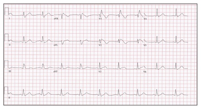
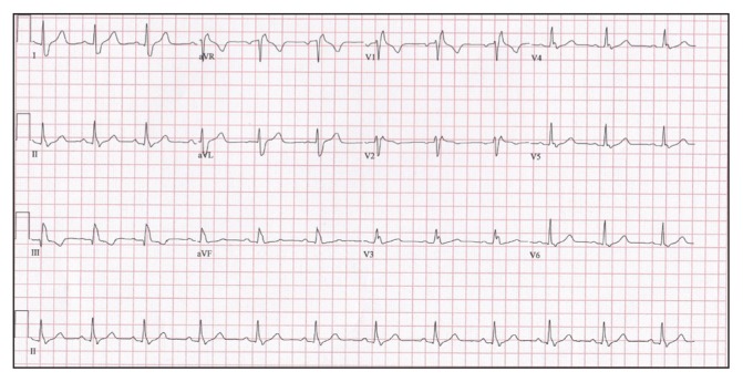
A middle-aged woman with a history of recently diagnosed seizure disorder and a family history of sudden cardiac death (SCD) in 3 first-degree maternal cousins (all of whom died in their early 40s) presented to the Emergency Department (ED) by ambulance in cardiac arrest. She was found unresponsive with agonal respirations by her husband, and was subsequently found to be in VF arrest by paramedics. The patient was treated with advanced cardiac life support protocol, including cardiopulmonary resuscitation, multiple defibrillatory shocks, and intravenous epinephrine boluses, with return of spontaneous circulation following ED arrival (50 minutes after she was first found down). Results of a postarrest ECG demonstrated changes suggestive of an acute anterior ST-elevation myocardial infarction and prompted the ED physician to consult interventional cardiology. The patient was endotracheally intubated in the ED and transported to the cardiac catheterization laboratory for emergent coronary angiography, which demonstrated no obstructive coronary artery disease. Results of a 12-lead ECG obtained following cardiac catheterization (Figure 1) demonstrated coved ST-segment elevation greater than or equal to 2 mm (0.2 mV) followed by a negative T wave in leads V1 and V2, consistent with type I BrS in the setting of the patient’s recent VF arrest. These findings were new compared with a previous ECG obtained 1 month earlier (Figure 2). The patient was extubated on hospital day 2 and received an implantable cardioverter-defibrillator (ICD) on hospital day 5 without accompanying electrophysiology studies, as her diagnosis of BrS was already confirmed. She was discharged home 4 days later. Two months following discharge, she returned to the ED after experiencing a syncopal episode. Inspection of the ICD at that time indicated the patient had experienced an episode of VF that was successfully terminated by a single shock of 40 joules. The patient was started on quinidine (324 mg twice daily) as an antidysrhythmic agent and was discharged from the ED without further syncopal episodes during the next 3 months. She was genetically tested for BrS, but her results were inaccessible during our review of her records. We do not know if her family members also underwent genetic testing.
Figure 1.
12-lead electrocardiogram obtained following coronary angiography on a middle-aged woman with recent ventricular fibrillation arrest and return of spontaneous circulation. The results demonstrate coved ST-segment elevation ≥ 2 mm (0.2 mV) followed by a negative T wave in leads V1 and V2; findings are consistent with Brugada type I syndrome (in setting of recent arrest).
Figure 2.
12-lead electrocardiogram for the same patient obtained 1 month before her cardiac arrest. The results demonstrate sinus rhythm with right bundle branch block.
DISCUSSION
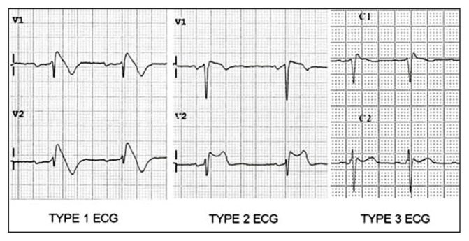
BrS is a sodium, calcium, or potassium channelopathy that was first described in 1992; it can be sporadic or familial, with a suspected autosomal-dominant inheritance pattern, and it is a major cause of sudden death in relatively young people with no evidence of structural heart disease.4–6 The estimated prevalence of BrS is 1 in 2000, and it is 8 to 10 times more prevalent in men than women.2,7 Women with BrS are also more commonly asymptomatic, have fewer spontaneous ECG changes, and a more favorable risk profile.2 These patients can present with syncope, seizures (often convulsive syncope), and nocturnal agonal breathing because of polymorphic ventricular tachycardia or VF.2,8 If these dysrhythmias are sustained, SCD may result. The BrS ECG is characterized by 3 types of repolarization patterns in leads V1 to V3, which may be dynamic and are often concealed (Figure 3).6,9 Inducers include certain medications (eg, sodium channel blockers, tricyclic antidepressants, and anesthetics), electrolyte imbalances (eg, hypokalemia, hyperkalemia, and hypercalcemia), and fever.4 The type 1 “coved” pattern requires an ST-segment elevation greater than or equal to 2 mm in 1 or more right precordial leads followed by a negative T wave (Figure 1).2 BrS can be diagnosed by this pattern and 1 or more of the following findings: Documented VF; polymorphic ventricular tachycardia; a family history of SCD; coved-type ECGs in family members; inducibility of ventricular tachycardia with programmed electrical stimulation; syncope; or nocturnal agonal respirations.10 The type 2 “saddleback” pattern is characterized by an ST-segment elevation greater than or equal to 0.5 mm in 1 or more right precordial leads followed by a trough displaying greater than or equal to 1 mm ST-segment elevation with a subsequent positive or biphasic T wave.2 The type 3 ECG pattern has either a saddleback or coved appearance with an ST-segment elevation of less than 1 mm (Figure 3).6,9 Types 2 and 3 are only diagnostic with conversion to type 1 following challenge with a sodium channel blocker.2,4
Figure 3.
The 3 electrocardiogram ST-segment elevation patterns that characterize Brugada syndrome.
Reprinted with kind permission from Josep Brugada and the E-Journal of the European Society of Cardiology Council for Cardiology Practice. Brugada J. Management of patients with a Brugada ECG pattern. E-journal of the ESC Council for Cardiology Practice 2009 Mar 17;7(24). Available from: www.escardio.org/Journals/E-Journal-of-Cardiology-Practice/Volume-7/Management-of-patients-with-a-Brugada-ECG-pattern.
In our case, the patient met diagnostic criteria for BrS on the basis of the 12-lead ECGs showing the hallmark coved-type ST elevation followed by an inverted T wave in leads V1 and V2 (Figure 1), nocturnal agonal respirations, and VF arrest. Her recent diagnosis of seizures, in fact, may have been recurrent convulsive syncope, commonly mistaken for a seizure disorder, a condition that raises suspicion for cardiac dysrhythmias.8 The mainstay of therapy for symptomatic patients with BrS is placement of an ICD.11 In addition to ICD placement, BrS can be managed with quinidine (up to 1–2 g daily), and catheter ablation for refractory ventricular arrhythmias.12,13 Asymptomatic patients displaying a Brugada ECG pattern (either spontaneously or after sodium channel blockade) should undergo electrophysiology studies if there is a family history of SCD thought secondary to BrS.10
Footnotes
Disclosure Statement
The author(s) have no conflicts of interest to disclose.
References
- 1.Milman A, Andorin A, Gourraud JB, et al. Age of first arrhythmic event in Brugada syndrome: Data from the SABRUS (survey on arrhythmic events in Brugada syndrome) in 678 patients. Circulation Arrhythm Electrophysiol. 2017 Dec;18:10. doi: 10.1161/CIRCEP.117.005222. [DOI] [PubMed] [Google Scholar]
- 2.Brugada J, Campuzano O, Arbelo E, Sarquella-Brugada G, Brugada R. Present status of Brugada syndrome: JACC state-of-the-art review. J Am Coll Cardiol. 2018 Aug 28;72(9):1046–59. doi: 10.1016/j.jacc.2018.06.037. [DOI] [PubMed] [Google Scholar]
- 3.Shimizu W. Update of diagnosis and management of inherited cardiac arrhythmias. Circ J. 2013;77:2867–72. doi: 10.1253/circj.CJ-13-1217. [DOI] [PubMed] [Google Scholar]
- 4.Nader A, Massumi A, Cheng J, et al. Inherited arrhythmic disorders: Long QT and Brugada syndromes. Tex Heart Inst J. 2007;34:67–75. [PMC free article] [PubMed] [Google Scholar]
- 5.Brugada P, Brugada J. Right bundle branch block, persistent ST-segment elevation and sudden cardiac death: A distinct clinical and electrocardiographic syndrome. A multicenter report. J Am Coll Cardiol. 1992 Nov 15;20:1391–6. doi: 10.1016/0735-1097(92)90253-J. [DOI] [PubMed] [Google Scholar]
- 6.Sarquella-Brugada G, Campuzano O, Arbelo E, Brugada J, Brugada R. Brugada syndrome: Clinical and genetic findings. Genet Med. 2016 Jan;18:3–12. doi: 10.1038/gim.2015.35. [DOI] [PubMed] [Google Scholar]
- 7.Gourraud J-B, Barc J, Thollet A, et al. The Brugada syndrome: A rare arrhythmia disorder with complex inheritance. Front Cardiovasc Med. 2016 Apr 25;3:9. doi: 10.3389/fcvm.2016.00009. [DOI] [PMC free article] [PubMed] [Google Scholar]
- 8.Sabu J, Regeti K, Mallappallil M, et al. Convulsive syncope induced by ventricular arrhythmia masquerading as epileptic seizures: Case report and literature review. J Clin Med Res. 2016;8(8):610–15. doi: 10.14740/jocmr2583w. [DOI] [PMC free article] [PubMed] [Google Scholar]
- 9.Brugada J. Management of patients with a Brugada ECG pattern. E-journal of the ESC Council for Cardiology Practice. 2009 Mar;17:7. (24). Available from: www.escardio.org/Journals/E-Journal-of-Cardiology-Practice/Volume-7/Management-of-patients-with-a-Brugada-ECG-pattern. [Google Scholar]
- 10.Brugada R, Campuzano O, Sarquella-Brugada G, Brugada P, Brugada J, Hong K. Brugada syndrome. In: Adam MP, Ardinger HH, Pagon RA, et al., editors. GeneReviews [Internet] Seattle, WA: University of Washington, Seattle; 2005. Mar 31, [Updated 2016 Nov 17] 1993–2019 [cited 2019 Aug 28]. Available from: www.ncbi.nlm.nih.gov/sites/books/NBK1517/ [Google Scholar]
- 11.Antzelevitch C. Brugada syndrome. Pacing Clin Electrophysiol. 2006 Oct;29(10):1130–59. doi: 10.1111/j.1540-8159.2006.00507.x. [DOI] [PMC free article] [PubMed] [Google Scholar]
- 12.Sieira J, Dendramis G, Brugada P. Pathogenesis and management of Brugada syndrome. Nat. Rev Cardiol. 2016 Dec;13:744–56. doi: 10.1038/nrcardio.2016.143. [DOI] [PubMed] [Google Scholar]
- 13.Al-Khatib SM, Stevenson WG, Ackerman MJ, et al. 2017 AHA/ACC/HRS guideline for management of patients with ventricular arrhythmias and the prevention of sudden cardiac death: A report of the American College of Cardiology/American Heart Association Task Force on Clinical Practice Guidelines and the Heart Rhythm Society. J Am Coll Cardiol. 2018 Sep 25;138:e272–391. doi: 10.1161/CIR.0000000000000549. [DOI] [PubMed] [Google Scholar]


