PLOS ONE logoLink to PLOS ONE
. 2019 Nov 8;14(11):e0224727. doi: 10.1371/journal.pone.0224727

The clinicopathological significance of Thrombospondin-4 expression in the tumor microenvironment of gastric cancer

Kenji Kuroda 1,2,3, Masakazu Yashiro 1,2,3,*, Tomohiro Sera 1,2,3, Yurie Yamamoto 2,3, Yukako Kushitani 1,2,3, Atsushi Sugimoto 1,2,3, Syuhei Kushiyama 1,2,3, Sadaaki Nishimura 1,2,3, Shingo Togano 1,2,3, Tomohisa Okuno 1,2,3, Tatsuro Tamura 1, Takahiro Toyokawa 1, Hiroaki Tanaka 1, Kazuya Muguruma 1, Masaichi Ohira 1
Editor: Masaru Katoh4
PMCID: PMC6839882  PMID: 31703077

Abstract

Introduction

Thrombospondin-4 [1] is an extracellular glycoprotein involved in wound healing and tissue remodeling. Although THBS4 is reportedly frequently expressed in solid tumors, there are few reports of the clinicopathological features of carcinomas with THBS4 expression. We evaluated the clinicopathologic significance of THBS4 expression in gastric carcinoma (GC).

Materials and methods

We retrospectively analyzed the cases of 584 GC patients. The expression of THBS4 in each tumor was evaluated by immunohistochemistry. We then divided the patients into the THBS4-high (n = 223, 38.2%) group and THBS4-low (n = 361, 61.8%) group. THBS4 expression in cancer-associated fibroblasts (CAFs), normal-associated fibroblasts (NFs) and gastric cancer cell lines was examined by western blotting.

Results

THBS4 is expressed on stromal cells with αSMA or Podoplanin expression in the GC microenvironment, but not expressed on cancer cells with cytokeratin expression. The western blot analysis results showed that CAFs (but not NFs and cancer cells) expressed THBS4. Compared to the THBS4-low expression status, the THBS4-high expression status was correlated with higher αSMA expression, higher invasion depth, lymph-node metastasis, lymphatic invasion, peritoneal cytology, peritoneal metastasis, larger tumor size, microscopic diffuse type, and the macroscopic diffuse infiltrating type. The THBS4-high group's 5-year overall survival rate was significantly poorer than that of the THBS4-low group. A multivariate analysis revealed that THBS4 expression was an independent prognostic factor.

Conclusion

THBS4 is expressed on CAFs in the gastric cancer microenvironment. THBS4 from CAFs is associated with the metastasis of cancer cells, and is a useful prognostic indicator for gastric cancer patients.

Introduction

Despite recent advances in diagnostic techniques and therapies for gastric cancer (GC), the prognosis of GC remains poor; GC is the fifth most common fatal carcinoma and the third leading morbidity in the world [2, 3]. To improve the prognosis of GC patients, new strategies based on its biological behavior are necessary. Tumor progression has been recognized as the product of evolving crosstalk between the cancer cells and the surrounding tissue or tumor stroma[4]. The elucidation of the characteristic features of tumor stroma could provide a new biological marker in the crosstalk.

Thrombospondin-4 (THBS4) is one of the extracellular secreted glycoproteins involved in wound healing and tissue remodeling by regulating the organization, repair, and remodeling of the extracellular matrix [57]. It was recently reported that THBS4 is frequently expressed in the tumor stroma of some types of solid cancers such as breast cancer and prostate cancer [8] [9]. By conducting a transcriptome-wide comparative analysis, Forester et al. [10] observed that THBS4 is a potent marker for diffuse-type gastric cancer, and they detected THBS4 expression in tumor stroma which showed positivity for α-Smooth muscle actin (αSMA) and vimentin.

Although it has been reported that THBS4 is highly expressed in cancer stroma, the significance of THBS4 in tumor stroma in the development of gastric cancer has been controversial. THBS4 was reported to stimulate the development of cancer [8, 1013], whereas other studies found that THBS4 acts as a tumor suppressor [14, 15]. Few investigations have examined the clinicopathological significance of THBS4, and we conducted the present study to evaluate the clinicopathological significance of THBS4 expression in gastric cancer, especially in tumor stroma.

Materials & methods

Patients and clinical materials

This study was retrospective analysis of 585 gastric cancer patients who received gastrectomy at Osaka City University Hospital. Gastric cancer tissues were obtained from each patient. The pathological diagnoses and classifications were made according to the Japanese classification of gastric carcinoma (14th edition) [15]. This study was approved by the Osaka City University Ethics Committee (approval number 924). Written informed consent for research was obtained from patients.

Immunohistochemical determination of THBS4, Podoplanin, αSMA and Cytokeratin

The immunohistochemical determination of THBS4, Podoplanin, αSMA and cytokeratin were examined as the manufacturer’s instructions. Shortly, slides were deparaffinized and activated by heating. After blocking endogenous peroxidase activity, the samples were incubated with anti-human THBS4 (R&D Systems, Minneapolis, MN: MAB2390; 1:200), anti-Pan-Keratin (Proteintech, Rosemont, IL: 26411-1-AP; 1:3000), anti-Podoplanin (Santa Cruz Biotechnology, Dallas, TX: sc-376695; 1:300) or anti-human αSMA (Leica Biosystems Newcastle, Newcastle, UK: NCL-L-SMA; 1:200). The samples were incubated with biotinylated second antibody. The samples were treated with streptavidin-peroxidase reagent, and counterstaining with Mayer’s hematoxylin. THBS4 expression was evaluated by intensity of staining and percentage of stained stromal cells, respectively: intensity was given scores 0–3 (0 = no, 1 = weak, 2 = moderate, 3 = intense), and the percentage of immunopositive stromal cells in all stroma cells was given scores 0–3 (0 = 0%, 1 = 1%-10%, 2 = 11%-50%, 3 = 51%-100%). The two scores were added to obtain the final result of 0–6. Expressions were considered THBS4-high when scores were 3 or more and THBS4-low when scores were 2 or less. Expressions were considered αSMA-high when scores were 5 or more and αSMA-low when scores were 4 or less. Two double-blinded independent observers who were unaware of clinical data and outcome evaluated, and when a discrepant evaluation between the two independent observers was found, the result was rechecked and discussed.

Gastric fibroblast cell lines and cancer cell lines

Three pairs of cancer associated fibroblast (CAF82, CAF105) and normal tissue fibroblast (NF82, NF105) were established from tumoral gastric wall and non-tumoral gastric wall, respectively. CAF82 and NF82, CAF105 and NF105 were from same patients. These fibroblasts were used from third passage to fifth passage in culture. CAFs and NFs were examined by αSMA staining, as previously reported [16]. MKN45 and NUGC4, which are diffuse and intestinal type gastric cancer cell lines, respectively, were used.

Immunostaining of fibroblasts

Fibroblasts were incubated into Lab-Tek II Chamber Slide System (Nunc, Naperville, IL) for 3 days. After washing with PBS and fixing with methanol for 10 min, fibroblasts were incubated with anti-podoplanin antibody (Santa Cruz Biotechnology, Dallas, TX: sc-376695; 1:300). The samples were incubated with biotinylated second antibody.

Western blot analysis

Cell lysates were collected after different treatments. After the protein concentration of each sample was adjusted, electrophoresis was carried out using 10% Tris/Gly gels (Invitrogen, Inc., Gaithersburg, MD). The protein bands obtained were transferred to an Immobilon-P Transfer membrane (Amersham, Aylesbury, UK). The membrane was kept in PBS-T (10 mM PBS and 0.05% Tween 20) supplemented with 5% bovine albumin (Sigma, St. Louis, MO) at room temperature for 1 h. Then, the membrane was placed in PBS-T solution containing each primary antibody: THBS4 (R&D Systems), β-actin (1:1000; Cell Signaling), and allowed to react at room temperature for 2 h. The levels of specific proteins in each lysate were detected by enhanced chemiluminescence using ECL plus (Amersham) followed by autoradiography.

Statistical analysis

Statistical analysis was performed using R for Mac OS X (version 3. 5. 2). The chi-square test was used to determine the significance of the difference between the covariates. Survival was measured from the date of surgery. Overall survival was calculated using Kaplan-Meier method, and survival curves were compared by log-rank test. The Cox proportional hazards model was used for multivariate analysis. A p-value <0.05 was defined as being statistically significant.

Results

Immunostaining findings of THBS4

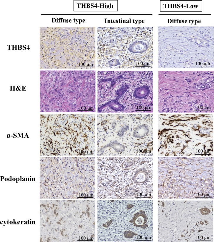
Fig 1 provides representative immunostaining patterns of THBS4. THBS4 was stained at the cytoplasm and the cell membrane of stroma cells, but cancer cells with cytokeratin expression did not express THBS4. Whereas, the THBS4 expression of the stromal cells was in agreement with the cells showing alpha-smooth muscle actin (αSMA) or Podoplanin expression. Of the total of 584 cases, 223 (38.2%) were THBS4-high.

Fig 1. Representative picture of THBS4 expression, H&E staining, αSMA expression, Podoplanin expression, and cytokeatin expression in diffuse and intestinal type of gastric cancer.

Fig 1

THBS4 was stained at the cytoplasm and cell membrane of stroma cells. The stromal cells with THBS4 expression were much to the cells showing αSMA or Podoplanin expression. cytokeratin was stained in cancer cells.

The relationship between clinicopathological features and the THBS4 expression of stromal cells

The clinicopathological characteristics of all cases according to their THBS4 expression in stroma are summarized in Table 1. Compared to the THBS4-low expression in stroma, THBS4-high expression in stroma was significantly associated with higher αSMA expression, macroscopic type 4 (p<0.001), greater tumor diameter (p<0.001), diffuse-type histology (p<0.001), microscopic undifferentiated-type, higher tumor invasion (p<0.001), lymph node metastasis (p<0.001), lymphatic invasion (p<0.001), peritoneal cytology (p<0.001), and peritoneal metastasis (p<0.001). Among Borrmann's type 4 cancer of 61 cases, 52 (85.2%) were THBS4-high. The results of subgroup analysis for type-4 or other type gastric cancer were shown in S1 and S2 Tables.

Table 1. Correlation between the expression of THBS4 in tumor stromal cells and clinicopathologic features in 584 patients with gastric carcinoma.

THBS4
High
(n = 223, 38.2%)
Low
(n = 361, 61.8%)
p value
Age (year-old)
    < 65 105 (47.1%) 151 (41.8%)
    ≥ 65 118 (52.9%) 210 (58.2%) 0.247
Gender
    Female 97 (43.5%) 162 (44.9%)
    Male 126 (56.5%) 199 (55.1%) 0.810
αSMA expression
    High 139 (62.3%) 136 (37.7%)
    Low 84 (37.7%) 225 (62.3%) <0.001
Macroscopic type
    type 4 52 (23.3%) 9 (2.5%)
    Other types 171 (76.7%) 352 (97.5%) <0.001
Tumor diameter
    < 50 103 (46.4%) 250 (69.3%)
    ≥ 50 120 (53.6%) 111 (30.7%) <0.001
Microscopic type
    Differentiated 81 (36.3%) 205 (56.8%)
    Undifferentiated 142 (63.7%) 156 (43.2%) <0.001
Depth of tumor invasion
    T1-2 73 (32.7%) 264 (73.1%)
    T3-4 150 (67.3%) 97 (26.9%) <0.001
Lymph node metastasis
    N0 83 (37.2%) 246 (68.1%)
    N1-3 140 (62.8%) 115 (31.9%) <0.001
Lymphatic invasion
    Absent 56 (25.1%) 204 (56.5%)
    Present 167 (74.9%) 157 (43.5%) <0.001
Venous invasion
    Absent 176 (84.2%) 305 (79.1%)
    Present 47 (15.8%) 56 (20.9%) 0.109
Ascites cytology
    Negative 174 (78.0%) 346 (95.8%)
    Positive 49 (22.0%) 15 (4.2%) <0.001
Peritoneal metastasis
    Absent 200 (89.7%) 349 (96.7%)
    Present 23 (10.3%) 12 (3.3%) 0.001
Hepatic metastasis
    Negative 216 (96.9%) 354 (98.1%)
    Positive 7 (3.1%) 7 (1.9%) 0.409
pStage
    I, II 99 (44.4%) 285 (78.9%)
    III, IV 124 (55.6%) 76 (21.1%) <0.001

Survival outcomes

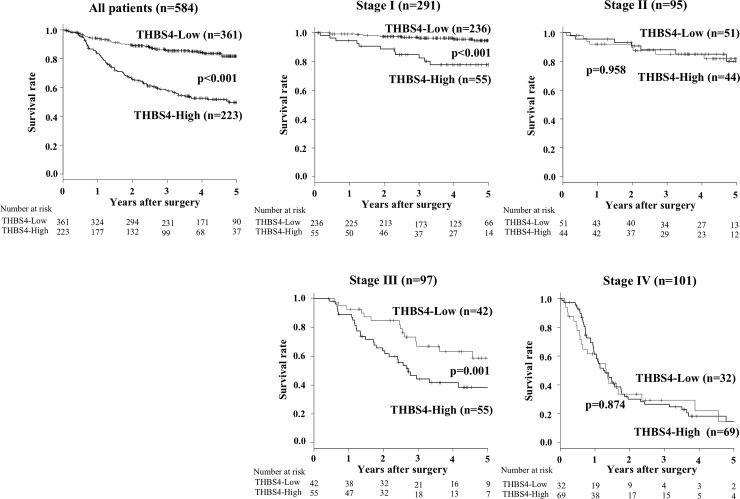
The 5-year overall survival rate of the patients in THBS4-high expression group (38.5%) was significantly poorer compared to that of the THBS4-low group (81.4%) (p<0.001; Fig 2). The results of subgroup analysis for type-4 or other type gastric cancer were shown in S1 Fig. Our analysis for each tumor stage revealed that the overall survival of the THBS4-high expression patients at Stage I and Stage III was significantly poorer than that of the patients with THBS4-low expression (p<0.001 and p = 0.001, respectively; Fig 2). The recurrence site according to pathological stages is shown in S2 Table. Hematogenous recurrence was frequent in patients at stage II patients, in compared with those at stage III.

Fig 2. The overall survival (OS) of the gastric cancer patients based on the THBS4 expression in their tumor stromal cells.

Fig 2

(A) The Kaplan-Meier survival curve indicates that the OS of all patients with high THBS4 expression in stromal cells was significantly worse than that of the patients with low THBS4 expression (p<0.001). (B–E) The Kaplan-Meier survival curve for each stage. The 5-year OS rates of the Stage I patients with high THBS4 expression were poorer than those of the Stage I patients with low THBS4 expression (p<0.001), and the same was true of the Stage III patients (p = 0.001).

Univariate and multivariate analyses

Table 2 provides the results of the univariate and multivariate analyses for overall survival. The univariate analysis revealed that poor survival was significantly correlated with high αSMA expression (p<0.001), high THBS4 expression (p<0.001), age ≥ 65 years old (p<0.001), macroscopic type 4 (p<0.001), microscopic undifferentiated type (p<0.001), high T stage (p<0.001), lymph node metastasis (p<0.001), lymphatic invasion (p<0.001), vascular invasion (p<0.001), ascites cytology positive (p<0.001), peritoneal metastasis (p<0.001), and hepatic metastasis (p<0.001).

Table 2. Univariate and multivariate Cox multiple regression analysis with respect to overall survival after surgery in 584 patients with gastric carcinoma.

Univariate analysis Multivariate analysis
Hazard Ratio (95% CI) p value Hazard Ratio (95% CI) p value
THBS4-high 3.43 (2.48–4.75) <0.001 1.53 (1.05–2.25) 0.028
Age ≥65 year-old 1.61 (1.16–2.23) 0.004 1.74 (1.23–2.46) 0.002
Female (vs. Male) 1.29 (0.93–1.79) 0.123
αSMA-high 2.37 (1.70–3.31) <0.001 1.27 (0.90–1.80) 0.180
Borrmann’s type 4 7.41 (5.22–10.5) <0.001 1.62 (1.05–2.50) 0.042
Tumor diameter ≥50 mm 6.27 (4.37–9.00) <0.001 1.49 (0.90–2.46) 0.088
Undifferentiated type 1.72 (1.24–2.37) 0.001 1.18 (0.81–1.70) 0.386
T3&4 (vs. T1&2) 6.72 (4.61–9.80) <0.001 1.41 (0.82–2.40) 0.211
N1-3 (vs. N0) 8.29 (5.50–12.5) <0.001 2.79 (1.62–4.80) <0.001
Lymphatic invasion 5.22 (3.42–7.95) <0.001 1.06 (0.61–1.83) 0.837
Vascular invasion 3.10 (2.22–4.32) <0.001 1.06 (0.73–1.53) 0.772
Cytology positive 7.42 (5.23–10.5) <0.001 1.91 (1.28–2.85) 0.001
Peritoneal metastasis 9.11 (5.97–13.9) <0.001 2.60 (1.64–4.12) <0.001
Hepatic metastasis 5.90 (3.18–11.0) <0.001 3.64 (1.89–7.02) <0.001

The multivariate analysis revealed that THBS4 expression, age ≥65 years, macroscopic type 4 (p<0.001), lymph node metastasis (p<0.001), cytology-positive status (p<0.001), peritoneal metastasis (p<0.001), and hepatic metastasis (p<0.001) were independent prognostic factors.

THBS4 expression on CAFs and NFs

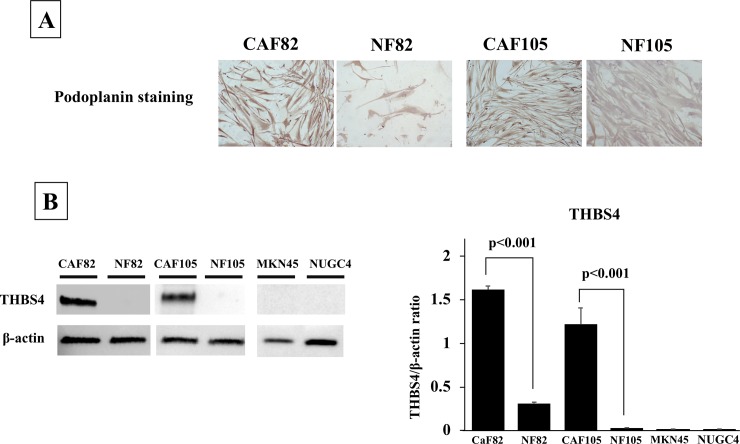
Fig 3A shows established CAFs and NFs stained with Podoplanin. The results of the western blot analysis revealed that the CAF82 and CAF105 significantly expressed THBS4 compared with NF82 and NF105, respectively (t-test, p<0.001 and p<0.001, respectively). MKN45 and NUGC4 did not. (Fig 3B).

Fig 3. The picture of CAFs and NFs stained with Podoplanin and the western blot analysis of THBS4.

Fig 3

(A), Representative picture of Podoplanin staining. Podoplanin was mainly stained at the cytoplasm fibroblasts. The expression level of Podoplanin was higher in CAFs, in compared with that in NFs. (B), THBS4 expression. CAFs expressed THBS4, but NFs and cancer cells did not.

Discussion

THBS4 was reported to be expressed not only in normal tissues but also in some types of solid cancers [8, 10, 17]. In the present study, we observed that gastric tumors (especially the stromal cells in tumor microenvironment) also express THBS4. In addition, the stromal cells expressing THBS4 also expressed αSMA or podoplanin, which is a marker of CAFs. In fact, the western blotting indicated that THBS4 was expressed from CAFs, but not from NFs and cancer cell lines. In case analysis, THBS4-high expression status was correlated with high αSMA expression. These findings suggest that in gastric cancer, THBS4 may be expressed by the gastric cancer-associated fibroblasts.

We reported that CAFs play an important role in the progression, growth, and spread of gastric cancer [18]. In present study, we showed in the results that THBS4-high expression in stroma was significantly associated with higher αSMA expression, higher tumor invasion, lymph node metastasis, lymphatic invasion, peritoneal cytology, and peritoneal metastasis. We also found that high THBS4 expression in tumor stroma was significantly poorer than that of the patients with low THBS4 expression. Moreover, multivariate analysis for overall survival showed that not αSMA expression on CAFs but THBS4 expression on CAFs was an independent prognostic factor. These findings indicated that THBS4 from CAFs might be associated with the invasion of cancer cells, and THBS4 from CAFs is associated with the metastasis of cancer cells.

By targeting the protein coding gene Kruppel-like factor 9 (KLF9) using gastric cancer cell lines, Chen et al. observed that THBS4 had positive effects on gastric cancer cells' proliferation and metastasis [1]. However, little is known about the molecular mechanism of THBS4 remains unclear. Muppala S et al. reported that THBS4 is upregulated in response to TGF-β and promotes the effect of TGF-β on angiogenesis, mediating cell proliferation and survival [10]. Forester et al. showed THBS4 transcription on CAFs is stimulated by incubation with conditioned medium derived from human diffuse gastric cancer lines [4]. It is speculated that TGF-β derived from cancer cells stimulated THBS4 expression on CAFs resulting in angiogenesis and cancer progression.

In particular, the stage I and stage III patients with high THBS4 expression had significantly poorer prognosis than the stage I and III patients with low THBS4 expression, respectively. High THBS4 expression in stromal cells was revealed as an independent prognostic factor for gastric cancer patients. THBS4 from CAFs might be a useful prognostic indicator for gastric cancer patients, especially for those with stage I or III cancer.

In contrast, no significant difference in prognosis was shown between the THBS4-high group and THBS4-low group at stage II. Liver metastasis is a relatively frequent recurrence pattern among patients with stage II gastric cancer [19], and THBS4 expression is not correlated with vascular invasion or liver metastasis, which might be one of the reasons for the absence of a difference in the prognosis between our two THBS4 expression groups at stage II. Additionally, effects of adjuvant chemotherapy might be caused especially at stage II. It was difficult to compare adjuvant regimen according to the stage, because the standard regimen for adjuvant chemotherapy was not established for the enrolled patients at the time. However, the prognosis of patients with lymph node metastasis which was significantly correlated with THBS4-high might be improved.

To the best of our knowledge, the present study is the first to investigate the clinicopathological significance of THBS4 in the gastric cancer microenvironment. Macroscopic type 4 and the histologic diffuse type were significantly associated with a high expression of THBS4 in our patient series. Remarkably, THBS4 is expressed in most stroma of Borrmann's type 4 cancer. THBS4 is involved in tissue remodeling [6]. Macroscopic type 4, diffusely infiltrating carcinoma, and scirrhous gastric carcinoma (also known as linitis plastica-type carcinoma) are characterized by cancer cell infiltration and proliferation accompanied by extensive stromal fibrosis and abundant extracellular matrix [20]. This typical histological finding of macroscopic type 4 might be regulated in part by the tissue remodeling activity of THBS4.

FGFR2 is overexpressed on macroscopic type-4 gastric cancer [21]. The secretion of FGF7 (a ligand of fibroblast growth factor receptor 2 [FGFR]2) by gastric fibroblasts is likely to contribute in a paracrine manner to the remarkable cell proliferation seen in scirrhous gastric cancer with FGFR2 overexpression [22]. Whereas Huang et al. reported that the downregulation of THBS4 promotes the FGFR2 signal of gastric cancer progression via the PI3K-AKT-mTOR pathway using non-type-4 gastric cancer cell lines [23]. It is necessary in the future to examine the effects of THBS4 on the PI3K-AKT-mTOR pathway by using type-4 gastric cancer cell lines with FGFR2 overexpression.

In conclusion, THBS4 is expressed on CAFs in the microenvironment of gastric cancer, especially in macroscopic type-4 gastric cancer. THBS4 from CAFs might be associated with the invasion of cancer cells, and THBS4 is also a useful prognostic indicator for gastric cancer patients, especially for those with stage I or stage III cancer.

Supporting information

S1 Fig. The Kaplan-Meier survival curve for Borrmann’s type 4 tumor and other type.

No significant difference in overall survival was shown between the THBS-high group and THBS-low group at Borrmann’s type 4. Whereas the prognosis of the patients with high THBS4 expression were poorer than that of the patients with low THBS4 expression at other type.

(TIFF)

S1 File. Data of clinicopathologic factors in 584 patients.

(XLSX)

S1 Raw Images. Original images for blots and gels.

(TIFF)

S2 Raw Images. Original images for blots and gels.

(TIFF)

S1 Table. Correlation between the expression of THBS4 in tumor stromal cells and clinicopathologic features in Borrmann’s type 4.

(DOCX)

S2 Table. Correlation between the expression of THBS4 in tumor stromal cells and clinicopathologic features in other macroscopic type.

(DOCX)

S3 Table. Recurrence pattern according to the stage.

(DOCX)

Data Availability

All relevant data are within the paper and its Supporting Information files.

Funding Statement

This study is partially supported by Japan Society for the Promotion of Science KAKENHI (Grant-in-Aid for Scientific Research B; Grant Number JP18H02883 (https://www.jsps.go.jp/english/e-grants/) to MY. The funder had no role in study design, data collection and analysis, decision to publish, or preparation of the manuscript. There was no additional external funding received for this study.

References

  • 1.Chen X, Huang Y, Wang Y, Wu Q, Hong S, Huang Z. THBS4 predicts poor outcomes and promotes proliferation and metastasis in gastric cancer. J Physiol Biochem. 2019;75(1):117–23. Epub 2019/02/13. 10.1007/s13105-019-00665-9 . [DOI] [PubMed] [Google Scholar]
  • 2.Global Burden of Disease Cancer C, Fitzmaurice C, Allen C, Barber RM, Barregard L, Bhutta ZA, et al. Global, Regional, and National Cancer Incidence, Mortality, Years of Life Lost, Years Lived With Disability, and Disability-Adjusted Life-years for 32 Cancer Groups, 1990 to 2015: A Systematic Analysis for the Global Burden of Disease Study. JAMA Oncol. 2017;3(4):524–48. Epub 2016/12/06. 10.1001/jamaoncol.2016.5688 [DOI] [PMC free article] [PubMed] [Google Scholar]
  • 3.Torre LA, Bray F, Siegel RL, Ferlay J, Lortet-Tieulent J, Jemal A. Global cancer statistics, 2012. CA Cancer J Clin. 2015;65(2):87–108. Epub 2015/02/06. 10.3322/caac.21262 . [DOI] [PubMed] [Google Scholar]
  • 4.Ishimoto T, Miyake K, Nandi T, Yashiro M, Onishi N, Huang KK, et al. Activation of Transforming Growth Factor Beta 1 Signaling in Gastric Cancer-associated Fibroblasts Increases Their Motility, via Expression of Rhomboid 5 Homolog 2, and Ability to Induce Invasiveness of Gastric Cancer Cells. Gastroenterology. 2017;153(1):191–204 e16. Epub 2017/04/10. 10.1053/j.gastro.2017.03.046 . [DOI] [PubMed] [Google Scholar]
  • 5.Muppala S, Xiao R, Krukovets I, Verbovetsky D, Yendamuri R, Habib N, et al. Thrombospondin-4 mediates TGF-beta-induced angiogenesis. Oncogene. 2017;36(36):5189–98. Epub 2017/05/10. 10.1038/onc.2017.140 [DOI] [PMC free article] [PubMed] [Google Scholar]
  • 6.Stenina-Adognravi O, Plow EF. Thrombospondin-4 in tissue remodeling. Matrix Biol. 2019;75–76:300–13. Epub 2017/11/16. 10.1016/j.matbio.2017.11.006 [DOI] [PMC free article] [PubMed] [Google Scholar]
  • 7.Adams JC, Lawler J. The thrombospondins. Cold Spring Harb Perspect Biol. 2011;3(10):a009712 Epub 2011/08/31. 10.1101/cshperspect.a009712 [DOI] [PMC free article] [PubMed] [Google Scholar]
  • 8.McCart Reed AE, Song S, Kutasovic JR, Reid LE, Valle JM, Vargas AC, et al. Thrombospondin-4 expression is activated during the stromal response to invasive breast cancer. Virchows Arch. 2013;463(4):535–45. Epub 2013/08/15. 10.1007/s00428-013-1468-3 . [DOI] [PubMed] [Google Scholar]
  • 9.Liu J, Cheng G, Yang H, Deng X, Qin C, Hua L, et al. Reciprocal regulation of long noncoding RNAs THBS4003 and THBS4 control migration and invasion in prostate cancer cell lines. Mol Med Rep. 2016;14(2):1451–8. Epub 2016/07/01. 10.3892/mmr.2016.5443 [DOI] [PMC free article] [PubMed] [Google Scholar]
  • 10.Forster S, Gretschel S, Jons T, Yashiro M, Kemmner W. THBS4, a novel stromal molecule of diffuse-type gastric adenocarcinomas, identified by transcriptome-wide expression profiling. Mod Pathol. 2011;24(10):1390–403. Epub 2011/06/28. 10.1038/modpathol.2011.99 . [DOI] [PubMed] [Google Scholar]
  • 11.Lin X, Hu D, Chen G, Shi Y, Zhang H, Wang X, et al. Associations of THBS2 and THBS4 polymorphisms to gastric cancer in a Southeast Chinese population. Cancer Genet. 2016;209(5):215–22. Epub 2016/05/11. 10.1016/j.cancergen.2016.04.003 . [DOI] [PubMed] [Google Scholar]
  • 12.Su F, Zhao J, Qin S, Wang R, Li Y, Wang Q, et al. Over-expression of Thrombospondin 4 correlates with loss of miR-142 and contributes to migration and vascular invasion of advanced hepatocellular carcinoma. Oncotarget. 2017;8(14):23277–88. Epub 2017/02/09. 10.18632/oncotarget.15054 [DOI] [PMC free article] [PubMed] [Google Scholar]
  • 13.Wu H, Zhang G, Li Z, Ma J, Han X, Xiang T, et al. Thrombospondin-4 expression as a prognostic marker in hepatocellular carcinoma. Gene. 2019;696:219–24. Epub 2019/02/26. 10.1016/j.gene.2019.02.049 . [DOI] [PubMed] [Google Scholar]
  • 14.Greco SA, Chia J, Inglis KJ, Cozzi SJ, Ramsnes I, Buttenshaw RL, et al. Thrombospondin-4 is a putative tumour-suppressor gene in colorectal cancer that exhibits age-related methylation. BMC Cancer. 2010;10:494 Epub 2010/09/18. 10.1186/1471-2407-10-494 [DOI] [PMC free article] [PubMed] [Google Scholar]
  • 15.van Doorn R, Zoutman WH, Dijkman R, de Menezes RX, Commandeur S, Mulder AA, et al. Epigenetic profiling of cutaneous T-cell lymphoma: promoter hypermethylation of multiple tumor suppressor genes including BCL7a, PTPRG, and p73. J Clin Oncol. 2005;23(17):3886–96. Epub 2005/05/18. 10.1200/JCO.2005.11.353 . [DOI] [PubMed] [Google Scholar]
  • 16.Fuyuhiro Y, Yashiro M, Noda S, Matsuoka J, Hasegawa T, Kato Y, et al. Cancer-associated orthotopic myofibroblasts stimulates the motility of gastric carcinoma cells. Cancer Sci. 2012;103(4):797–805. Epub 2012/02/11. 10.1111/j.1349-7006.2012.02209.x . [DOI] [PMC free article] [PubMed] [Google Scholar]
  • 17.Dakhova O, Ozen M, Creighton CJ, Li R, Ayala G, Rowley D, et al. Global gene expression analysis of reactive stroma in prostate cancer. Clin Cancer Res. 2009;15(12):3979–89. Epub 2009/06/11. 10.1158/1078-0432.CCR-08-1899 [DOI] [PMC free article] [PubMed] [Google Scholar]
  • 18.Yashiro M, Hirakawa K. Cancer-stromal interactions in scirrhous gastric carcinoma. Cancer Microenviron. 2010;3(1):127–35. Epub 2011/01/07. 10.1007/s12307-010-0036-5 [DOI] [PMC free article] [PubMed] [Google Scholar]
  • 19.Song J, Lee HJ, Cho GS, Han SU, Kim MC, Ryu SW, et al. Recurrence following laparoscopy-assisted gastrectomy for gastric cancer: a multicenter retrospective analysis of 1,417 patients. Ann Surg Oncol. 2010;17(7):1777–86. Epub 2010/02/13. 10.1245/s10434-010-0932-4 . [DOI] [PubMed] [Google Scholar]
  • 20.Japanese Gastric Cancer A. Japanese classification of gastric carcinoma: 3rd English edition. Gastric Cancer. 2011;14(2):101–12. Epub 2011/05/17. 10.1007/s10120-011-0041-5 . [DOI] [PubMed] [Google Scholar]
  • 21.Yashiro M, Shinto O, Nakamura K, Tendo M, Matsuoka T, Matsuzaki T, et al. Synergistic antitumor effects of FGFR2 inhibitor with 5-fluorouracil on scirrhous gastric carcinoma. Int J Cancer. 2010;126(4):1004–16. Epub 2009/07/22. 10.1002/ijc.24763 . [DOI] [PubMed] [Google Scholar]
  • 22.Nakazawa K, Yashiro M, Hirakawa K. Keratinocyte growth factor produced by gastric fibroblasts specifically stimulates proliferation of cancer cells from scirrhous gastric carcinoma. Cancer Res. 2003;63(24):8848–52. . [PubMed] [Google Scholar]
  • 23.Huang T, Liu D, Wang Y, Li P, Sun L, Xiong H, et al. FGFR2 Promotes Gastric Cancer Progression by Inhibiting the Expression of Thrombospondin4 via PI3K-Akt-Mtor Pathway. Cell Physiol Biochem. 2018;50(4):1332–45. Epub 2018/10/26. 10.1159/000494590 . [DOI] [PubMed] [Google Scholar]

Decision Letter 0

Masaru Katoh

6 Aug 2019

PONE-D-19-19270

The Clinicopathological Significance of Thrombospondin-4 Expression in the Tumor Microenvironment of Gastric Cancer

PLOS ONE

Dear Dr. Yashiro,

Thank you for submitting your manuscript to PLOS ONE. After careful consideration, we feel that it has merit but does not fully meet PLOS ONE’s publication criteria as it currently stands. Therefore, we invite you to submit a revised version of the manuscript that addresses the points raised during the review process.

(1) Immunostaining of Podoplanin (PDPN) as a widely accepted marker of cancer associated fibroblasts (CAFs)

(2) Detailed methodological description of CAFs and normal-associated fibroblasts (NAFs) and presentation of their images

(3) Explanation about THBS4 expression in stroll cells but not cancer cells in primary gastric cancer, as well as presentation of more clear images of THBS4 immunostaining, and results of THBS4 immunostaining

(4) Presentation and statistical analysis related to Western blot analysis

(5) Refinement of Kaplan-Meier analysis

(6) Additional experiments or discussion on the functions of thrombospondin-4 (THBS4) in CAFs

(7) Other issues pointed out by Reviewers

We would appreciate receiving your revised manuscript by Sep 20 2019 11:59PM. When you are ready to submit your revision, log on to https://www.editorialmanager.com/pone/ and select the 'Submissions Needing Revision' folder to locate your manuscript file.

If you would like to make changes to your financial disclosure, please include your updated statement in your cover letter.

To enhance the reproducibility of your results, we recommend that if applicable you deposit your laboratory protocols in protocols.io, where a protocol can be assigned its own identifier (DOI) such that it can be cited independently in the future. For instructions see: http://journals.plos.org/plosone/s/submission-guidelines#loc-laboratory-protocols

Please include the following items when submitting your revised manuscript:

  • A rebuttal letter that responds to each point raised by the academic editor and reviewer(s). This letter should be uploaded as separate file and labeled 'Response to Reviewers'.

  • A marked-up copy of your manuscript that highlights changes made to the original version. This file should be uploaded as separate file and labeled 'Revised Manuscript with Track Changes'.

  • An unmarked version of your revised paper without tracked changes. This file should be uploaded as separate file and labeled 'Manuscript'.

Please note while forming your response, if your article is accepted, you may have the opportunity to make the peer review history publicly available. The record will include editor decision letters (with reviews) and your responses to reviewer comments. If eligible, we will contact you to opt in or out.

We look forward to receiving your revised manuscript.

Kind regards,

Masaru Katoh, M.D., Ph.D.

Academic Editor

PLOS ONE

Journal Requirements:

1.    When submitting your revision, we need you to address these additional requirements.

Please ensure that your manuscript meets PLOS ONE's style requirements, including those for file naming. The PLOS ONE style templates can be found at

http://www.journals.plos.org/plosone/s/file?id=wjVg/PLOSOne_formatting_sample_main_body.pdf and http://www.journals.plos.org/plosone/s/file?id=ba62/PLOSOne_formatting_sample_title_authors_affiliations.pdf

2. Thank you for stating the following in the Acknowledgments Section of your manuscript:

This study is partially founded by KAKENHI Grant-in-Aid for Scientific Research, Nos. 18H02883 (M.Y.).

We note that you have provided funding information that is not currently declared in your Funding Statement. However, funding information should not appear in the Acknowledgments section or other areas of your manuscript. We will only publish funding information present in the Funding Statement section of the online submission form.

Please remove any funding-related text from the manuscript and let us know how you would like to update your Funding Statement. Currently, your Funding Statement reads as follows:

The author(s) received no specific funding for this work.

Additionally, because some of your funding information pertains to [commercial funding//patents], we ask you to provide an updated Competing Interests statement, declaring all sources of commercial funding.

In your Competing Interests statement, please confirm that your commercial funding does not alter your adherence to PLOS ONE Editorial policies and criteria by including the following statement: "This does not alter our adherence to PLOS ONE policies on sharing data and materials.” as detailed online in our guide for authors  http://journals.plos.org/plosone/s/competing-interests.  If this statement is not true and your adherence to PLOS policies on sharing data and materials is altered, please explain how.

Please include the updated Competing Interests Statement and Funding Statement in your cover letter. We will change the online submission form on your behalf.

3.  PLOS ONE now requires that authors provide the original uncropped and unadjusted images underlying all blot or gel results reported in a submission’s figures or Supporting Information files. This policy and the journal’s other requirements for blot/gel reporting and figure preparation are described in detail at https://journals.plos.org/plosone/s/figures#loc-blot-and-gel-reporting-requirements and https://journals.plos.org/plosone/s/figures#loc-preparing-figures-from-image-files. When you submit your revised manuscript, please ensure that your figures adhere fully to these guidelines and provide the original underlying images for all blot or gel data reported in your submission. See the following link for instructions on providing the original image data: https://journals.plos.org/plosone/s/figures#loc-original-images-for-blots-and-gels.

  In your cover letter, please note whether your blot/gel image data are in Supporting Information or posted at a public data repository, provide the repository URL if relevant, and provide specific details as to which raw blot/gel images, if any, are not available. Email us at plosone@plos.org if you have any questions.

4.  In your Data Availability statement, you have not specified where the minimal data set underlying the results described in your manuscript can be found. PLOS defines a study's minimal data set as the underlying data used to reach the conclusions drawn in the manuscript and any additional data required to replicate the reported study findings in their entirety. All PLOS journals require that the minimal data set be made fully available. For more information about our data policy, please see http://journals.plos.org/plosone/s/data-availability.

Upon re-submitting your revised manuscript, please upload your study’s minimal underlying data set as either Supporting Information files or to a stable, public repository and include the relevant URLs, DOIs, or accession numbers within your revised cover letter. For a list of acceptable repositories, please see http://journals.plos.org/plosone/s/data-availability#loc-recommended-repositories. Any potentially identifying patient information must be fully anonymized.

Important: If there are ethical or legal restrictions to sharing your data publicly, please explain these restrictions in detail. Please see our guidelines for more information on what we consider unacceptable restrictions to publicly sharing data: http://journals.plos.org/plosone/s/data-availability#loc-unacceptable-data-access-restrictions. Note that it is not acceptable for the authors to be the sole named individuals responsible for ensuring data access.

We will update your Data Availability statement to reflect the information you provide in your cover letter.

[Note: HTML markup is below. Please do not edit.]

Reviewers' comments:

Reviewer's Responses to Questions

Comments to the Author

1. Is the manuscript technically sound, and do the data support the conclusions?

The manuscript must describe a technically sound piece of scientific research with data that supports the conclusions. Experiments must have been conducted rigorously, with appropriate controls, replication, and sample sizes. The conclusions must be drawn appropriately based on the data presented.

Reviewer #1: Partly

Reviewer #2: Partly

Reviewer #3: Yes

Reviewer #4: Partly

**********

2. Has the statistical analysis been performed appropriately and rigorously?

Reviewer #1: Yes

Reviewer #2: Yes

Reviewer #3: Yes

Reviewer #4: Yes

**********

3. Have the authors made all data underlying the findings in their manuscript fully available?

The PLOS Data policy requires authors to make all data underlying the findings described in their manuscript fully available without restriction, with rare exception (please refer to the Data Availability Statement in the manuscript PDF file). The data should be provided as part of the manuscript or its supporting information, or deposited to a public repository. For example, in addition to summary statistics, the data points behind means, medians and variance measures should be available. If there are restrictions on publicly sharing data—e.g. participant privacy or use of data from a third party—those must be specified.

Reviewer #1: Yes

Reviewer #2: No

Reviewer #3: Yes

Reviewer #4: Yes

**********

4. Is the manuscript presented in an intelligible fashion and written in standard English?

PLOS ONE does not copyedit accepted manuscripts, so the language in submitted articles must be clear, correct, and unambiguous. Any typographical or grammatical errors should be corrected at revision, so please note any specific errors here.

Reviewer #1: Yes

Reviewer #2: Yes

Reviewer #3: Yes

Reviewer #4: Yes

**********

5. Review Comments to the Author

Please use the space provided to explain your answers to the questions above. You may also include additional comments for the author, including concerns about dual publication, research ethics, or publication ethics. (Please upload your review as an attachment if it exceeds 20,000 characters)

Reviewer #1: This manuscript is to evaluate that THBS4 expressed in CAFs is a prognostic marker to predict poor prognosis in gastric cancer. They proved that THBS4 was expressed in stromal cells expressing alpha-SMA in gastric cancer, and positive THBS4 expression is correlated with poor prognosis of gastric cancer patients. The results are clear, but for publication in Plos One, following points should be fixed.

1. Authors determined the expression of THBS4 in gastric cancer tissues according to the intensity and the percentage of stained cancer cells and stromal cells (Page 4, Line 71). However, they said that it was only expression in stromal cells. How did authors calculate the percentage of positive THBS cells? Was the percentage meaning the ratio of positive stromal cells in all cells including cancer cells and stromal cells?

2. In figure 1, the positive staining of THBS4 look like too diffuse in the entire tissues. Can authors show the clearer photo for THBS4 staining?

3. The gastric cancer generally showed histologic heterogeneity according to the classification. In figure 1, I recommend that authors show the representative photos for THBS4 expression in two different histological subtypes (diffuse and intestinal type).

4. In figure 1, the staining photo of cytokeratin, cancer cell specific maker, should be added to support that THBS4 was not stained in cancer cells.

5. In figure 3, authors just showed the expression of THBS4 in NAF and CAF. I recommend authors that the results should be added the expression of THBS of several gastric cancer cell lines.

6. Authors described in abstract and conclusion that “THBS4 from CAFs is associated with the metastasis of cancer cells” and “THBS4 from CAFs might be associated with the invasion of cancer cells”. However, they did not show the any data for proof of concept. Authors should add the experimental data to prove them, or the descriptions in abstract and conclusion should be toned down.

7. Authors emphasized that THBS4 is especially expression on CAFs of macroscopic type-4 gastric cancer. It would be better to add the results of subgroup analysis for type-4 or other type gastric cancer.

8. Authors insisted that non-significance of stage II would be explained as recurrent pattern and adjuvant chemotherapy. To supper this hypothesis, recurrent pattern or adjuvant regimen according to the stage should be added.

Reviewer #2: The authors investigated the clinicopathological significance of stromal thrombospondin-4 expression in the tumor microenvironment of gastric cancer, and found that THBS4 was expressed on CAFs. Stromal THBS4 was associated with the metastasis of cancer cells. The results are interesting and promising. However, this article suffers from several major flaws:

1. The authors used α-SMA to indicate the location of CAFs. However, α-SMA is a microfilament protein with contractile ability, mainly expressed at myoepithelial cells. Although α-SMA expression could also appear in the transformation of fibroblasts to myofibroblasts around cancer cells, the positive rate of α-SMA in CAFs is not high, and some of muscle tissues may be unexpectedly positive. I recommend the authors to add podoplanin (PDPN) in immunostaining assays, which is a widely accepted marker for CAFs with a relative high expression level.

2. Primary culture was performed to obtain CAFs and NAFs in this research. However, there was no demonstrations or photos to show the status of cells in order to verify the results. And the isolations and purifications of CAFs and NAFs were not mentioned either.

3. Inconsistent data were found between abstract and main text (e.g. positive rate of THBS4 expression).

4. For WB assay, the authors did not elucidate the sample size and necessary statistical analysis (t-test). A solitary result cannot clarify the real world.

5. According to the clinical data, THBS4 in stroma has obvious correlations with tumor invasion and lymph node metastasis. Since the primary CAFs have been cultured successfully, the authors should perform gene function experiments (e.g. migration & invasion) subsequently and explore the potential mechanisms to explain the phenomena they observed.

6. Are there any references to support the IHC evaluating method of THBS4 expression? If yes, please cite the articles, otherwise the authors should state the reason why score 3 was the watershed.

7. Terms were not used properly in this article. NAF was not common in the description of normal fibroblast. And definitions of positive and high were confused.

Reviewer #3: Dear editors,

In this study, the authors mainly investigated the clinicopathological value of THBS4

In gastric cancer based on the retrospective cohort from their own center. The research methods were statistical analyses of clinical data, including chi-square test, Cox proportional hazards regression models, as well as survival curves. The endpoint parameter was overall survival (OS). The article was well written, and focused on practical clinical aspects. While some problems still existed and should be improved.

1. The exact correlation of α-SMA or CAFs and THBS4 should be explained more apparently both in Introduction and Discussion.

2. The qualities of all K-M curves were poor. We believe this problem should be focused since key results of this study were reflected herein. Some normative forms could be referenced, for instance, ANNALS OF ONCOLOGY.

3. Information from Figure 3 was too simple to explain exact relation between the expression of THBS4 and α-SMA or CAFs.

4. References should be updated by using articles in recent 5 years.

Reviewer #4: The authors Kuroda et.al have identified THBS4 as a marker of CAFs in gastric cancer. IHC analysis on 584 GC patients specimen shows statistical significance when THBS4 expression is compared with the clinico-pathological characteristics of the patients. However, I have some comments with respect to the methodology of CAFs and NAFs isolation for western blot analysis. The authors have not elaborated on the specific markers that were used to identify these from the samples.

**********

6. PLOS authors have the option to publish the peer review history of their article (what does this mean?). If published, this will include your full peer review and any attached files.

If you choose “no”, your identity will remain anonymous but your review may still be made public.

Do you want your identity to be public for this peer review? For information about this choice, including consent withdrawal, please see our Privacy Policy.

Reviewer #1: No

Reviewer #2: No

Reviewer #3: No

Reviewer #4: Yes: Kakoli Das

[NOTE: If reviewer comments were submitted as an attachment file, they will be attached to this email and accessible via the submission site. Please log into your account, locate the manuscript record, and check for the action link "View Attachments". If this link does not appear, there are no attachment files to be viewed.]

While revising your submission, please upload your figure files to the Preflight Analysis and Conversion Engine (PACE) digital diagnostic tool, https://pacev2.apexcovantage.com/. PACE helps ensure that figures meet PLOS requirements. To use PACE, you must first register as a user. Registration is free. Then, login and navigate to the UPLOAD tab, where you will find detailed instructions on how to use the tool. If you encounter any issues or have any questions when using PACE, please email us at figures@plos.org. Please note that Supporting Information files do not need this step.

PLoS One. 2019 Nov 8;14(11):e0224727. doi: 10.1371/journal.pone.0224727.r002

Author response to Decision Letter 0


26 Sep 2019

Reviewer #1

Thank you very much for the careful review of the Reviewer #1. We corrected several points according to the descriptions by the Reviewer #1, as described below. We indicated the changes point by point and highlighted them in the revised paper.

1. Authors determined the expression of THBS4 in gastric cancer tissues according to the intensity and the percentage of stained cancer cells and stromal cells (Page 4, Line 71). However, they said that it was only expression in stromal cells. How did authors calculate the percentage of positive THBS cells? Was the percentage meaning the ratio of positive stromal cells in all cells including cancer cells and stromal cells?

We calculated the percentage of positive THBS cells by the ratio of stained stromal cells in all stromal cells. We added the comments in the materials and methods. (on page4 line 22-26)

2. In figure 1, the positive staining of THBS4 look like too diffuse in the entire tissues. Can authors show the clearer photo for THBS4 staining?

We replaced the picture with the clearer photo for THBS4 staining in Figure 1.

3. The gastric cancer generally showed histologic heterogeneity according to the classification. In figure 1, I recommend that authors show the representative photos for THBS4 expression in two different histological subtypes (diffuse and intestinal type).

We added the picture of THBS4 expression in two different histological subtypes including diffuse type and intestinal type. We added some comments in the manuscript. (on page 7 line 12-13)

4. In figure 1, the staining photo of cytokeratin, cancer cell specific maker, should be added to support that THBS4 was not stained in cancer cells.

We evaluated the expression of cytokeratin as cancer cell specific maker in the gastric cancer tissues. Cancer cells were positive for cytokeratin staining, but negative for THBS4 staining. The pictures of cytokeratin were added in Figure 1. We added these comments in the manuscript. (on page 2 line 15) (on page 4 line 17) (on page 7 line 5-6) (on page 7 line 12) (on page 7 line 15)

5. In figure 3, authors just showed the expression of THBS4 in NAF and CAF. I recommend authors that the results should be added the expression of THBS4 of several gastric cancer cell lines.

In accordance with the Reviewer’s comment, we added western blot of THBS4 of two gastric cancer cell lines, MKN45 and NUGC4 in Figure 3B. We added some comments in the manuscript. (on page 2 line 13) (on page 2 line 17) (on page 5 line 9-10) (on page 11 line 12) (on page 11 line 18) (on page 12 line 3-7)

6. Authors described in abstract and conclusion that “THBS4 from CAFs is associated with the metastasis of cancer cells” and “THBS4 from CAFs might be associated with the invasion of cancer cells”. However, they did not show the any data for proof of concept. Authors should add the experimental data to prove them, or the descriptions in abstract and conclusion should be toned down.

We showed in the results that THBS4-high expression in stroma was significantly correlated with higher tumor invasion (p<0.001), lymph node metastasis (p<0.001), lymphatic invasion (p<0.001), peritoneal cytology (p<0.001), and peritoneal metastasis (p<0.001). Taken together, we found that the prognosis of patients with high THBS4 expression in tumor stroma was significantly poorer than that of patients with low THBS4 expression. These findings indicated that THBS4 from CAFs might be associated with the invasion and metastasis of cancer cells. We added these comments in the discussion. (on page 12 line 12-20)

7. Authors emphasized that THBS4 is especially expression on CAFs of macroscopic type-4 gastric cancer. It would be better to add the results of subgroup analysis for type-4 or other type gastric cancer.

We performed the subgroup analysis of type-4 or other types gastric cancer. No significant difference of clinic-pathological factors was found between THBS4-high group and THBS4-low group in Bormann’s type 4 cancer (S1 Fig and S1 Table). In contrast, the other types gastric cancer had significant differences between the two groups. (S1 Fig and S2 Table). THBS4 might be associated with Bormann’s type 4 cancer, but not associated with malignant progression of Bormann’s type 4 cancer. We added these comments in the manuscript. (on page 7 line 25-28) (on page 9 line 6-7)

8. Authors insisted that non-significance of stage II would be explained as recurrent pattern and adjuvant chemotherapy. To supper this hypothesis, recurrent pattern or adjuvant regimen according to the stage should be added.

As reviewer pointed out, we added S3 Table which shows recurrence site according to the stage. Hematogenous recurrence was frequent in patients at stage II patients, in compared with those at stage III. In contrast, it was difficult to compare adjuvant regimen according to the stage, because the standard regimen for adjuvant chemotherapy was not established for the enrolled patients at the time. We added these comments in the text.

(on page 9 line 10; on page 9 line 13)

Reviewer #2

Thank you very much for the careful review of the Reviewer #2. We correct several points according to the descriptions by the Reviewer #2, as follows.

1. The authors used α-SMA to indicate the location of CAFs. However, α-SMA is a microfilament protein with contractile ability, mainly expressed at myoepithelial cells. Although α-SMA expression could also appear in the transformation of fibroblasts to myofibroblasts around cancer cells, the positive rate of α-SMA in CAFs is not high, and some of muscle tissues may be unexpectedly positive. I recommend the authors to add podoplanin (PDPN) in immunostaining assays, which is a widely accepted marker for CAFs with a relative high expression level.

I agree with the Reviewer’s comment, we added the immunostaining experiment of podoplanin which evaluated the for CAF in tumor. The picture of podoplanin staining was added in Fig 1. And we commented the Podoplanin immunostaining in the text. (on page 2 line 15) (on page 4 line 11-20) (on page 7 line 8) (on page 7 line 15) (on page 12 line 5).

2. Primary culture was performed to obtain CAFs and NAFs in this research. However, there was no demonstrations or photos to show the status of cells in order to verify the results. And the isolations and purifications of CAFs and NAFs were not mentioned either.

We added representative pictures of CAFs and NFs by Podoplanin staining in Fig 3A. The method for the primary culture of CAFs and NFs was added in the manuscript. CAFs and NFs were defined by αSMA staining, as previously reported. (on page 5 line 4-9) (on page 5 line 12-16)

1. Fuyuhiro Y, Yashiro M, Noda S, Matsuoka J, Hasegawa T, Kato Y, et al. Cancer-associated orthotopic myofibroblasts stimulates the motility of gastric carcinoma cells. Cancer Sci. 2012;103(4):797-805.

2. Fuyuhiro Y, Yashiro M, Noda S, Kashiwagi S, Matsuoka J, Doi Y, et al. Myofibroblasts are associated with the progression of scirrhous gastric carcinoma. Exp Ther Med. 2010;1(4):547-51.

3. Inconsistent data were found between abstract and main text (e.g. positive rate of THBS4 expression).

We corrected them. (on page 2 line 11)

4. For WB assay, the authors did not elucidate the sample size and necessary statistical analysis (t-test). A solitary result cannot clarify the real world.

We added statistical analysis of western blot of THBS4 in CAFs and NFs in Fig 3B. We added the comments in the results. (on page 11 line 9-12) (on page 11 line 17-18).

5. According to the clinical data, THBS4 in stroma has obvious correlations with tumor invasion and lymph node metastasis. Since the primary CAFs have been cultured successfully, the authors should perform gene function experiments (e.g. migration & invasion) subsequently and explore the potential mechanisms to explain the phenomena they observed.

We agree that additional experiments on function of THBS4 would be valuable, as reviewer commented. We plan to investigate THBS4 function in CAFs, and submit as a new paper of in vitro experiment in the near future. Then, we added these comments in the discussion, as follows. Muppala S et al. reported that THBS4 is upregulated in response to TGFβ and promotes the effect of TGFβ on angiogenesis, mediating cell proliferation and survival [3]. Forester et al. showed THBS4 transcription on CAFs was stimulated by incubation with conditioned medium derived from human diffuse gastric cancer lines [4]. It was speculated that TGF-β derived from cancer cells stimulated THBS4 expression on CAFs resulting in cancer proliferation and progression. We added these comments in the text. (on page 12 line 23-29)

3. Muppala S, Xiao R, Krukovets I, Verbovetsky D, Yendamuri R, Habib N, et al. Thrombospondin-4 mediates TGF-beta-induced angiogenesis. Oncogene. 2017;36(36):5189-98.

4. Forster S, Gretschel S, Jons T, Yashiro M, Kemmner W. THBS4, a novel stromal molecule of diffuse-type gastric adenocarcinomas, identified by transcriptome-wide expression profiling. Mod Pathol. 2011;24(10):1390-403.

6. Are there any references to support the IHC evaluating method of THBS4 expression? If yes, please cite the articles, otherwise the authors should state the reason why score 3 was the watershed.

We cited a reference which supports our IHC evaluating method of Thrombospondin4 (THBS4), as described below.

5. Huang T, Liu D, Wang Y, Li P, Sun L, Xiong H, et al. FGFR2 Promotes Gastric Cancer Progression by Inhibiting the Expression of Thrombospondin4 via PI3K-Akt-Mtor Pathway. Cell Physiol Biochem. 2018;50(4):1332-45.

7. Terms were not used properly in this article. NAF was not common in the description of normal fibroblast. And definitions of positive and high were confused.

We corrected NAF to NF. We also corrected “positive” to” high”. (on page 2 line 13) (on page 7 line 8-9) (on page 11 line 8)

Reviewer #3: Dear editors,

In this study, the authors mainly investigated the clinicopathological value of THBS4

In gastric cancer based on the retrospective cohort from their own center. The research methods were statistical analyses of clinical data, including chi-square test, Cox proportional hazards regression models, as well as survival curves. The endpoint parameter was overall survival (OS). The article was well written, and focused on practical clinical aspects. While some problems still existed and should be improved.

1. The exact correlation of α-SMA or CAFs and THBS4 should be explained more apparently both in Introduction and Discussion.

THBS4-high expression in stroma was significantly associated with higher αSMA expression. Forester et al. showed THBS4 is expressed on CAFs which are stimulated by diffuse gastric cancer cells. These findings suggest that CAFs with THBS4 expression might be activated by diffuse gastric cancer cells. Moreover, multivariate analysis for overall survival showed that not αSMA expression on CAFs but THBS4 expression on CAFs was an independent prognostic factor. CAFs with THBS4 expression might be associated with malignant potential of gastric cancer cells. We added these comments in the text. (Table 1) (Table 2) (on page 3 line 14-16) (on page 4 line 28-29) (on page 7 line 22) (on page 10 line 8) (on page 12 line 7-8) (on page 12 line 12-20)

Forster S, Gretschel S, Jons T, Yashiro M, Kemmner W. THBS4, a novel stromal molecule of diffuse-type gastric adenocarcinomas, identified by transcriptome-wide expression profiling. Mod Pathol. 2011;24(10):1390-403.

2. The qualities of all K-M curves were poor. We believe this problem should be focused since key results of this study were reflected herein. Some normative forms could be referenced, for instance, ANNALS OF ONCOLOGY.

In accordance with the Reviewer’s comment, we renew the Kaplan-Meier curve referencing ANNALS OF ONCOLOGY. Graph size and line style were changed and censors were added.

3. Information from Figure 3 was too simple to explain exact relation between the expression of THBS4 and α-SMA or CAFs.

We added the comments of Fig 3 more in detail, as follows. (on page 11 line 14-18)

Figure legends

Fig 3. The picture of CAFs and NFs stained with Podoplanin and the western blot analysis of THBS4. (A), Representative picture of Podoplanin staining. Podoplanin was mainly stained at the cytoplasm fibroblasts. The expression level of Podoplanin was higher in CAFs, in compared with that in NFs. (B). THBS4 expression. CAFs expressed THBS4, but NFs and cancer cells did not.

4. References should be updated by using articles in recent 5 years.

We updated references using recent articles.

Reviewer #4

Thank you very much for the careful review of the Reviewer #4. We correct several points according to the descriptions by the Reviewer #4, as follows.

I have some comments with respect to the methodology of CAFs and NAFs isolation for western blot analysis. The authors have not elaborated on the specific markers that were used to identify these from the samples.

We used the specific markers, Podoplanin, to identify CAFs and NFs in Figure 3A. We also added the comments in the materials and methods and the results. (on page 5 line 12-16)

Attachment

Submitted filename: Response to Reviewers.docx

Decision Letter 1

Masaru Katoh

22 Oct 2019

The Clinicopathological Significance of Thrombospondin-4 Expression in the Tumor Microenvironment of Gastric Cancer

PONE-D-19-19270R1

Dear Dr. Yashiro,

We are pleased to inform you that your manuscript has been judged scientifically suitable for publication and will be formally accepted for publication once it complies with all outstanding technical requirements.

Within one week, you will receive an e-mail containing information on the amendments required prior to publication. When all required modifications have been addressed, you will receive a formal acceptance letter and your manuscript will proceed to our production department and be scheduled for publication.

Shortly after the formal acceptance letter is sent, an invoice for payment will follow. To ensure an efficient production and billing process, please log into Editorial Manager at https://www.editorialmanager.com/pone/, click the "Update My Information" link at the top of the page, and update your user information. If you have any billing related questions, please contact our Author Billing department directly at authorbilling@plos.org.

If your institution or institutions have a press office, please notify them about your upcoming paper to enable them to help maximize its impact. If they will be preparing press materials for this manuscript, you must inform our press team as soon as possible and no later than 48 hours after receiving the formal acceptance. Your manuscript will remain under strict press embargo until 2 pm Eastern Time on the date of publication. For more information, please contact onepress@plos.org.

With kind regards,

Masaru Katoh, M.D., Ph.D.

Academic Editor

PLOS ONE

Additional Editor Comments (optional):

Reviewers' comments:

Reviewer's Responses to Questions

Comments to the Author

1. If the authors have adequately addressed your comments raised in a previous round of review and you feel that this manuscript is now acceptable for publication, you may indicate that here to bypass the “Comments to the Author” section, enter your conflict of interest statement in the “Confidential to Editor” section, and submit your "Accept" recommendation.

Reviewer #1: All comments have been addressed

Reviewer #2: All comments have been addressed

Reviewer #3: All comments have been addressed

**********

2. Is the manuscript technically sound, and do the data support the conclusions?

The manuscript must describe a technically sound piece of scientific research with data that supports the conclusions. Experiments must have been conducted rigorously, with appropriate controls, replication, and sample sizes. The conclusions must be drawn appropriately based on the data presented.

Reviewer #1: Yes

Reviewer #2: Yes

Reviewer #3: Yes

**********

3. Has the statistical analysis been performed appropriately and rigorously?

Reviewer #1: Yes

Reviewer #2: Yes

Reviewer #3: Yes

**********

4. Have the authors made all data underlying the findings in their manuscript fully available?

The PLOS Data policy requires authors to make all data underlying the findings described in their manuscript fully available without restriction, with rare exception (please refer to the Data Availability Statement in the manuscript PDF file). The data should be provided as part of the manuscript or its supporting information, or deposited to a public repository. For example, in addition to summary statistics, the data points behind means, medians and variance measures should be available. If there are restrictions on publicly sharing data—e.g. participant privacy or use of data from a third party—those must be specified.

Reviewer #1: Yes

Reviewer #2: Yes

Reviewer #3: Yes

**********

5. Is the manuscript presented in an intelligible fashion and written in standard English?

PLOS ONE does not copyedit accepted manuscripts, so the language in submitted articles must be clear, correct, and unambiguous. Any typographical or grammatical errors should be corrected at revision, so please note any specific errors here.

Reviewer #1: Yes

Reviewer #2: Yes

Reviewer #3: Yes

**********

6. Review Comments to the Author

Please use the space provided to explain your answers to the questions above. You may also include additional comments for the author, including concerns about dual publication, research ethics, or publication ethics. (Please upload your review as an attachment if it exceeds 20,000 characters)

Reviewer #1: Authors revised manuscript according to reviewers' comments.

It is acceptable to be published in PlosOne

Reviewer #2: The authors answered most of the questions I raised, and modified the mistakes. I am satisfied with the revised version.

Reviewer #3: (No Response)

**********

7. PLOS authors have the option to publish the peer review history of their article (what does this mean?). If published, this will include your full peer review and any attached files.

If you choose “no”, your identity will remain anonymous but your review may still be made public.

Do you want your identity to be public for this peer review? For information about this choice, including consent withdrawal, please see our Privacy Policy.

Reviewer #1: No

Reviewer #2: No

Reviewer #3: No

Acceptance letter

Masaru Katoh

29 Oct 2019

PONE-D-19-19270R1

The Clinicopathological Significance of Thrombospondin-4 Expression in the Tumor Microenvironment of Gastric Cancer

Dear Dr. Yashiro:

I am pleased to inform you that your manuscript has been deemed suitable for publication in PLOS ONE. Congratulations! Your manuscript is now with our production department.

If your institution or institutions have a press office, please notify them about your upcoming paper at this point, to enable them to help maximize its impact. If they will be preparing press materials for this manuscript, please inform our press team within the next 48 hours. Your manuscript will remain under strict press embargo until 2 pm Eastern Time on the date of publication. For more information please contact onepress@plos.org.

For any other questions or concerns, please email plosone@plos.org.

Thank you for submitting your work to PLOS ONE.

With kind regards,

PLOS ONE Editorial Office Staff

on behalf of

Dr. Masaru Katoh

Academic Editor

PLOS ONE

Associated Data

    This section collects any data citations, data availability statements, or supplementary materials included in this article.

    Supplementary Materials

    S1 Fig. The Kaplan-Meier survival curve for Borrmann’s type 4 tumor and other type.

    No significant difference in overall survival was shown between the THBS-high group and THBS-low group at Borrmann’s type 4. Whereas the prognosis of the patients with high THBS4 expression were poorer than that of the patients with low THBS4 expression at other type.

    (TIFF)

    S1 File. Data of clinicopathologic factors in 584 patients.

    (XLSX)

    S1 Raw Images. Original images for blots and gels.

    (TIFF)

    S2 Raw Images. Original images for blots and gels.

    (TIFF)

    S1 Table. Correlation between the expression of THBS4 in tumor stromal cells and clinicopathologic features in Borrmann’s type 4.

    (DOCX)

    S2 Table. Correlation between the expression of THBS4 in tumor stromal cells and clinicopathologic features in other macroscopic type.

    (DOCX)

    S3 Table. Recurrence pattern according to the stage.

    (DOCX)

    Attachment

    Submitted filename: Response to Reviewers.docx

    Data Availability Statement

    All relevant data are within the paper and its Supporting Information files.


    Articles from PLoS ONE are provided here courtesy of PLOS

    RESOURCES