. 2019 Oct 30;8:e49910. doi: 10.7554/eLife.49910

Figure 2. SCGN is expressed neuroendocrine cells.

(a) Immunohistochemistry (IHC) staining for SCGN in rectal biopsies obtained from a healthy individual. 40x magnification. (b) IHC staining for SCGN in rectal biopsies obtained from P2, one of the probands in the index family. 40x magnification. (c) SCGN and chromogranin A (CGA) immunofluorescence staining of murine colonic epithelium (scale bar 200 µm – inset 20 µm). (d) Morphometric quantification of SCGN and CGA staining patterns in epithelial cells of three sites of the murine gut. Cell population percentages are shown. SB: Small bowel (e) SCGN and synaptophysin (SYP) immunofluorescence staining in subepithelial cells of the murine large intestine (scale bar 200 µm – inset 20 µm). (f) SCGN and CGA immunofluorescence staining in murine pancreatic islets (scale bar 200 µm - inset 100 µm).

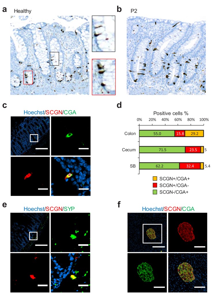
Figure 2—source data 1. Source data for Figure 2D.
DOI: 10.7554/eLife.49910.011

Figure 2.

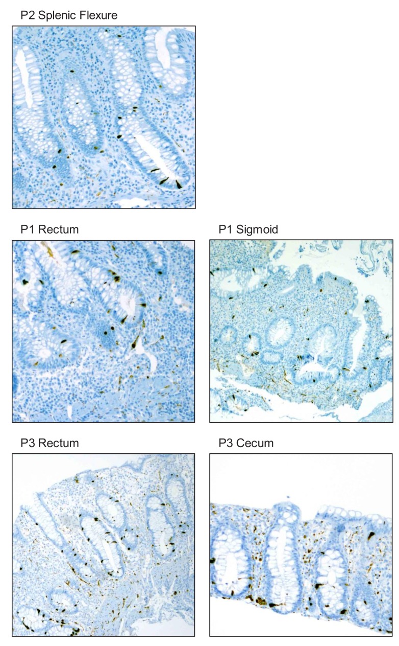
Figure 2—figure supplement 1. Immunohistochemistry (IHC) staining for SCGN in colonic biopsies obtained from probands P1, P2, and P3. 40x magnification.

Figure 2—figure supplement 1.

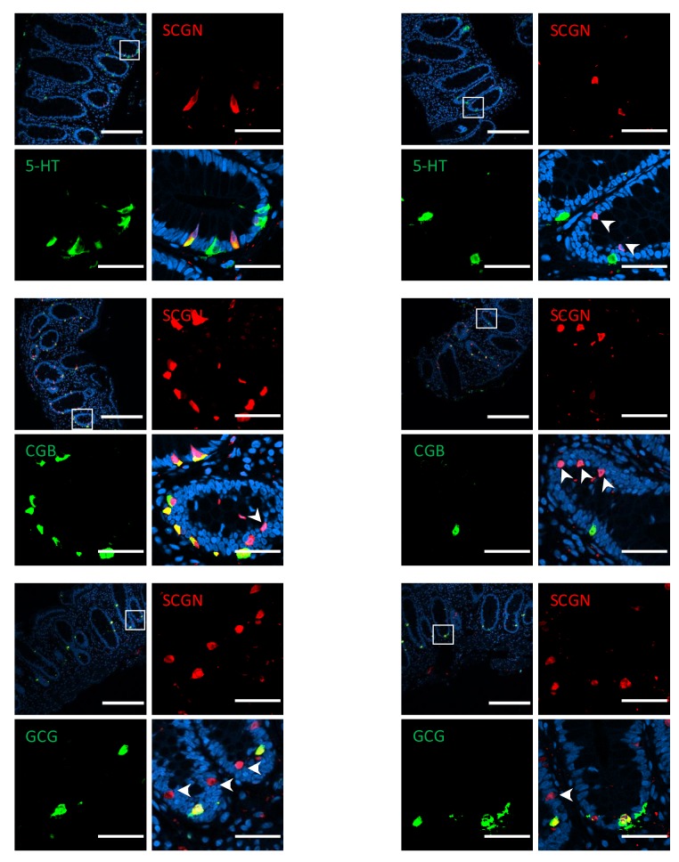
Figure 2—figure supplement 2. Immunofluorescence staining for markers of EEC lineage in healthy human colon (scale bar 200µ - inset 40 µm).

Figure 2—figure supplement 2.

Arrows indicate cells that are exclusively positive for SCGN. 5-HT, 5-hydroxytryptophan (serotonin); CGB, chromogranin B; GCG, glucagon precursor peptide.
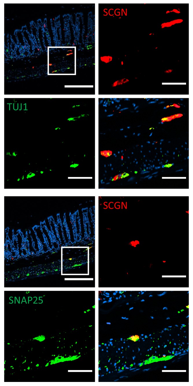
Figure 2—figure supplement 3. Immunofluorescence staining for neuronal markers in murine colon of C57BL/6 mice (scale bar 200 µm - inset 50 µm).

Figure 2—figure supplement 3.

TUJ1, neuron-specific class III β-tubulin; SNAP25, synaptosomal nerve-associated protein 25.