TO THE EDITOR:
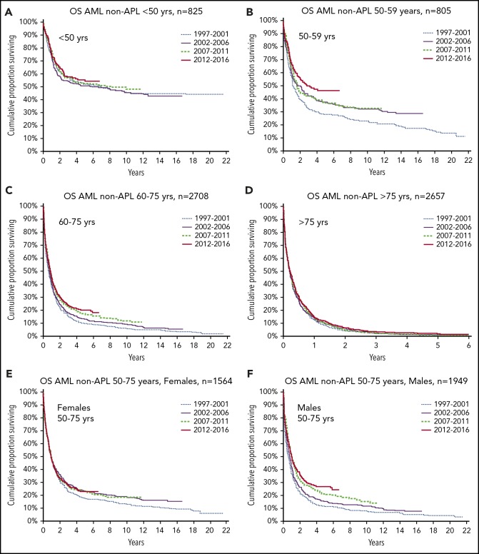
Acute myeloid leukemia (AML) is a devastating disease, globally causing 85 000 deaths per year in 2016, a number predicted to double in 2040, with years of life lost estimated to be 2.6 million.1 We aimed to analyze changes in epidemiology, management, and outcome of AML during 20 years with no new drugs2,3 using the Swedish AML Registry (N = 7708, of whom 6994 had AML [excluding acute promyelocytic leukemia] diagnosed from 1997-2016, with a median follow-up of 8.1 years; approved by ethics review), with a focus on development over time by sex and age. Overall, there was improved survival over time (log-rank P < .00018), but the improvement was restricted to the age group of 50 to 75 years (P < .00001), whereas no improvement was seen in younger (P = .67) or older patients (P = .50; Figure 1). Furthermore, survival strongly improved in middle-age men (P < .00001), but not in women (P = .69). Male AML patients now have equal survival to female patients.
Figure 1.
Overall survival (OS) of AML (non–acute promyelocytic leukemia [APL]) by time period, age at diagnosis, and sex. Age <50 years (n = 825, P = .67) (A); age 50 to 59 years (n = 805, P = .003) (B); age 60 to 75 years (n = 2708, P = .00002) (C); age >75 years (n = 2657, P = .50) (D); women age 50 to 75 years (n = 1564, P = .69) (E); and men age 50 to 75 years (n = 1949, P < .00001) (F). Lines represent the 4 time periods, distinguished by their length of follow-up.
OS was strongly associated with age4 (supplemental Materials, available on the Blood Web site). Analysis of relative survival5 yielded almost identical results (data not shown) because of the poor survival of AML patients. For the entire 20-year period from 1997 to 2016, female and male patients had similar OS (P = .89). Within the age group of 50 //tcgqto 75 years, women had better survival than men (P = .0009). Unexpectedly, we found that OS improved with time only in male patients (male patients, P < .00001; female patients, P = .69; Figure 1). Women age 50 to 75 years had better survival than men during the first half of the study (1997-2001, P = .00004; 2002-2006, P = .0027), whereas there was no survival difference by sex during the latter two time periods (P = .68 and P = .82, respectively). Cox regression analysis showed as expected strong correlation with AML type (de novo vs secondary)6 and Eastern Cooperative Oncology Group (ECOG)/World Health Organization performance status (PS),4 but also with year of diagnosis and sex (Table 1). By multivariable Cox regression using ECOG PS and AML type as covariates, we analyzed hazard ratio by sex, comparing survival in the first period with survival in the following three periods (supplemental Materials), confirming the previous findings.
Table 1.
Cox regression analysis of influence of baseline factors on OS
| n | Univariate | Multivariate | |||||
|---|---|---|---|---|---|---|---|
| Hazard ratio | 95% CI | P | Hazard ratio | 95% CI | P | ||
| Year of diagnosis, per y | 6994 | 0.988 | 0.984-0.993 | <.001 | 0.991 | 0.987-0.996 | <.001 |
| Age, per y | 6993 | 1.052 | 1.050-1.054 | <.001 | 1.045 | 1.042-1.047 | <.001 |
| Sex | |||||||
| Female | 3370 | 1 | 1 | ||||
| Male | 3624 | 1.025 | 0.975-1.079 | .335 | 1.067 | 1.013-1.124 | .015 |
| AML type | |||||||
| De novo | 4833 | 1 | 1 | ||||
| tAML | 685 | 1.564 | 1.438-1.702 | <.001 | 1.535 | 1.409-1.673 | <.001 |
| MDS/AML | 902 | 1.741 | 1.616-1.876 | <.001 | 1.474 | 1.364-1.593 | <.001 |
| MPN/AML | 368 | 2.218 | 1.988-2.474 | <.001 | 1.935 | 1.730-2.165 | <.001 |
| ECOG/WHO PS | |||||||
| 0 | 1270 | 1 | 1 | ||||
| 1 | 2971 | 1.415 | 1.312-1.526 | <.001 | 1.254 | 1-162-1.353 | <.001 |
| 2 | 1238 | 2.405 | 2.204-2.624 | <.001 | 1.826 | 1.670-1.996 | <.001 |
| 3 | 837 | 4.355 | 3.957-4.793 | <.001 | 2.989 | 2.707-3.301 | <.001 |
| 4 | 437 | 6.590 | 5.869-7400 | <.001 | 5.176 | 4.598-5.827 | <.001 |
Multivariate analysis included parameters with a minimum of missing data (ie, those listed, but not cytogenetic data).
CI, confidence interval; MDS, myelodysplastic syndrome; MPN, myeloproliferative neoplasia; tAML, therapy-related AML.
We then characterized the AML disease in patients age 50 to 75 years through the study periods. Survival over time improved more with intermediate- than high-risk cytogenetics7 (n = 1649, P = .0048 and n = 730, P = .099, respectively). Men had improved survival over time in all genetic risk groups (favorable: n = 136, P = .0025; intermediate: n = 900, P = .0003; high risk: n = 434, P = .003), whereas no improvement was seen among women (favorable: n = 138, P = .13; intermediate: n = 748, P = .6; high risk: n = 296, P = .75, respectively). Furthermore, the improvement with time was stronger in de novo than in secondary AML (n = 2291, P < .00001 and n = 1222, P = .015, respectively). Patients with good PS (ECOG 0-2) improved over time (n = 2969, P = .002), in contrast to those with poor PS (ECOG 3-4: n = 544, P = .27).
ECOG PS at diagnosis improved over time up to year 2011 but was thereafter stable. This was most evident among the oldest patients, with no difference by sex in any time period.
More than 80% of patients up to age 70 years were eligible for intensive therapy, with lower proportions in older ages. Overall, 60% of patients received intensive therapy, with a slightly increasing frequency by time in patients up to age 75 years. Complete remission (CR) rates from intensive treatment improved in all age groups over time. Women age 50 to 75 years had higher CR rates than men during the first decade, but there was no difference by sex thereafter. The early death (ED) rate was mainly dependent on PS and age,4 but choice of therapy may also have played a role. Patients not administered intensive therapy had much higher ED rates, irrespective of age.8 From 2008, some patients were selected for hypomethylating agents as primary therapy. Allogeneic stem cell transplantation (alloSCT) was applied with time to a higher proportion of patients of both sexes, especially those age 50 to 69 years.
We here report a broad analysis of diagnostic findings, management, and outcome of almost 7000 AML patients diagnosed during a 20-year period, comprising the overall AML population in Sweden, with a long and complete follow-up. OS improved significantly in the total population; however, this improvement was found to be restricted to men in the age group of 50 to 75 years, who showed a continuous improvement by time, leading to an abrogation of the sex difference9 in survival, which is established in AML10,11 as well as in other cancers.11-13 Thus, the strong survival disadvantage for male vs female patients seen in the first half of the study period changed to equal outcome by sex. The data are strong and demonstrated by different statistical methods, including Cox regression with PS and AML type as covariates, but the causes of change cannot be defined with certainty.
In 2004, we formed a national AML group, providing national guidelines that have been generally implemented, with recurrent evaluations regularly reported to and discussed with all clinicians. Treatment protocols are age dependent, but with no difference according to sex. However, the opportunities for alloSCT have improved through reduced-intensity conditioning and donor registries, with greater donor availability and quality of tissue typing; this has clearly most benefitted patients in the age group of 50 to 75 years, although no sex preference could be identified. Supportive care has improved, with better transfusions and oral antifungals, and the ED and CR rates have improved with time, more for male than for female patients, which might have contributed to the observed survival results.
In addition, changes in general health status might have had an influence. ECOG PS improved during the period, but mainly among the elderly, and there was no sex difference. Globally, women live longer than men, both overall14 and among those with cancer.10-13 The survival gap between the sexes ranges from 6.1 years among countries with a high or middle sociodemographic index to 2.9 years among countries with a low index, with a minor decrease since 1990.14 The sex gap in life expectancy in Sweden was greatest in 1978 and decreased from 5.2 years in 1997 to 3.6 years in 2014.15 This was mainly due to reduced mortality from cardiovascular diseases, with greater improvement in men than in women. There are important differences by sex in smoking habits.16,17 Smoking-related deaths from lung cancer and chronic respiratory diseases and deaths from falls and mental diseases decreased in men but increased in women age >60 years, but most of the increase was in women age >80 years.15
Unfortunately, no survival improvement was seen among older AML patients, despite more use of hypomethylating agents.18 Also, we observed no survival improvement in young patients, in whom toxicity was already managed and alloSCT used whenever possible; new treatment strategies are now needed.
In conclusion, survival improved by time in the age group of 50 to 75 years. Men had an ongoing improvement, but women started from a higher level, and survival had already stabilized in 2002. There were changes by time in PS, ED, CR, and alloSCT rates, although these changes did not fully explain our survival results. In contrast to the general population and other studied cancer populations,12 male and female AML patients now have the same life expectancy. We believe that the health status of the population, including smoking habits, has been of importance in the equalization of survival. Results from the current classic treatment options might have reached a plateau, and we look forward to a new era with molecular diagnostics and new therapies.
Supplementary Material
The online version of this article contains a data supplement.
Acknowledgments
The Swedish AML Registry is supported by the Swedish Association of Local Authorities and Regions and Region Skåne and Regionalt Cancercentrum Syd, and the research was supported by the Swedish Cancer Society.
Authorship
Contribution: G.J. designed study, performed analyses, and wrote manuscript; O.H. performed statistics; and all authors critically reviewed and approved the manuscript.
Conflict-of-interest disclosure: The authors declare no competing financial interests.
A complete list of the members of the Swedish AML Registry appears in the supplemental appendix.
Correspondence: Gunnar Juliusson, Department of Hematology, Skåne University Hospital, S-221 85 Lund, Sweden; e-mail: gunnar.juliusson@med.lu.se.
REFERENCES
- 1.Foreman KJ, Marquez N, Dolgert A, et al. Forecasting life expectancy, years of life lost, and all-cause and cause-specific mortality for 250 causes of death: reference and alternative scenarios for 2016-40 for 195 countries and territories. Lancet. 2018;392(10159):2052-2090. [DOI] [PMC free article] [PubMed] [Google Scholar]
- 2.Yates JW, Wallace HJ Jr., Ellison RR, Holland JF. Cytosine arabinoside (NSC-63878) and daunorubicin (NSC-83142) therapy in nonlymphocytic leukemia. Cancer Chemother Rep. 1973;57(4):485-488. [PubMed] [Google Scholar]
- 3.Wei AH, Tiong IS. Midostaurin, enasidenib, CPX-351, gemtuzumab ozogamicin, and venetoclax bring new hope to AML. Blood. 2017;130(23):2469-2474. [DOI] [PubMed] [Google Scholar]
- 4.Juliusson G, Antunovic P, Derolf A, et al. Age and acute myeloid leukemia: real world data on decision to treat and outcomes from the Swedish Acute Leukemia Registry. Blood. 2009;113(18):4179-4187. [DOI] [PubMed] [Google Scholar]
- 5.Bower H, Andersson TML, Björkholm M, Dickman PW, Lambert PC, Derolf Å. Continued improvement in survival of acute myeloid leukemia patients: an application of the loss in expectation of life. Blood Cancer J. 2016;6(2):e390. [DOI] [PMC free article] [PubMed] [Google Scholar]
- 6.Hulegårdh E, Nilsson C, Lazarevic V, et al. Characterization and prognostic features of secondary acute myeloid leukemia in a population-based setting: a report from the Swedish Acute Leukemia Registry. Am J Hematol. 2015;90(3):208-214. [DOI] [PubMed] [Google Scholar]
- 7.Döhner H, Estey E, Grimwade D, et al. Diagnosis and management of AML in adults: 2017 ELN recommendations from an international expert panel. Blood. 2017;129(4):424-447. [DOI] [PMC free article] [PubMed] [Google Scholar]
- 8.Juliusson G, Lazarevic V, Hörstedt AS, Hagberg O, Höglund M; Swedish Acute Leukemia Registry Group . Acute myeloid leukemia in the real world: why population-based registries are needed. Blood. 2012;119(17):3890-3899. [DOI] [PMC free article] [PubMed] [Google Scholar]
- 9.Özdemir BC, Csajka C, Dotto GP, Wagner AD. Sex differences in efficacy and toxicity of systemic treatments: an undervalued issue in the era of precision oncology. J Clin Oncol. 2018;36(26):2680-2683. [DOI] [PubMed] [Google Scholar]
- 10.Hossain MJ, Xie L. Sex disparity in childhood and young adult acute myeloid leukemia (AML) survival: Evidence from US population data. Cancer Epidemiol. 2015;39(6):892-900. [DOI] [PMC free article] [PubMed] [Google Scholar]
- 11.Mohammadi M, Cao Y, Glimelius I, Bottai M, Eloranta S, Smedby KE. The impact of comorbid disease history on all-cause and cancer-specific mortality in myeloid leukemia and myeloma - a Swedish population-based study. BMC Cancer. 2015;15:850. [DOI] [PMC free article] [PubMed] [Google Scholar]
- 12.Radkiewicz C, Johansson ALV, Dickman PW, Lambe M, Edgren G. Sex differences in cancer risk and survival: a Swedish cohort study. Eur J Cancer. 2017;84:130-140. [DOI] [PubMed] [Google Scholar]
- 13.Socialstyrelsen. Cancer mortality in Sweden. http://www.socialstyrelsen.se/publikationer2018/2018-12-51. Accessed March 2019.
- 14.GBD 2017 Mortality Collaborators Global, regional, and national age-sex-specific mortality and life expectancy, 1950-2017: a systematic analysis for the Global Burden of Disease Study 2017. Lancet. 2018;392(10159):1684-1735. [DOI] [PMC free article] [PubMed] [Google Scholar]
- 15.Sundberg L, Agahi N, Fritzell J, Fors S. Why is the gender gap in life expectancy decreasing? The impact of age- and cause-specific mortality in Sweden 1997-2014. Int J Public Health. 2018;63(6):673-681. [DOI] [PMC free article] [PubMed] [Google Scholar]
- 16.GBD 2017 Risk Factor Collaborators Global, regional, and national comparative risk assessmenmt of 84 behavioural, environmental and occupational, and metabolic risks or clusters of risks for 195 countries and territories, 1990-2017: a systematical analysis for the Global Burden of Disease Study 2017 [published corrections appear in Lancet. 2019;393(10167):132 and Lancet. 2019;393(10190):e44]. Lancet. 2018;392(10159):1923-1994. [DOI] [PMC free article] [PubMed] [Google Scholar]
- 17.World Health Organization WHO global report on trends in prevalence of tobacco smoking 2015. http://www.who.int/tobacco/publications/surveillance/reportontrendstobaccosmoking/en/. Accessed March 2019.
Associated Data
This section collects any data citations, data availability statements, or supplementary materials included in this article.


