

Abstract
A 22-year-old male patient presented with right infra-auricular sinus since birth with preauricular swelling and discharge on and off since age of 3 years. He had no history of previous surgery. Patient noted frequent swelling and foul smelling discharge inferior to right tragus through a small puncta every 3–6 monthly, resulting in abscess formation and scaring around 1–1.5 cms below and anterior to puncta site. The infra tragal sinus tract and the cyst were completely removed surgically by infra-auricular approach. The histopathology report was suggestive of an infra-auricular preauricular sinus. No recurrence was noted on follow up to 2 years. We report this case for its rarity since less than 5 cases have been reported in literature.
Keywords: Preauricular sinus, Excision, Abscess
Introduction
Preauricular sinuses were first described by Heusinger [1]. Preauricular sinuses are congenital malformations that result from defective or incomplete fusion of the six auricular hillocks [2]. One-third of the patients remain asymptomatic and require no treatment [2]. However, once a preauricular sinus becomes infected, its excision is mandatory.
We present the case of one patient that had recurrent infrauricular sinus and abscesses for which he was managed medically on outpatient basis. Clinical examination revealed an infra-auricular pit. Surgical excision of the entire tract had resolved the problem and the patient has been asymptomatic ever since during our 2 years follow-up.
Case Report
22-year-old patient presented to our clinic with complaints of recurrent swelling and discharge, incrustation from the right ear located inferior to the tragus with an abscess located inferior to right tragus and lobule. These symptoms started since childhood, was treated on outpatient basis with oral antibiotics. There was no history of any ear pain or hearing loss. Patient gives history of frequent swelling and foul smelling discharge inferior to right tragus through a small opening site (puncta) every 3–6 monthly, resulting in abscess formation and scaring around 1–1.5 cms below and anterior to puncta site. On examination a soft, painless swelling with a sinus opening was located inferior to the right tragus, 1–2 cm below (Fig. 1). He had no other congenital ear anomaly apart from this one.
Fig. 1.

Soft, painless swelling with a sinus opening located inferior to the right tragus, 1–2 cm below and puncta 3 mm below tragus
The right sided facial nerve status was normal, oral cavity examination was normal, no signs of cervical lymphadenopathy. No additional pathologies were observed in opposite side ear. Nose, throat and systemic examinations was normal. The routine blood investigation and relevant radiological investigations done were also normal.
The patient underwent surgery under local anesthesia. Based on the diagnosis of preauricular sinus, an 23G needle, was inserted as a probe inside the sinus tract and fixed at the sinus entrance.
An elliptical incision was performed around the sinus site for the dissection of the tract. Infra-auricular approach was preferred for a wide exposure, further the elliptical incision was extended inferiorly to the tragus (2–4 cm below) and the resection was performed up to the right parotid area. The entire tract was followed until its complete removal along with puncta and inferior part of tragal cartilage to which puncta attached (Fig. 2).
Fig. 2.
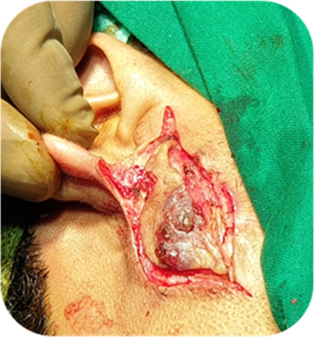
Infra-auricular approach, elliptical incision given around the sinus site
The infra tragal sinus tract and the cyst were totally removed (Fig. 3). The operated site wound was closed in layers after adequately securing hemostasis, no surgical drain was kept.
Fig. 3.
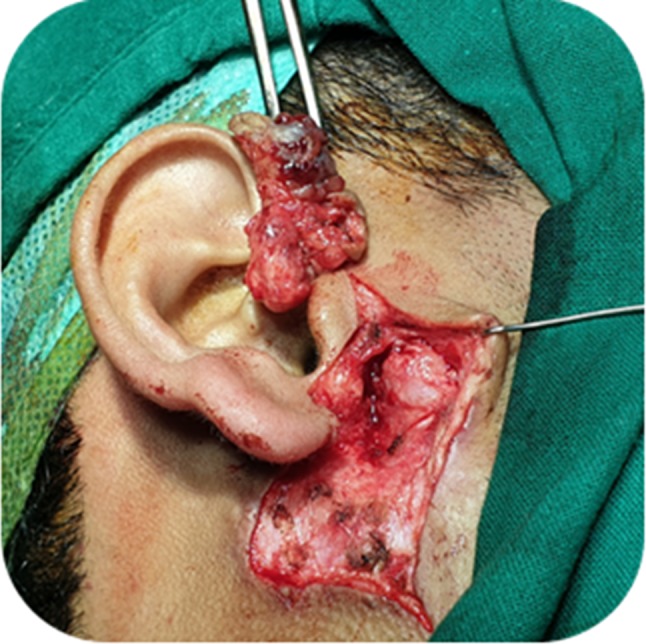
Dissection of infra tragal sinus
The patient was given oral prophylactic antibiotics and analgesics after the surgery. The patient was discharged the next day, postoperative status was uneventful. The histopathology report (Fig. 4) was suggestive of an infra tragal preauricular sinus. No recurrence noted on follow up to 2 year.
Fig. 4.

Histopathology: ×10 and ×40 haematoxylin and eosin stain of right infra-tragal sinus
Discussion
The external ear develops from six mesenchymal proliferations (hillocks): three from the caudal edge of the 1st branchial arch and other three from the cephalic edge of the 2nd branchial arch. The tragus and the anterior crus of the helix margin develop from the 1st arch, and the rest of the auricle is formed from the 2nd arch [3]. The three predominant theories for the development of preauricular sinuses are:
Possible incomplete fusion of the 1st arch hillocks;
Isolation of ectodermal folds during auricle formation;
Defective closure of the most dorsal part of the first branchial cleft [4].
Preauricular sinuses and anomalies of the first branchial cleft can have similar presentations. Preauricular sinuses usually present with an opening anterior to the ascending limb of the helix. Occasionally, they occur along the lateral or posterior surface of the helicine crus and the superior posterior margin of the helix, the tragus, or the lobule [5].
In the current case, an infra-auricular sinus was present, an elliptical incision is made in the skin around the sinus pit and the sinus is opened. The sinus is viewed and followed from both outside and inside. Each subsequent branching tract is opened and followed until every dead end is identified and excised.
Conclusion
We report this case for its rarity, since less than 5 cases have been reported in literature till date.
Author’s Contribution
AN: performed surgery, prepared the manuscript and patient preoperative and postoperative management and follow up. DKR: preparation of the manuscript, literature review and review of manuscript.
Compliance with Ethical Standards
Conflict of interest
The authors declare that they have no conflict of interests.
Ethical Approval
All procedures performed in studies involving human participants were in accordance with the ethical standards of the institutional and/or national research committee and with the 1964 Helsinki declaration and its later amendments or comparable ethical standards.
Informed Consent
Informed consent was obtained from all individual participants included in the study.
Footnotes
Publisher's Note
Springer Nature remains neutral with regard to jurisdictional claims in published maps and institutional affiliations.
Contributor Information
Ahilasamy Nagalingeswaran, Email: nahilasamy@yahoo.com.
Rajendran Dinesh Kumar, Email: dinuraj1186@gmail.com.
References
- 1.Heusinger HK. Hals–Kiemen Finstein von Noch Nicht Beobacheter Form. Virchows Arch. 1864;29(3–4):358–380. doi: 10.1007/BF01937182. [DOI] [Google Scholar]
- 2.Martin-Granizo R, Pérez-Herrero MC, Sánchez-Cuéllar A. Methylene blue staining and probing for fistula resection: application in a case of bilateral congenital preauricular fistulas. Int J Oral Maxillofac Surg. 2002;31(4):439–441. doi: 10.1054/ijom.2001.0062. [DOI] [PubMed] [Google Scholar]
- 3.Choi SJ, et al. The variant type of preauricular sinus: postauricular sinus. Laryngoscope. 2007;117(10):1798–1802. doi: 10.1097/MLG.0b013e3180caa1ca. [DOI] [PubMed] [Google Scholar]
- 4.Emery PJ, Salama NY. Congenital preauricular sinus. A study of 31 cases seen over a 10 year period. Int J Pediatr Otorhinolaryngol. 1981;3(3):205–212. doi: 10.1016/0165-5876(81)90004-5. [DOI] [PubMed] [Google Scholar]
- 5.Yeo SW, et al. The preauricular sinus: factors contributing to recurrence after surgery. Am J Otolaryngol Head Neck Med Surg. 2006;27(6):396–400. doi: 10.1016/j.amjoto.2006.03.008. [DOI] [PubMed] [Google Scholar]