Significance
Six decades after DNA structure was first revealed, fundamental questions remain open. How is the entwined embrace of double-stranded nucleic acids formed or disrupted? How does the energetics underlying this process influence nucleic-acid processing machineries? By combining simulations and experiments, our work addresses these questions and reveals that asymmetric base-pair dynamics drives the stepwise separation of nucleic acid duplexes, predicts the unwinding efficiency of helicases, and intimately relates the intrinsic dynamics of base pairs to the enzymatic mechanism evolved for their opening. Taken together, our data suggest a layer of regulation of the genetic material encoded in the “unwindability” of the double helix.
Keywords: double helix, nucleic acids, simulations, experiments, unwindability
Abstract
The opening of a Watson–Crick double helix is required for crucial cellular processes, including replication, repair, and transcription. It has long been assumed that RNA or DNA base pairs are broken by the concerted symmetric movement of complementary nucleobases. By analyzing thousands of base-pair opening and closing events from molecular simulations, here, we uncover a systematic stepwise process driven by the asymmetric flipping-out probability of paired nucleobases. We demonstrate experimentally that such asymmetry strongly biases the unwinding efficiency of DNA helicases toward substrates that bear highly dynamic nucleobases, such as pyrimidines, on the displaced strand. Duplex substrates with identical thermodynamic stability are thus shown to be more easily unwound from one side than the other, in a quantifiable and predictable manner. Our results indicate a possible layer of gene regulation coded in the direction-dependent unwindability of the double helix.
Since its discovery, the structure of the DNA double helix (1, 2), with its striking implications for DNA replication and DNA recombination (3), has provided a means to probe and understand the molecular biology of “genetic material” (4). Soon after the Watson–Crick (W-C) model was proposed, it was recognized that DNA strand separation was critical to DNA function (5), thus motivating the quest for conditions that would disrupt the W-C hydrogen bonds so as to separate the 2 strands of the DNA double helix (3). However, the way the elementary steps of these conformational transitions affect nucleic-acid processing machineries is still not fully understood.
The properties of DNA and RNA double helices have shaped the structure and mechanisms of proteins endowed with the ability of opening base pairs to perform chemical modification (e.g., methyltransferases) or to allow DNA and RNA remodeling (e.g., helicases). Chemical modifications are usually achieved by the specific flipping out, also named extrusion, of the target nucleobase (6, 7). These types of base-flipping processes have been the focus of intense computational work (8, 9), and NMR studies have further shown the sequence dependence of the flipping rate of internal base pairs in relevant biological contexts (10, 11). More generally, local thermal fluctuations of base pairs (12–15) can transiently separate double-stranded (ds) nucleic acids, and this phenomenon affects the binding, assembly, and translocation of gene-expression machines (16–18). In this respect, A·T- or A·U-rich segments show increased breathing (decreased stability) than G·C-rich segments (14, 15) and are, for instance, more readily unwound by helicases—ATP-fueled motor proteins capable of separating the duplex (16) with a sequence-dependent stepping velocity (19) and processivity (20).
Complementary to base extrusion, the iteration of base-pair opening events at the junction between single-stranded (ss) and ds nucleic acids, also referred to as unzipping, complies with a different set of steric and torsional constraints and approximates the biological process of helix opening by helicases, as suggested by X-ray crystallography (16, 21) and single-molecule experiments (19, 22, 23). The separation of ds nucleic acids is assumed to follow the classical zipper model (24), where base-pair opening occurs as a concerted process with an equivalence (or symmetry) between 2 complementary nucleobases. This assumption has been largely based on the difficulty to observe and characterize the “invisible” intermediate states—for instance, those ss/ds junctions with only 1 of the 2 nucleobases flipped out (25–27)—which are too few and whose duration is too short for experimental determination. In this context, computer simulations could bring about a major productivity leap, providing thermodynamic and kinetic information on transition intermediates that might escape spectroscopic detection. Indeed, the large amount of molecular dynamics (MD) work on the melting of base pairs in various conditions (13, 26–37) suggests that base-pair opening is an asynchronous process, with one base unstacking significantly before the other. However, systematic studies on how the nucleic acid sequence can affect base-pair opening intermediates during the unzipping of ss/ds junctions are lacking. Furthermore, according to the classical zipper model, a G·C repeat, for example, 5′-Gn-3′/3′-Cn-5′, will be equally unzipped by a helicase regardless of whether the helicase tracks on the 5′-Gn-3′ or 5′-Cn-3′ strand. This is a paradigm that has long influenced the understanding of the nucleic-acid processing apparatus. However, the evidence that base-pair opening can be asynchronous and that the intrinsic dynamics of nucleic acids, even at the single base-pair level, can modulate the function of the genetic material (17–19, 38, 39) posits the challenge of questioning how the mechanism of duplex separation, resolved with atomistic spatiotemporal resolution, can impact the function of nucleic-acid processing machineries.
Here, by combining simulations and experiments, we study how RNA and DNA duplexes unwind and how the energetics underlying the elementary step of base-pair opening influences the function of helicases. First, we present a high-throughput structure-based MD approach to provide a comprehensive view of the unwinding mechanism of double helices. By analyzing thousands of base-pair opening and closing events, we uncover a systematic asymmetry in the dynamics of a W-C base pair that makes 1 of the 2 nucleobases more likely to flip out. Such asymmetric dynamics is encoded in the sequence and affects the intermediate states populated by dsRNA and dsDNA during opening. As a result, base unpairing is, systematically, a stepwise asymmetric process with 1 of the 2 nucleobases being the weak point from which the base pair breaks. Based on this observation, we then explore the impact of asymmetric nucleobase dynamics in the context of helicase unwinding, whereby we predict enhanced unwinding efficiency for pyrimidine-rich substrates on the displaced strand. Finally, using biochemical and fluorescence-based assays, we test our hypothesis by measuring the unwinding of various designed substrates by helicase enzymes. The experiments confirm the predictions and corroborate a model that intimately relates the asymmetric dynamics of base pairs to the unwinding mechanism of helicases. Our results demonstrate that one duplex portion can be more easily unwound from one side than from the other. Taken together, our data suggest a layer of gene regulation encoded in the direction-dependent “unwindability” of the double helix.
Results
Extensive Simulations Show Different Pathways of Base-Pair Opening in dsDNA and dsRNA.
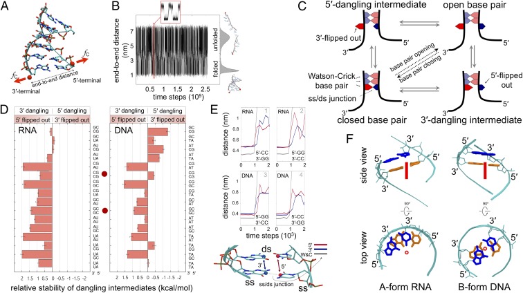
By simulating ds separation and annealing in short duplexes, we monitored thousands of base-pair opening and closing events (Fig. 1 A and B) and systematically analyzed the nucleobase dynamics of the 16 nearest-neighbor base-pair combinations (Methods and SI Appendix). We observed 2 elementary steps during the opening of each base pair (Fig. 1C); hereafter we omit to mention the reverse base-pair closing events, which are observed with statistically identical probabilities in our equilibrium simulation. These steps are: 1) the unpairing and unstacking (also referred to as “flip out”) of a nucleobase, either at the 5′ or 3′ terminus of a ss/ds junction, resulting in a dangling-base intermediate; and 2) the unstacking of the opposite nucleobase, completing the base-pair opening event. We quantified the preference of each nearest-neighbor base-pair combination to follow the pathway passing through either a 3′- or 5′-dangling intermediate (Fig. 1D). In other words, we quantified the 5′ or 3′ flip-out events that lead to the opening of a W-C base pair at ss/ds junctions. Two different scenarios emerged when comparing dsRNA to dsDNA (Fig. 1 D and E).
Fig. 1.
Modeling the formation and rupture of double helices by mimicking constant-force optical-tweezer experiments (59). (A) Ribbon molecular graphics representation of the ds nucleic acid model and schematic of the reaction coordinate. Red spheres show the 3′ and 5′ hydroxyl groups of the terminal base pair, defining the end-to-end distance, where the external constant force, fC, was applied (red arrows; Methods). (B) Time series of the hopping between folded and unfolded states, zoomed in the red inset. Histogram of the end-to-end distance is on the right, together with sample conformations. (C) Schematic of the stepwise mechanism of base-pair opening/closing. The bases in the ss portion are not shown for sake of clarity. (D) Free-energy difference between the unbiased population of 5′- and 3′-dangling intermediates (SI Appendix, Methods) for the base pair at the bottom of each nearest-neighbor combination shown on the vertical axis. Positive values correspond to higher population of 3′-dangling intermediates. Red dots highlight the base-pair combinations explicitly discussed in the main text. Bars indicate SE from bootstrapping (60). (E) Time evolution of the base-pair opening process at a ss/ds junction in RNA and DNA. The distances used to detect W-C pairing (gray), 5′-stacking (red), and 3′-stacking (blue) of the closing base pair are shown. Data are averaged over windows of 300 time steps. (F) Side and top views of adjacent W-C base pairs in RNA and DNA duplexes. The major axis of the helix is shown as a red circle and a red line in the top and side view, respectively. Sugar-phosphate backbone is in cyan sticks and ribbons. Adjacent base pairs are colored in blue and orange to highlight overlap extension.
In RNA duplexes, the nucleobase at the 5′ terminus of a ss/ds junction consistently showed higher propensity to flip out when compared to the complementary W-C base at the 3′ terminus (Fig. 1 D, Left). That is, the opening of a RNA base pair occurs with higher probability through a 3′-dangling intermediate rather than a 5′-dangling intermediate, thus generating a conserved asymmetry (or directionality) in the opening mechanism. In sharp contrast, in DNA duplexes, both 5′ and 3′ flipped-out intermediates were significantly populated at ss/ds junctions, and their relative population was exclusively modulated by the sequence (Fig. 1 D, Right). For example, the energetics of unwinding favors the stepwise opening of the ss/ds junction 5′-NNCCNN-3′/5′-NNGGNN-3′ through the 3′-dangling intermediate 5′-NNCCNN-3′/5′-NNGGNN-3′ (superscript denotes flipped-out bases) both in RNA and DNA. In the time series plots (Fig. 1E), this preference is manifested by the early increment of the stacking distance at the 5′ terminus (red line in panels 1 and 4 of Fig. 1E). If the 2 strands are swapped 5′-NNGGNN-3′/5′-NNCCNN-3′, the preference changes to the 5′-dangling intermediate in DNA 5′-NNGGNN-3′/5′-NNCCNN-3′, whereas it remains unchanged in RNA (panels 2 and 3 in Fig. 1E).
The geometric features of A-type and B-type helices provide a structural interpretation of our results (Fig. 1F). In B-DNA, both the 5′ and 3′ ends of a ss/ds junction are equally buried in the helix, and the displacement probability depends only on the sequence. Vice versa, in A-RNA, the bases at the 5′ end of a ss/ds junction are less buried in the neighboring environment, thus facilitating displacement events that lead to 5′ flipped-out intermediates.
The overall dynamics of base-pair opening described here is similar to that observed in accurate atomistic MD simulations (13, 26–37). However, water and ion effects as well as noncanonical structures (13, 30, 37) acting as kinetic traps are by construction omitted from our structure-based model. Nevertheless, these results are consistent with the population of stacked conformers observed in ultrafast spectroscopy experiments on short duplexes (40) and with the analysis of structural databases (25) and can be related to the stabilization provided by dangling ends, although the latter comparison should be interpreted with caution (SI Appendix, Discussion) (26, 40, 41). Even though the asymmetric intermediate species involved in the opening of a base pair can be difficult to measure experimentally (42), we hypothesized that these intermediates could impact nucleic-acid processing machines. In the next sections, we thus explore the possible consequences of asymmetric base-pair opening on the function of helicases—which, in turn, are a proxy of base-pair dynamics (17–19).
Relating the Asymmetric Dynamics of Base Pairs to the Unwinding Efficiency of Helicases: The Unwindability Index.
To examine whether the observed asymmetry influences the function of helicases, we started by analyzing base-pair dynamics in the context of superfamily (SF) 1 and 2 helicases (Fig. 2) (43), monomeric or dimeric nontoroidal enzymes comprising the minimal building-block domains necessary for helicase activity. From a general standpoint, SF1 and SF2 helicases unwind the duplexes by first loading onto an overhanging terminal region and then translocating toward the duplex along this loading strand, thereby peeling off the complementary bases. In particular, the migration of the tracking strand through a specific protein tunnel facilitates, actively or passively, the displacement of the complementary strand by steric exclusion (16, 43).
Fig. 2.
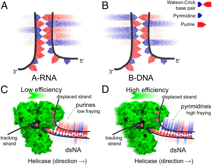
Pictorial representation of nucleic acids unwinding catalyzed by SF1 and SF2 helicases. The flip-out probability of nucleobases at ss/ds junctions is depicted with fading arrows. High color intensity corresponds to a high flip-out probability. (A) In A-RNA, the flip out of bases at the 5′-end is consistently favored over the flip out of the complementary base at the 3′-end. Vice versa, in B-DNA (B), the direction of base-pair opening depends on the sequence only. We postulate that helicase unwinding efficiency is low when purines are on the displaced strand (C) and high when pyrimidines are on the displaced strand (D). Helicase structure (NS3; Protein Data Bank ID code 3O8R) (61) rendered with Visual Molecular Dynamics (62).
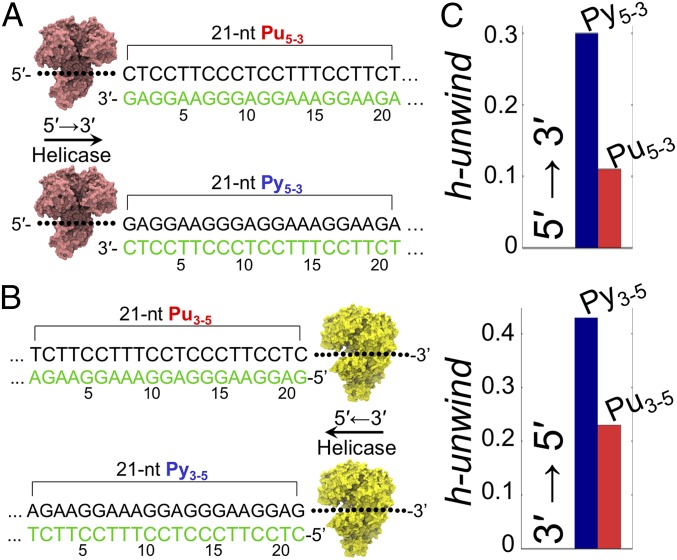
A key feature of the helicase unwinding model that we propose below is that the population of stacked nucleobases at the displaced strand represents, by direct or indirect interaction with the helicase, a bottleneck for the unwinding activity. Note that the MD simulations presented herein show that purines have lower propensity to flip out than pyrimidines (see, for example, the CC/GG and GG/CC combinations highlighted by a red dot in Fig. 1D). In the helicase context, we surmise that the lower flipping propensity of purines at the displaced terminus may result in lower unwinding efficiency. That is, the action of helicases would be impeded by the purines at the displaced strand. In this scenario, duplex opening would thus show a low and high efficiency when purines and pyrimidine are displaced, respectively (Fig. 2 C and D). To provide a quantitative prediction of unwinding efficiencies, here we heuristically introduce a “helix unwindability” index (“h-unwind”) that allows direct comparison of simulations and experiments (SI Appendix, Methods). H-unwind is inversely related to the population of stacked nucleobases at the displaced strand—thus, the higher the h-unwind value, the higher the helicase unwinding efficiency. Two sets of DNA constructs were then designed, and the corresponding h-unwind was computed (Fig. 3). The constructs contained a 21-nucleotide (nt) duplex region with the displaced strand consisting entirely of purines (Pu5-3 for 5′ → 3′ processing helicases and Pu3-5 for 3′ → 5′ helicases) or pyrimidines (Py5-3 for 5′ → 3′ helicases and Py3-5 for reverse polarity; Fig. 3 A and B). Although the 2 sets of DNA had the same duplex portion (with swapped strands), the h-unwind allows to differentiate them and predicts Py5-3, Py3-5 to be more efficiently unwound than Pu5-3, Pu3-5 by a detectable amount (Fig. 3C). We next moved from the in silico predictions to in vitro assays.
Fig. 3.
The h-unwind predicts a direction-dependent unwinding efficiency of helicases. (A) DNA duplexes Pu5-3 and Py5-3 have a 21-nt sequence in the displaced strand (in green) that is homopurine and homopyrimidine, respectively. (B) Pu3-5 and Py3-5 have a homopurine and homopyrimidine sequence in the displaced strand (in green), respectively. (C) Duplexes with homopyrimidines in the displaced strand (Py5-3 and Py3-5) have higher h-unwind values and are thus predicted to be more efficiently unwound than the homopurine analogs (Pu5-3 and Pu5-3).
Pyrimidines on the Displaced Strand of DNA Duplexes Facilitate Helicase Unwinding.
The 2 sets of DNA constructs (Py5-3, Pu5-3 and Py3-5, Pu3-5, described in the previous section) were combined with a 21-nt ss overhang and were labeled with indocarbocyanine (Cy3) fluorophore and the 4-([4-(dimethylamino)phenyl]azo)benzoic acid succinimidyl ester (Dab) quencher (44) at duplex termini (Fig. 4 and SI Appendix, Table S1 for complete sequences). Before measuring the efficiency of helicase enzymes to unwind such constructs, we carried out melting experiments (SI Appendix, Fig. S2) to confirm that both the fluorescence labeling and Pu/Py strand swapping had no impact on duplex thermodynamic stability—a fundamental feature for the aims of the helicase unwinding assays described below.
Fig. 4.
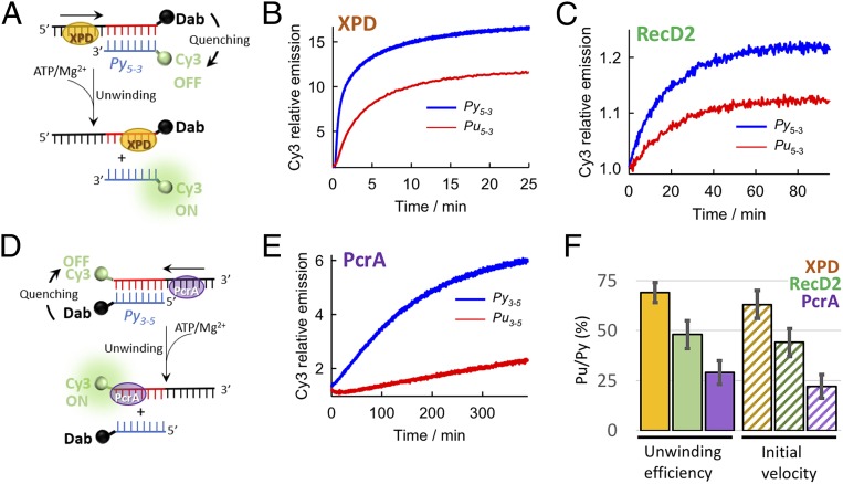
Proof of concept experiments show that duplexes with homopyrimidines on the displaced strand (Py5-3, Py3-5) are more easily unwound by helicases than the homopurine homologs (Pu5-3, Pu3-5). Schematic of the fluorescence quenching assay used to investigate duplex unwinding by helicases with 5′ → 3′ (A) or 3′ → 5′ (D) directionality. ATP addition initiates unwinding and results in strand separation with recovery of Cy3 emission, which is otherwise quenched by Dab in the duplex. Normalized variation in fluorescence intensity of Cy3 as a function of time for Py5-3-XPD (blue) and Pu5-3-XPD (red) (B), Py5-3-RecD2 (blue) and Pu5-3-RecD2 (red) (C), and Py3-5-PcrA (blue) and Pu3-5-PcrA (red) (E). (F) Percentage of unwinding amplitude and initial velocity (pattern) observed for homopurine sequences in the displaced strand relative to homopyrimidine sequences for the 3 helicases investigated.
We then assessed the unwinding activity of 3 DNA helicases, XPD, RecD2, and PcrA (Fig. 4). XPD and RecD2 helicases are members of SF2 and SF1, respectively, and both translocate along DNA with a 5′ → 3′ polarity (43). To probe the general validity of our asymmetric model, we also analyzed the unwinding activity of SF1 helicase PcrA, which moves along DNA in a 3′ → 5′ direction (45).
The XPD helicase unwinds DNA during nucleotide excision repair (46). The unwinding time courses showed a progressive increase in Cy3 emission, indicative of separation of annealed strands (Fig. 4A), reaching a plateau in a similar timescale (∼25 min) for both substrates (Fig. 4B). However, the relative increase in Cy3 emission, reflecting the amount of ss product formed, was significantly lower for Pu5-3, which reached only 64% of the signal observed for Py5-3 (Fig. 4F). The relative difference in unwinding efficiency between Pu5-3 and Py5-3 was further confirmed by native polyacrylamide gel electrophoresis (PAGE) using DNA constructs carrying only the Cy3 fluorophore (SI Appendix, Fig. S3). Comparable differences in relative unwinding efficiencies were also obtained when using a 10-fold molar excess of a 21-nt unstructured and noncomplementary trap sequence that sequesters excess helicase protein in solution (SI Appendix, Fig. S4 A and B). Similar unwinding efficiencies were observed in the presence of either a 12-nt (SI Appendix, Fig. S4C) or a 21-nt (SI Appendix, Fig. S4D) complementary to the Cy3-labeled displaced strand that prevents duplex reannealing. Oligonucleotide trapping sequences are listed in SI Appendix, Table S4.
We then used the SF1 helicase RecD2 to unwind Pu5-3 and Py5-3. RecD2 is a homolog of the RecD subunit of the bacterial RecBCD enzyme involved in dsDNA repair (47). Here, the unwinding activity led to an increase in Cy3 emission until reaching a plateau at ∼60 min (Fig. 4C). The fluorescence intensity profiles of Pu5-3 unwound by RecD2 exhibited only a 48% recovery of the Cy3 signal level detected for Py5-3 (Fig. 4F). Native PAGE using only Cy3-labeled constructs confirmed a 2-fold lower unwinding efficiency for Pu5-3 compared to Py5-3. (SI Appendix, Fig. S5). Thus, RecD2 unwinds DNA duplexes containing homopyrimidine sequences (Py5-3) in the displaced strand more efficiently than homopurine sequences (Pu5-3), and with a slightly more pronounced bias toward homopyrimidine sequences in the displaced strand than XPD (Fig. 4F).
Next, we tested the unwinding efficiency of the Bacillus stearothermophilus PcrA helicase, which shows 3′ → 5′ directionality (Fig. 4D) (45). Unwinding of Py3-5 resulted in an ∼6-fold increase in Cy3 emission. In contrast, Pu3-5 displayed only a 2-fold increase at identical unwinding conditions (Fig. 4E and SI Appendix, Fig. S7). From these values, the unwinding of Pu3-5 was estimated to be only 29% of that of Py3-5, and this was also confirmed by native PAGE (SI Appendix, Fig. S6). In vitro unwinding by PcrA under multiple turnover conditions is a slow process, and a timescale of ∼300 min at room temperature is comparable to that previously reported using a similar 10-fold excess of PcrA over substrate (48).
For the 3 enzymes, a calculation of the relative initial unwinding velocities for the 2 types of substrates revealed a similar trend to that observed for the unwinding efficiencies (Fig. 4F). Furthermore, XPD unwinding in the presence of 3 different trapping strands confirmed that DNA substrates carrying a homopurine sequence in the displaced strand are unwound with initial velocities that are between 60 to 40% lower than for homopyrimidine sequences (SI Appendix, Fig. S4A and Table S4). Finally, binding experiments also ruled out the possibility that the observed bias is induced by a differential affinity of the helicases for ss homopyrimidine or homopurine tracking strands (SI Appendix, Table S5).
The above observations are in agreement with the results reported by Taylor et al. (49) for the processive NPH-II RNA helicase. According to those results, a 3-fold increment of unwinding amplitude is observed when homopyrimidines are located on the 5′ displaced strand. Importantly, sequences with identical thermodynamic stability also show this behavior (49). Whereas Taylor et al.’s results could be regarded as an unusual set of observations, our analysis and experiments suggest that the molecular events underlying such a purine/pyrimidine bias can be straightforwardly related to the intrinsic dynamics of the duplex and are thus universal.
Taken together, the experimental characterization of the 3 model helicases confirms h-unwind predictions and demonstrates that the constructs with the homopyrimidine sequence at the displaced strand, namely Py5-3, Py3-5, were always more efficiently unwound, regardless the helicase type and directionality.
Biological Implications
It is known that the sequence-dependent structure (50, 51) and stability (52) of DNA may contribute to modulate the activity of nucleic-acid processing machines (16–18, 39). However, asynchronous base-pair opening had never been tested in the context of helicase unwinding, and systematic studies were lacking on how the nucleic acid sequence could affect the base-pair intermediates during unzipping of ss/ds junctions. Our results linking asymmetric base-pair dynamics to unwinding efficiency support a mechanism where helicases are sensitive to the sequence composition in a way related not only to the stability of the base pair but also to the stability of the asymmetric intermediates involved during opening—thus further expanding the structural and functional complexity of the double helix. Specifically, helicases can more easily unwind substrates with pyrimidine-rich displaced strands, whether RNA (49) or DNA. Such purine/pyrimidine discrimination directly suggests that the sequence of nucleic acid duplexes contributes to regulating, as well as targeting, helicase activity in an orientation-dependent manner. That is, helicases can more easily unwind duplex portions if they proceed from one side than the other—hence bringing about an unanticipated level of gene regulation. For instance, the direction-dependent sequence bias may influence the efficiency with which sense and antisense strands of a genomic region are processed. Quantifying and knowing the most efficient unwinding direction of a specific duplex region should influence our thinking about how a specific helicase works and how it is used within the cell (SI Appendix, Discussion).
From an evolutionary perspective, the intrinsic dynamics of nucleic acids may represent a biasing factor on the evolution of helicase mechanisms (53, 54). For RNA, the relative stability of dangling intermediates shown in Fig. 1D conveys that the opening of base pairs in A-RNA naturally follows a specific directionality, namely passing through 5′ flipped-out intermediates. Therefore, one may argue that helicases evolved to unwind RNA would prevalently show one specific unwinding directionality. On the other hand, the opening of base pairs in B-DNA includes either 3′ or 5′ flipped-out intermediates (depending on the sequence), and one may argue that this bimodality may have left a signature in the evolution of DNA helicases. It is therefore intriguing to note that, among well-characterized RNA helicases, 3′ → 5′ directionality seems to have prevailed along evolution, as they are overrepresented when compared to DNA helicases, which, in contrast, show a more balanced ratio between 3′ → 5′ and 5′ → 3′ directionality (SI Appendix, Table S8). We speculate that the directionality selected for RNA and DNA helicases along evolution reflects the different intrinsic dynamics of RNA and DNA base pairs.
Overall, the sequence bias and direction-dependent efficiency of unwinding reported here may confer an additional layer of complexity for the evolutionary fine-tuning of genome function and cell-cycle regulation.
Conclusions
By gaining access in a systematic manner to the atomistic spatiotemporal details of the unwinding process, we show that the opening of a base pair follows a stepwise mechanism whose directionality is modulated by the sequence and that, being controlled by the extension of stacking interactions, differs in A- and B-helices. Furthermore, we reveal that the general unzipping model stating the unwinding preference of helicases solely dependent on the thermodynamic stability of the substrate should be amended to include both the directionality of the helicase and the strand-specific nucleobase dynamics of the double helix. However, note that the simulations and experiments presented herein were performed on relatively short duplexes and that the model should be further extended in order to consider the probability of bubble formations in longer duplexes. A key feature of the unwinding model is that the population of stacked nucleobases at the displaced strand represents a bottleneck (or barrier) for helicase activity. As we have shown, the experimentally observed purine/pyrimidine bias finds a direct explanation in the different kinetics of base-pair opening rather than in the stability of the base pair itself. From a functional perspective, such a bias may represent yet another level in the fine-regulation of genome function. Our results offer motivation and a challenge for the design of future experimental research on the mechanisms of helicases and of duplex opening, and ultimately on the modulation of gene expression by the direction-dependent unwindability of the target sequence. We envision that the asymmetric unwinding model will enable characterization of the influence of the intrinsic dynamics of nucleic acids on the functioning of other helicase superfamilies and nucleic-acid processing machineries, thus furthering our understanding of gene regulation.
Methods
Computational Model and Unwinding Simulations.
We studied hexameric A-type RNA and B-type DNA canonical duplexes represented with an all-atoms structure-based Hamiltonian generated with SMOG (55). Multiple opening and closure events of RNA and DNA duplexes were achieved by applying a constant force, fC = 14 pN, between the 3′ and 5′ hydroxyl groups of the terminal base pair (Fig. 1A) and running Langevin dynamics with GROMACS (56) and PLUMED (57). The bias in the population distribution due to the application of the external constant force fC was removed by applying the reweighting procedure described in SI Appendix, Methods. Note that, although the exact mechanism of base-pair opening could depend on the choice of the biased variable, the reweighting procedure ensures that the computed relative stability of the 5′ and 3′-dangling intermediates (Fig. 1D) is not dependent on this choice. Further analysis of the pathway population sampled during the opening of base pairs is reported in SI Appendix, Fig. S1 and the related SI Appendix, Supplementary Text. Full methodological details are in SI Appendix. PLUMED input files are available on PLUMED-NEST (www.plumed-nest.org) as plumID:19.074.
Protein Expression and Purification, DNA Labeling and Unwinding.
XPD from Thermoplasma acidophilum, PcrA from B. stearothermophilus (gift from Mark S. Dillingham, School of Biochemistry, University of Bristol, University Walk, Clifton, United Kingdom), and RecD2 from Deinococcus radiodurans (gift from Dale Wigley, Faculty of Medicine, Department of Infectious Disease, London, United Kingdom) were expressed and purified as described in SI Appendix, Methods. Labeled and unlabeled DNA strands (SI Appendix, Table S1) were purchased from IDT. Dry DNA pellets were resuspended in 50 mM tris(hydroxymethyl)aminomethane⋅HCl (pH 7.5) to a final concentration of 100 μM and stored at −20 °C. The DNA constructs were hybridized and purified as reported elsewhere (58) (SI Appendix, Methods). DNA-unwinding assays were performed using the kinetic scan option of a Cary Eclipse spectrophotometer (Agilent Technologies LDA UK Ltd.). Assays were carried out with excitation at 547 nm and emission collected at 565 nm. ATP concentration was 1, 5, and 0.1 mM for XPD, RecD2, and PcrA unwinding assays, respectively. Full experimental details are in SI Appendix, Methods. The research data underpinning this publication can be accessed at https://doi.org/10.17630/84c3a74e-eb89-4b37-a3ed-cd1cf0feeae3.
Supplementary Material
Acknowledgments
We thank Anna Marie Pyle for enlightening suggestions and Dan Tawfik for critically reading the manuscript and inspiring discussions. Biljana Petrovic Stojanovska is acknowledged for expression and purification of PcrA and XPD and Tanya Yates at IRB Barcelona for editorial support. We are grateful to the Lafontaine laboratory, Université de Sherbrooke (UdeS), and the numerous colleagues who have shared their comments throughout the development of this work. F.C. thanks Laurène Bastet, Gaston Giroux, and the Bibliothèque Roger-Maltais at UdeS for providing infrastructures and support. F.C. acknowledges sabbatical funding (2013 to 2015) from Romano Colizzi and Maria Gaudio in Taranto, Italy, and has received support by the European Union’s Horizon 2020 Research and Innovation Programme under Marie Skłodowska-Curie Grant 752415. C.P.-G. thanks the Engineering and Physical Sciences Research Council (EPSRC) and the University of St. Andrews for financial support. Work in M.F.W. and J.C.P.’s laboratories was supported by Grant 091825/Z/10/Z from the Wellcome Trust. G.B.’s laboratory has received funding from the European Research Council (ERC) under the European Union’s Seventh Framework Programme (FP/2007-2013)/ERC Grant 306662, S-RNA-S.
Footnotes
The authors declare no competing interest.
This article is a PNAS Direct Submission. H.M.A.-H. is a guest editor invited by the Editorial Board.
Data deposition: Research data from experiments and simulations can be accessed at https://doi.org/10.17630/84c3a74e-eb89-4b37-a3ed-cd1cf0feeae3. The PLUMED input files to reproduce the simulations results reported in this paper have been deposited on PLUMED-NEST (plumID:19.074).
This article contains supporting information online at www.pnas.org/lookup/suppl/doi:10.1073/pnas.1901086116/-/DCSupplemental.
References
- 1.Watson J. D., Crick F. H., Molecular structure of nucleic acids; a structure for deoxyribose nucleic acid. Nature 171, 737–738 (1953). [DOI] [PubMed] [Google Scholar]
- 2.Franklin R. E., Gosling R. G., Molecular configuration in sodium thymonucleate. Nature 171, 740–741 (1953). [DOI] [PubMed] [Google Scholar]
- 3.Alberts B., DNA replication and recombination. Nature 421, 431–435 (2003). [DOI] [PubMed] [Google Scholar]
- 4.Cobb M., 60 years ago, Francis Crick changed the logic of biology. PLoS Biol. 15, e2003243 (2017). [DOI] [PMC free article] [PubMed] [Google Scholar]
- 5.Watson J. D., Crick F. H., Genetical implications of the structure of deoxyribonucleic acid. 1953. JAMA 269, 1967–1969 (1993). [PubMed] [Google Scholar]
- 6.Klimasauskas S., Kumar S., Roberts R. J., Cheng X., HhaI methyltransferase flips its target base out of the DNA helix. Cell 76, 357–369 (1994). [DOI] [PubMed] [Google Scholar]
- 7.Nakae S., et al. , Structure of the EndoMS-DNA complex as mismatch restriction endonuclease. Structure 24, 1960–1971 (2016). [DOI] [PubMed] [Google Scholar]
- 8.Priyakumar U. D., MacKerell A. D. Jr, Computational approaches for investigating base flipping in oligonucleotides. Chem. Rev. 106, 489–505 (2006). [DOI] [PubMed] [Google Scholar]
- 9.Cao L., Lv C., Yang W., Hidden conformation events in DNA base extrusions: A generalized-ensemble path optimization and equilibrium simulation study. J. Chem. Theory Comput. 9, 3756–3768 (2013). [DOI] [PMC free article] [PubMed] [Google Scholar]
- 10.Roy N. S., et al. , Enhanced basepair dynamics pre-disposes protein-assisted flips of key bases in DNA strand separation during transcription initiation. Phys. Chem. Chem. Phys. 20, 9449–9459 (2018). [DOI] [PubMed] [Google Scholar]
- 11.Kim W., et al. , Base-pair opening dynamics of primary miR156a using NMR elucidates structural determinants important for its processing level and leaf number phenotype in Arabidopsis. Nucleic Acids Res. 45, 875–885 (2017). [DOI] [PMC free article] [PubMed] [Google Scholar]
- 12.Galindo-Murillo R., Roe D. R., Cheatham T. E. 3rd, On the absence of intrahelical DNA dynamics on the μs to ms timescale. Nat. Commun. 5, 5152 (2014). [DOI] [PMC free article] [PubMed] [Google Scholar]
- 13.Zgarbová M., Otyepka M., Šponer J., Lankaš F., Jurečka P., Base pair fraying in molecular dynamics simulations of DNA and RNA. J. Chem. Theory Comput. 10, 3177–3189 (2014). [DOI] [PubMed] [Google Scholar]
- 14.von Hippel P. H., Johnson N. P., Marcus A. H., Fifty years of DNA “breathing”: Reflections on old and new approaches. Biopolymers 99, 923–954 (2013). [DOI] [PMC free article] [PubMed] [Google Scholar]
- 15.Frank-Kamenetskii M. D., Prakash S., Fluctuations in the DNA double helix: A critical review. Phys. Life Rev. 11, 153–170 (2014). [DOI] [PubMed] [Google Scholar]
- 16.Pyle A. M., Translocation and unwinding mechanisms of RNA and DNA helicases. Annu. Rev. Biophys. 37, 317–336 (2008). [DOI] [PubMed] [Google Scholar]
- 17.Jose D., Weitzel S. E., von Hippel P. H., Breathing fluctuations in position-specific DNA base pairs are involved in regulating helicase movement into the replication fork. Proc. Natl. Acad. Sci. U.S.A. 109, 14428–14433 (2012). [DOI] [PMC free article] [PubMed] [Google Scholar]
- 18.Phelps C., Lee W., Jose D., von Hippel P. H., Marcus A. H., Single-molecule FRET and linear dichroism studies of DNA breathing and helicase binding at replication fork junctions. Proc. Natl. Acad. Sci. U.S.A. 110, 17320–17325 (2013). [DOI] [PMC free article] [PubMed] [Google Scholar]
- 19.Qi Z., Pugh R. A., Spies M., Chemla Y. R., Sequence-dependent base pair stepping dynamics in XPD helicase unwinding. eLife 2, e00334 (2013). [DOI] [PMC free article] [PubMed] [Google Scholar]
- 20.Pincus D. L., Chakrabarti S., Thirumalai D., Helicase processivity and not the unwinding velocity exhibits universal increase with force. Biophys. J. 109, 220–230 (2015). [DOI] [PMC free article] [PubMed] [Google Scholar]
- 21.Lee J. Y., Yang W., UvrD helicase unwinds DNA one base pair at a time by a two-part power stroke. Cell 127, 1349–1360 (2006). [DOI] [PMC free article] [PubMed] [Google Scholar]
- 22.Schlierf M., Wang G., Chen X. S., Ha T., Hexameric helicase G40P unwinds DNA in single base pair steps. eLife 8, e42001 (2019). [DOI] [PMC free article] [PubMed] [Google Scholar]
- 23.Cheng W., Arunajadai S. G., Moffitt J. R., Tinoco I. Jr, Bustamante C., Single-base pair unwinding and asynchronous RNA release by the hepatitis C virus NS3 helicase. Science 333, 1746–1749 (2011). [DOI] [PMC free article] [PubMed] [Google Scholar]
- 24.Pörschke D., Eigen M., Co-operative non-enzymic base recognition. 3. Kinetics of the helix-coil transition of the oligoribouridylic–Oligoriboadenylic acid system and of oligoriboadenylic acid alone at acidic pH. J. Mol. Biol. 62, 361–381 (1971). [DOI] [PubMed] [Google Scholar]
- 25.Mohan S., et al. , Mechanism of RNA double helix-propagation at atomic resolution. J. Phys. Chem. B 113, 2614–2623 (2009). [DOI] [PubMed] [Google Scholar]
- 26.Colizzi F., Bussi G., RNA unwinding from reweighted pulling simulations. J. Am. Chem. Soc. 134, 5173–5179 (2012). [DOI] [PubMed] [Google Scholar]
- 27.Xu X., Yu T., Chen S.-J., Understanding the kinetic mechanism of RNA single base pair formation. Proc. Natl. Acad. Sci. U.S.A. 113, 116–121 (2016). [DOI] [PMC free article] [PubMed] [Google Scholar]
- 28.Kundu S., Mukherjee S., Bhattacharyya D., Melting of polymeric DNA double helix at elevated temperature: A molecular dynamics approach. J. Mol. Model. 23, 226 (2017). [DOI] [PubMed] [Google Scholar]
- 29.Wang Y., Wang Z., Wang Y., Liu T., Zhang W., The nearest neighbor and next nearest neighbor effects on the thermodynamic and kinetic properties of RNA base pair. J. Chem. Phys. 148, 045101 (2018). [DOI] [PubMed] [Google Scholar]
- 30.Pinamonti G., Paul F., Noé F., Rodriguez A., Bussi G., The mechanism of RNA base fraying: Molecular dynamics simulations analyzed with core-set Markov state models. J. Chem. Phys. 150, 154123 (2019). [DOI] [PubMed] [Google Scholar]
- 31.Cieplak P., Cheatham T. E., Kollman P. A., Molecular dynamics simulations find that 3′ phosphoramidate modified DNA duplexes undergo a B to A transition and normal DNA duplexes an A to B transition. J. Am. Chem. Soc. 119, 6722–6730 (1997). [Google Scholar]
- 32.Hagan M. F., Dinner A. R., Chandler D., Chakraborty A. K., Atomistic understanding of kinetic pathways for single base-pair binding and unbinding in DNA. Proc. Natl. Acad. Sci. U.S.A. 100, 13922–13927 (2003). [DOI] [PMC free article] [PubMed] [Google Scholar]
- 33.Wong K.-Y., Pettitt B. M., The pathway of oligomeric DNA melting investigated by molecular dynamics simulations. Biophys. J. 95, 5618–5626 (2008). [DOI] [PMC free article] [PubMed] [Google Scholar]
- 34.Perez A., Orozco M., Real-time atomistic description of DNA unfolding. Angew. Chem. Int. Ed. Engl. 49, 4805–4808 (2010). [DOI] [PubMed] [Google Scholar]
- 35.Izanloo C., Parsafar G. A., Abroshan H., Akbarzadeh H., Denaturation of Drew-Dickerson DNA in a high salt concentration medium: Molecular dynamics simulations. J. Comput. Chem. 32, 3354–3361 (2011). [DOI] [PubMed] [Google Scholar]
- 36.Wang Y., Gong S., Wang Z., Zhang W., The thermodynamics and kinetics of a nucleotide base pair. J. Chem. Phys. 144, 115101 (2016). [DOI] [PubMed] [Google Scholar]
- 37.Galindo-Murillo R., et al. , Assessing the current state of amber force field modifications for DNA. J. Chem. Theory Comput. 12, 4114–4127 (2016). [DOI] [PMC free article] [PubMed] [Google Scholar]
- 38.Delagoutte E., von Hippel P. H., Helicase mechanisms and the coupling of helicases within macromolecular machines. Part I: Structures and properties of isolated helicases. Q. Rev. Biophys. 35, 431–478 (2002). [DOI] [PubMed] [Google Scholar]
- 39.Ngo T. T. M., Zhang Q., Zhou R., Yodh J. G., Ha T., Asymmetric unwrapping of nucleosomes under tension directed by DNA local flexibility. Cell 160, 1135–1144 (2015). [DOI] [PMC free article] [PubMed] [Google Scholar]
- 40.Liu J. D., Zhao L., Xia T., The dynamic structural basis of differential enhancement of conformational stability by 5′- and 3′-dangling ends in RNA. Biochemistry 47, 5962–5975 (2008). [DOI] [PubMed] [Google Scholar]
- 41.Kara M., Zacharias M., Stabilization of duplex DNA and RNA by dangling ends studied by free energy simulations. Biopolymers 101, 418–427 (2014). [DOI] [PubMed] [Google Scholar]
- 42.Bothe J. R., et al. , Characterizing RNA dynamics at atomic resolution using solution-state NMR spectroscopy. Nat. Methods 8, 919–931 (2011). [DOI] [PMC free article] [PubMed] [Google Scholar]
- 43.Fairman-Williams M. E., Guenther U.-P., Jankowsky E., SF1 and SF2 helicases: Family matters. Curr. Opin. Struct. Biol. 20, 313–324 (2010). [DOI] [PMC free article] [PubMed] [Google Scholar]
- 44.Martinez-Senac M. M., Webb M. R., Mechanism of translocation and kinetics of DNA unwinding by the helicase RecG. Biochemistry 44, 16967–16976 (2005). [DOI] [PubMed] [Google Scholar]
- 45.Soultanas P., Dillingham M. S., Wiley P., Webb M. R., Wigley D. B., Uncoupling DNA translocation and helicase activity in PcrA: Direct evidence for an active mechanism. EMBO J. 19, 3799–3810 (2000). [DOI] [PMC free article] [PubMed] [Google Scholar]
- 46.Rudolf J., Rouillon C., Schwarz-Linek U., White M. F., The helicase XPD unwinds bubble structures and is not stalled by DNA lesions removed by the nucleotide excision repair pathway. Nucleic Acids Res. 38, 931–941 (2010). [DOI] [PMC free article] [PubMed] [Google Scholar]
- 47.Shadrick W. R., Julin D. A., Kinetics of DNA unwinding by the RecD2 helicase from Deinococcus radiodurans. J. Biol. Chem. 285, 17292–17300 (2010). [DOI] [PMC free article] [PubMed] [Google Scholar]
- 48.Niedziela-Majka A., Chesnik M. A., Tomko E. J., Lohman T. M., Bacillus stearothermophilus PcrA monomer is a single-stranded DNA translocase but not a processive helicase in vitro. J. Biol. Chem. 282, 27076–27085 (2007). [DOI] [PubMed] [Google Scholar]
- 49.Taylor S. D., Solem A., Kawaoka J., Pyle A. M., The NPH-II helicase displays efficient DNA x RNA helicase activity and a pronounced purine sequence bias. J. Biol. Chem. 285, 11692–11703 (2010). [DOI] [PMC free article] [PubMed] [Google Scholar]
- 50.Hunter C. A., Sequence-dependent DNA structure. The role of base stacking interactions. J. Mol. Biol. 230, 1025–1054 (1993). [DOI] [PubMed] [Google Scholar]
- 51.Balaceanu A., et al. , Modulation of the helical properties of DNA: Next-to-nearest neighbour effects and beyond. Nucleic Acids Res. 47, 4418–4430 (2019). [DOI] [PMC free article] [PubMed] [Google Scholar]
- 52.SantaLucia J., Jr, A unified view of polymer, dumbbell, and oligonucleotide DNA nearest-neighbor thermodynamics. Proc. Natl. Acad. Sci. U.S.A. 95, 1460–1465 (1998). [DOI] [PMC free article] [PubMed] [Google Scholar]
- 53.Turner D. H., Bevilacqua P. C., “Thermodynamic considerations for evolution by RNA” in The RNA World, Gesteland R. F., Cech T. R., Atkins J. F., Eds. (Cold Spring Harbor Laboratory Press, 1993), 2nd ed, pp. 447–464. [Google Scholar]
- 54.Tokuriki N., Tawfik D. S., Protein dynamism and evolvability. Science 324, 203–207 (2009). [DOI] [PubMed] [Google Scholar]
- 55.Noel J. K., Whitford P. C., Sanbonmatsu K. Y., Onuchic J. N., SMOG@ctbp: Simplified deployment of structure-based models in GROMACS. Nucleic Acids Res. 38, W657–W661 (2010). [DOI] [PMC free article] [PubMed] [Google Scholar]
- 56.Hess B., Kutzner C., van der Spoel D., Lindahl E., GROMACS 4: Algorithms for highly efficient, load-balanced, and scalable molecular simulation. J. Chem. Theory Comput. 4, 435–447 (2008). [DOI] [PubMed] [Google Scholar]
- 57.Bonomi M., et al. ; PLUMED consortium , Promoting transparency and reproducibility in enhanced molecular simulations. Nat. Methods 16, 670–673 (2019). [DOI] [PubMed] [Google Scholar]
- 58.Constantinescu-Aruxandei D., Petrovic-Stojanovska B., Penedo J. C., White M. F., Naismith J. H., Mechanism of DNA loading by the DNA repair helicase XPD. Nucleic Acids Res. 44, 2806–2815 (2016). [DOI] [PMC free article] [PubMed] [Google Scholar]
- 59.Liphardt J., Onoa B., Smith S. B., Tinoco I. Jr, Bustamante C., Reversible unfolding of single RNA molecules by mechanical force. Science 292, 733–737 (2001). [DOI] [PubMed] [Google Scholar]
- 60.Di Palma F., Colizzi F., Bussi G., Using reweighted pulling simulations to characterize conformational changes in riboswitches. Methods Enzym. 553, 139–162 (2015). [DOI] [PubMed] [Google Scholar]
- 61.Appleby T. C., et al. , Visualizing ATP-dependent RNA translocation by the NS3 helicase from HCV. J. Mol. Biol. 405, 1139–1153 (2011). [DOI] [PMC free article] [PubMed] [Google Scholar]
- 62.Humphrey W., Dalke A., Schulten K., VMD: Visual molecular dynamics. J. Mol. Graph. 14, 33–38, 27–28 (1996). [DOI] [PubMed] [Google Scholar]
Associated Data
This section collects any data citations, data availability statements, or supplementary materials included in this article.


