

Abstract
Background
Peritoneal loose bodies are rare lesions that are usually found as an incidental finding during abdominal surgery. Large loose bodies, measuring more than 5 cm, are rare and only a few cases are reported in the literature. Peritoneal loose bodies are usually infarcted appendices epiploicae, which become detached and appear as a peritoneal loose body in the abdominal cavity.
Case Presentation
We report here the first case, in the local Ethiopian context, of a giant “egg-like” loose peritoneal body measuring 7 × 6 cm found in a 50-year-old man who presented with a cramping abdominal pain and features of abdominal obstruction. The current hypothesis as regards these bodies and the diagnostic challenges is discussed.
Conclusion
Small peritoneal loose bodies are common but giant and symptomatic ones', like the one discussed here, are very rare and a diagnostic challenge. And, in the context of intestinal obstruction, a high index of suspicion is needed in order to diagnose them.
Keywords: peritoneal loose body, peritoneal, giant egg like
Introduction
Peritoneal loose bodies are rare lesions that are usually found incidentally during laparotomy. Few cases have ever been reported. Of these most are no larger than a pea. Giant loose bodies, which are greater than 5cm in diameter, are very rare findings.
It is generally agreed upon that these loose bodies arise from epiploic appendices via sequential processes of torsion, infraction, saponification and calcification. In most cases peritoneal loose bodies are asymptomatic, as they are found incidentally during investigation for other complaints. Abdominal imaging and if needed biopsy result will suffice for the diagnosis. We report a case of symptomatic giant peritoneal loose body in a fifty years old patient and discuss on possible manifestations, pathogenesis and management of such cases.
Case Presentation
A 50-years-old male patient came to the emergency room with a complaint of abdominal pain of four days durations which was crampy in character associated with failure to pass faeces and flatus of two days duration.
On physical examination the patient pulse rate was 114 /min, respiratory rate was 20 /min and the blood pressure was 120/70mmhg. Abdominal examination revealed distended abdomen with visible peristalsis and tenderness at the epigastric area, and hyperactive bowel sound. There was formed stool on examining gloved finger during per-rectal examination.
On investigation WBC was 16,000/ml with neutrophil percentage of 84%, hematocrit was 44 %, renal and liver function were all within normal range. Erect abdominal X-ray showed multiple air fluid levels (Figure 1).
Figure 1.
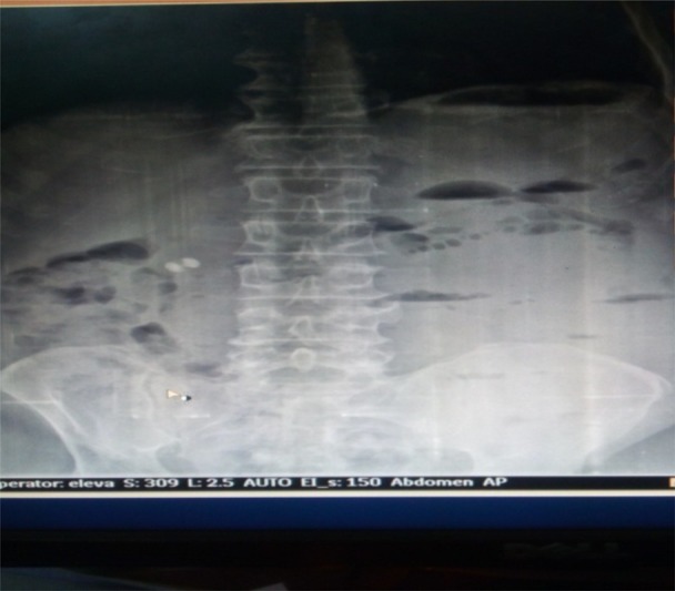
Erect x-ray showing multiple air fluid level
With preoperative diagnosis of abdominal obstruction, patient was optimized for exploration and exploratory laparotomy was done. upon exploration Intraoperative finding revealed extensive interloop non-obstructing adhesion in addition to obstructing band which was 30cm from ileocecal valve arising from antimesenric side of the small bowel(Figure 2).
Figure 2.
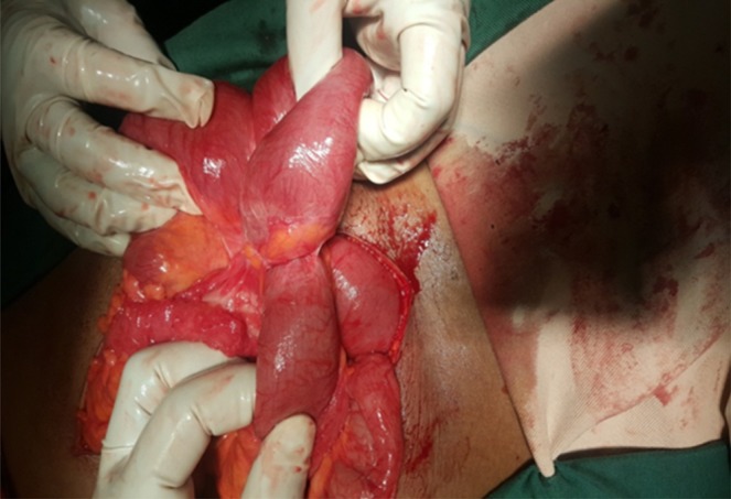
Intraoperative interloop adhesion
There was a free whitish circular hard mass on the right paracolic gutter (Figure 3). Adhesiolysis was done and mass was removed.
Figure 3.
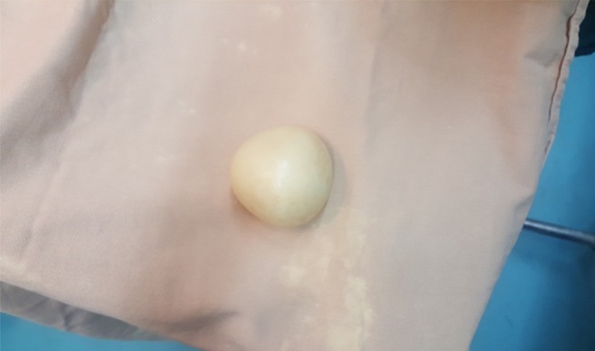
Macrograph of the giant egg-like loose body.
The loose body removed was whitish, oval shaped, with a smooth surface, hard in consistency, and measuring 7.5x6x5cm, and cut-section showed a homogenous grey white solid area. Microscopy showed well circumscribed tissue consisting of concentrically arranged fibrous tissue with scattered degenerating cells over the periphery (Figure 4).
Figure 4.

Micrograph at high magnification (x400) showing haphazardly arranged degenerating cells.
Discussion
Giant loose bodies within the peritoneum are very rare and only a few cases have been reported in literature (1–4). The exact pathogenetic mechanism behind the formation of these loose bodies is not fully understood; but, according to many published articles, the current hypothesis holds that these bodies are formed as a result of a sequential process which starts with torsion of an epiploica, followed by ischemia, saponification and calcification (2–4). The attachment or pedicle becomes gradually atrophied and finally detaches from the colon surface thus becoming a loose body. And, once an appendix epiploica gets saponified and calcified, the exudative serum fluid - which is rich in protein, accumulates around it and, because of the increased temperature in the peritoneal cavity, it gives the appearance of a boiled egg. The size of the peritoneal body increases with time because of a gradual deposition of body serum at the periphery (3–5).
Peritoneal loose bodies are usually small and asymptomatic and found during routine exploration of the abdomen for some other pathology at autopsy. And, when symptomatic, they are usually difficult to diagnose (1–4). When symptomatic, they usually present with various symptoms due to compression effects like abdominal pain and intestinal obstruction (5).
The most common form of presentation in symptomatic patients is lower abdominal pain and features of intestinal obstruction, as is the case in our patient. If a patient presents with features of intestinal obstruction and X-ray films shows multiple air-fluid levels and a calcified lesion in the abdomen, which moves with a change in position of the patient, there should be a high index of suspicion for diagnosis of a giant loose peritoneal body. Additional imaging modalities that can help in the diagnosis are CT and MRI scans, which can be useful for differentiating these from other lesions. However, it is very difficult to differentiate between these loose bodies and other abdominal benign lesions with calcification, like soft tissue neoplasm's or tuberculous granulomas. Mechanisms how patients with loose bodies in the peritoneum develop obstruction is not known yet.
In our case, because the patient presented with acute intestinal obstruction and an X-ray of his abdomen only showed multiple air-fluid levels and no calcified lesions, our patient was directly taken up for an urgent laparotomy without waiting for CT or MRI scans. Intraoperative finding of multiple adhesions in the peritoneum explains his preoperative obstructive symptoms but whether the adhesions are due to the presence of loose body or not is not fully understood. As discussed earlier, compression effect secondary to the mass might have aggravated the pain.
Peritoneal loose bodies are rare and, in most of the cases, small in size. However, giant loose bodies are very rare and only a few cases have been reported in the literature. The current hypothesis on their development is uncertain.
Pre-operative diagnosis of these lesions is difficult and a high index of suspicion should be kept in any symptomatic patient with a mobile lesion in the abdomen or a calcified lesion in the pelvis on X-ray.
No specific treatment is required in asymptomatic patients, however if these entities become associated with complications like intestinal obstruction, or if there is an abdominal mass of obscure origin, or when diagnosis is in doubt, then exploration is required.
References
- 1.Farmlett EJ, Fishman EK, Jones B, Siegelman SS. Torsion of lipoma of appendix epiploica: CT evaluation. Journal of Computer Assisedt Tomography. 1985;9(2):366–368. doi: 10.1097/00004728-198503000-00027. [DOI] [PubMed] [Google Scholar]
- 2.Takada A, Moriya Y, Muramatsu Y, Sagae T. A case of giant peritoneal loose bodies mimicking calcified leiomyoma originating from the rectum. Japanese journal of Clinical Oncology. 1998;28(7):441–442. doi: 10.1093/jjco/28.7.441. [DOI] [PubMed] [Google Scholar]
- 3.Nomura H, Hata F, Yasoshima T, Kuwahara S, Naohara T, Nishimori H, et al. Giant peritoneal loose body in the pelvic cavity: report of a case. Surgery Today. 2003;33(10):791–793. doi: 10.1007/s00595-003-2573-8. [DOI] [PubMed] [Google Scholar]
- 4.Ortiz FE, Arias VJ, Ruano CA. Loose intraperitoneal body. Boletines y Trabajos. Sociedad de Cirugia de Buenos Aries. 1962;46:690–701. [PubMed] [Google Scholar]
- 5.Gayer G, Petrovitch I. CT diagnosis of a large peritoneal loose body: a case Report and review of literature. The British Journal of Radiol. 2011;84:83–85. doi: 10.1259/bjr/98708052. [DOI] [PMC free article] [PubMed] [Google Scholar]