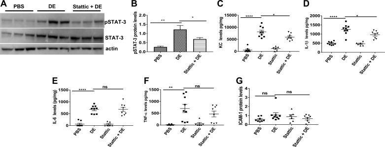
Natarajan K, Meganathan V, Mitchell C, Boggaram V. Organic dust induces inflammatory gene expression in lung epithelial cells via ROS-dependent STAT-3 activation. Am J Physiol Lung Cell Mol Physiol 317: L127–L140, 2019. First published May 1, 2019; doi:10.1152/ajplung.00448.2018.—In Fig. 6A, the actin panel was incorrect in that it inadvertently included two extra bands (the last 2 bands). The corrected figure is shown below with the appropriate bands in the actin panel. The figure legend remains the same, and the correction does not affect the conclusions of the figure or the study.
Fig. 6.
Effects of Stattic, a STAT-3 inhibitor, on STAT-3 activation and induction of inflammatory mediators in mouse lungs. Female C57BL/6 mice received vehicle or Stattic via intraperitoneal injections 16 and 1 h before intranasal administration of 50 μl of PBS or 20% dust extract (DE). After 3 h, mice were euthanized, and lungs were cleared of blood by perfusing with PBS and homogenized. A and B: levels of pSTAT-3, STAT-3, and actin were determined by Western blot analysis, and pSTAT-3 level was normalized to STAT-3, which had been normalized to actin. Data shown are means ± SE (n = 5 for PBS, n = 6 for DE and Stattic + DE). *P < 0.05, **P < 0.01 according to one-way analysis of variance using Tukey’s multiple-comparison test. C–F: levels of keratinocyte chemoattractant (KC), IL-1β, IL-6, and TNF-α were determined by ELISA and normalized to total protein levels. G: ICAM-1 levels were determined by Western blot analysis and normalized to actin levels. Data shown are means ± SE (n = 6–9). *P < 0.05, **P < 0.01, ****P < 0.0001; ns, not significant according to one-way analysis of variance using Tukey’s multiple-comparison test.


