

Abstract
Tracheal and subglottic stenosis are chronic inflammatory processes which can occur as a result of several possible aetiologies, most commonly as a result of prolonged intubation. All consecutive cases of subglottic and tracheal stenosis, secondary to prolonged intubation treated endoscopically over a period of 2 years were reviewed. The surgical approach consisted of radial incision and ablation using Holmium YAG laser, balloon dilatation and topical instillation of mitomycin C through flexible fiberoptic bronchoscope. Ventilation throughout was maintained through LMA. Laser fiber delivered through working channel of bronchoscope. CRA balloon passed through adopter of LMA. Every patient followed for 1 year with 1, 3, 6 months and 1 year interval. Serial balloon dilatation and mitomycin C instillation done in patients during follow up visit. Thirteen patients who underwent airway intervention during study period were studied for clinical outcome. Average follow up was 1 year. Etiology for airway stenosis in all patients of study group was intubation injury. Average frequency of balloon dilatation required was three. Average tracheal lumen achieved at the end of 1 year in our study group was 70%. Symptomatic improvement observed in all patients. Average PEFR achieved was up to 60% of predicted value. Benign subglottic and tracheal stenosis can be safely and effectively managed with flexible bronchoscopy, holmium YAG lasar ablation, balloon dilatation and Mitomycin-C after securing the airway with LMA for general anaesthesia and optimal ventilation.
Keywords: Tracheal stenosis, Laryngeal mask airway, Laser ablation, Mitomycin-C, Bronchoscopic balloon dilatation
Introduction
Tracheal and subglottic stenosis are chronic inflammatory processes which can occur as a result of several possible aetiologies, most commonly as a result of prolonged intubation [1] and other causes such as chemical or thermal injury, following direct trauma after bacterial or mycobacterial infections, inflammatory diseases such as sarcoidosis and Wegener granulomatosis, after radiotherapy and finally idiopathic causes [2]. After damaging the mucosa, inflammatory process will follow and it will activate the fibroblasts which will participate in the wound healing process and lead to scar formation and contraction at the stenosis site [3, 4]. Airway can be re-established by means of mechanical dilatation with bronchoscope, balloon dilatation, laser ablation or with combination of different methods [2]. High rate of re-stenosis ranging from 40 to 70% are seen after endoscopic treatment of tracheal stenosis. Treatments studied to reduce relapses and for better results include steroids and mitomycin-C (MMC) [5].
Optimal airway access for intraoperative ventilation and surgical procedure is one of the important goals to manage airway stenosis. To achieve this, we combine laryngeal mask airway (LMA) ventilation technique with flexible bronchoscopic delivery of laser through LMA to stenotic site, followed by balloon dilatation and subsequent instillation of Mitomycin-C. Use of LMA along with flexible fiberoptic bronchoscope (FFB) has opened new possibilities to effectively visualise and manage laryngeo-tracheal region. LMA and FFB in combination with laser has been described to successfully diagnose and treat airway lesions [6–8].
Lasers quickly began to be used after the work done by Noyori, Ritter, Campbell and Koester in retinal surgery [9, 10]. Lasers are being used in many surgical fields ranging from ophthalmology, urology to otolaryngology.
Bronchoscopic balloon dilatation (BBD) is a simple, safe, efficient and inexpensive adjunct in combined treatment for severe post intubational tracheal stenosis.
Mitomycin-C is a naturally occurring antibiotic which is isolated from Streptomyces casepitosis. It is usually used as an anti-neoplastic agent by inhibiting DNA synthesis, by cross-linking action on DNA and by suppressing RNA and protein synthesis. The mechanism of action is not well understood. Studies have shown that mitomycin-C can inhibit fibroblast proliferation at low concentrations with even 5 min of exposure [11–13].
In the present study, we describe the use of a multi-modality treatment, which consisted of laser therapy, balloon dilatation and subsequent local application of MMC for the dilatation of the stenosis using a video assisted flexible bronchoscopy through a LMA for optimal intra-operative ventilation.
Materials and Methods
This was a retrospective review of the patients with subglottic and tracheal stenosis, secondary to intubation injury for the duration of 2 years. All patients were offered endoscopic treatment using described protocol and was followed up accordingly. Every patient was evaluated with computed tomography (CT) of chest and virtual bronchoscopy which was followed by video-assisted flexible fiberoptic bronchoscopy to assess site, length and consistency of stenosis, as use of laser and its concomitant results largely depended upon the same.
Surgical Protocol
All endoscopic interventions were done by the same interventional respiratory physician in the endoscopy unit after informed and written consent. Every patient was evaluated with flexible bronchoscopy to visualise and assess the stenosis. CT chest virtual bronchoscopy protocol was performed in every patient to assess length, consistency and single or multi-level stenotic segment. The procedure was performed under general anaesthesia with spontaneous ventilation via LMA after positioning it over the laryngeal inlet and securing the airway by inflating the cuff.
Holmium YAG laser was delivered thorough the working channel of the bronchoscope to achieve optimal laser ablation and incision of the stenosis. Delivered laser fibre energy is optimised depending on the consistency and fibrosis of the stenotic segment. More the fibrosis, more energy is required for better outcome. Average laser delivery power setting was 8 W (optimal range is 1.6–10 W) [14] and the average total energy used was 2.93 kJ (optimal range is 0.026–6 kJ) [14]. Care was taken to follow routine laser safety precautions prior to its use. To minimise the risk of fire in the airway, the FiO2 was reduced to 21% during laser ablation [15]. Use of laser was halted when patients showed desaturation and was adequately oxygenated with 100% FiO2 [16].
Under direct visualization, radial incisions and ablation of stenosis were performed using Holmium YAG laser fibre. More energy was delivered through laser fibre at the site of fibrosis. Usual laser settings used were 7.5 W power and 1.5 kJ energy. Laser fibre energy was carefully manipulated to minimize the injury to the membranous part of trachea. Laser fibre and bronchoscope was removed after the optimal laser ablation.
Controlled radial expansion (CRE) balloon was passed through the LMA and was guided over the stenotic segment under bronchoscopic view. Bronchoscope was passed through the LMA beside the balloon catheter for better guidance of the balloon dilatation. CRE balloon used in our practice depends on size, length and diameter of the stenosis and normal diameter of the lumen. In our practice, the sizes of the balloon used are the following:
Boston Scientific Corporation [17] balloon sizes that are used in our centre are as follows:
1. 8, 9, 10 mm causing 3, 5.5,9 atmospheric pressures respectively
2. 10, 11, 12 mm causing 3, 5 and 8 atmospheric pressures respectively
3. 12, 13.5 and 15 mm causing 3, 4.5 and 8 atmospheric pressures respectively
4. 15, 16.5,18 mm causing 3, 4.5 and 7 atmospheric pressures respectively
5. 18, 19, 20 mm causing 3, 4.5 and 6 mm atmospheric pressures respectively.
BBD was done under bronchoscopic view and optimally dilated balloon was kept for 45–60 s with simultaneous vigilant watch on haemodynamics. The balloon was inflated using sterile water as contrast medium and fluoroscopic control was not required because balloon positioning could be easily controlled visually with a bronchoscope with no risk of inappropriate balloon placement. BBD was repeated until the stenosis was optimally dilated. Minimum two and maximum of three attempts BBD were done in our practice.
After visualising the dilated trachea, CRE balloon was again inflated and 3 ml of 0.2 mg/mml of mitomycin C was introduced through the working channel of the bronchoscope with visual guidance to prevent re-stenosis due to fibrosis [18]. After 45–60 s, solution was aspirated; and the procedure was repeated twice. The deflated balloon and bronchoscope was then removed from the adaptor of LMA after visualisation of the tracheobronchial tree.
After the above intervention, the patient was awaken from the general anaesthesia and was given broad spectrum antibiotics and steroids for 5 days post procedure.
Results
A total of 13 patients (7 males and 6 females) with median age of 28 years were enrolled in the study from 2016 to 2017. All patients presented with signs and symptoms of upper airway obstruction including shortness of breath, stridor, cough, dyspnea and wheezing. Flow-volume curve of every patient showed typical box pattern suggesting fixed airway obstruction.
The aetiology in all patients was secondary to prolonged mechanical ventilation through endotracheal tube or tracheostomy tube shown in Table 1. Mean age in our study group was 28 years. Almost 70% patients were of the age group 15–30 years.
Table 1.
Aetiology for prolonged mechanical ventilation
| Aetiology | ET tube | Tracheostomy tube | Total |
|---|---|---|---|
| Organophosphorous compound poisoning (OPC) | 3 | 2 | 5 |
| Snake bite | 2 | 0 | 2 |
| Road traffic accident (RTA) | 2 | 4 | 6 |
| Total | 7 | 6 | 13 |
In our study 5 patients (38%) required prolonged ventilation for respiratory failure due to consumption of OPC. Rest of the patients etiology for ventilation was snake bite (2 patients) and Poly-trauma due to RTA (6 patients).
Patients were graded according to Myer-Cotton scale for tracheal stenosis [19]. All patients had Grade II to Grade III obstruction. The location, size and degree of stenosis of tracheal lesions are described below (Table 2).
Table 2.
The location, size and degree of stenosis of tracheal lesions
| Location (no. of cases) | Length (no. of cases) | Degree of stenosis (no. of cases) |
|---|---|---|
| Subglottic (3) | < 1 cm(1) | 60–70% (1) |
| Upper trachea (4) | 1–3 cm(8) | 70–80% (4) |
| Mid and lower trachea (6) | > 3 cm(4) | > 80% (8) |
In our study group, out of 13 patients with tracheal stenosis, 3 were in subglotic region, 4 in upper trachea and 6 in mid and lower trachea. Almost equal number of web like (7 cases) and complex type (6 cases) of stenosis was observed in our study group. Eight patients had length of stenosis around 1–3 cm, 4 patients with length of more than 3 cm and single patient had length less than 1 cm.
All patients had more than 60% of stenosis. Most patients (8 cases) were showing critical stenosis of more than 80%. Four showed 70–80% luminal narrowing and one case showed 60–70% luminal narrowing.
All patients in our study group were followed up regularly with interval of 1, 3, 6 months and 1 year. During every visit, patient underwent flexible video assisted fiberoptic bronchoscopy to see for residual stenosis in tracheal lumen. Laser ablation and balloon dilatation were offered in patients who showed more than 50% re-stenosis. One of the patient who showed complex and multilevel stenosis was referred for surgical intervention in view of poor outcome after 3 months and one patient was lost for follow up after 3 months.
In our study we monitored peak expiratory flow rate (PEFR) during every visit in all patients (Table 3). Eleven out of 13 patients showed significant improvement in PEFR. One patient was lost for follow up after 3 months.
Table 3.
Peak expiratory flow rates on follow up
| Time duration | PEFR Less than 100 Lit/min | PEFR between 100 and 150 L/m | PEFR between 150 and 200 L/m | PEFR more than 200 L/m | Total |
|---|---|---|---|---|---|
| Pre-op PEFR | 06 | 01 | 00 | 00 | 07b |
| After 1 month PEFR | 01 | 02 | 02 | 08 | 13 |
| After 3 month PEFR | 02 | 00 | 03 | 08 | 13 |
| After 6 month PEFR | 00 | 00 | 04 | 07 | 11a |
| After 1 year PEFR | 00 | 00 | 04 | 07 | 11a |
aOne patient was lost for follow up after 3 months and 1 patient was referred for surgical intervention after 3 months
bSix patients were on tracheostomy tube and could not perform PEFR manoeuvre
Pre-treatment bronchoscopic image showing grade 3 tracheal stenosis (Fig. 1), Bronchoscopic view immediately after laser ablation, balloon dilatation and Mitomycin-C instillation (Fig. 2) and bronchoscopic view 1 year post treatment of a patient (Fig. 3) is illustrated below:
Fig. 1.
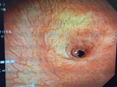
Pre Treatment
Fig. 2.
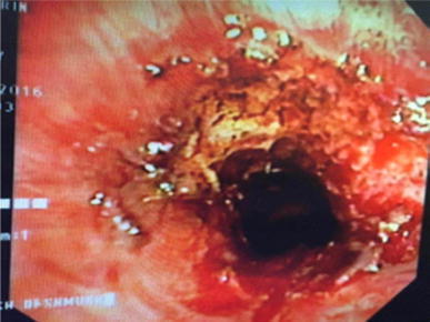
Post surgical intervention
Fig. 3.
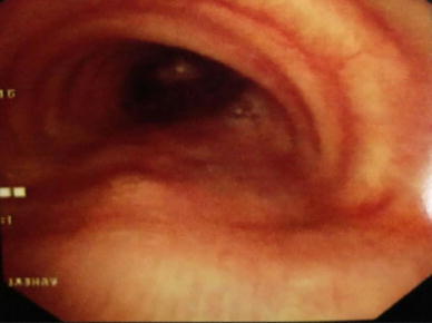
One year post treatment
Out of 13 patients whom we offered Holmium YAG laser and serial balloon dilatation, 11 patients showed significant improvement in symptoms and PEFR.
In eleven out of 13 patients more than 65% luminal opening achieved at the end of 1 year. The overall success rate was 80% at the end of 1 year. There were no intra-operative or peri-operative death observed in our study.
Discussion
In our study the intubation injury and prolonged intubation is the dominant cause observed and this was in concordance with the study done by Nopawan et al. [16] in which 6 out of 11 cases showed intubation injury as the dominant case. Respiratory failure and prolonged intubation secondary to OPC consumption was the most common cause. Benign subglottic and tracheal stenosis secondary to prolonged intubation has an increasing trend due to the increase in number of ICUs in the developing countries that admits critically ill patients and start treatment without following necessary guidelines of prevention of intubation complications.
This study demonstrates optimal management of benign subglottic and tracheal stenosis with Holmium YAG laser and balloon dilatation. This is prospective study included 13 patients. Our outcomes are based on symptom control, degree of tracheal lumen achieved and PEFR improvement. Alleviation of symptom of shortness of breath and noisy breathing were observed immediately post procedure in all patients in our study. These results are comparable to previous study reports using CO2 laser and bronchoscopic dilatation with rigid or flexible bronchoscopy.
In our study we used Holmium YAG laser which in previous studies seldom used as compared to CO2 laser in bronchoscopic management of tracheal stenosis. Use of Holmium YAG laser in Otolaryngology and Pulmonary Medicine was not routinely demonstrated. Holmium YAG laser is popularized in the field of Urology than other specialities. The Holmium YAG laser has a 2100-nm wavelength in the infrared spectrum with up to a 0.5 mm depth of penetration. The energy can be delivered through a variety of small diameter disposable and re-usable delivery devices which allows for precise delivery of laser energy. It has unique feature of minimal penetration depth, variable coagulability and cutting edge. We found it safe and effective in radial incisions and ablation of stenosis in our study group patients. Holmium YAG laser was used as per laser safety protocol in our study.
We observed that laser ablation is relatively easy with lesion at subglottic than lower tracheal level. Length of stenosis is inversely proportional to percent of dilatation achieved and optimal outcome. More than two level stenosis with length of the stenosis more than 3 cms show poor outcome in our study. Single level stenosis with length of less than 2 cms show excellent outcome in our study.
Segmental tracheomalacia post procedure can be seen with stenosis with larger length and extensive granulation with pliable mucosa. In these patients we observed poor outcomes after serial ablations and balloon dilatation. Surgical resection and end to end anastomosis are prefered modes of treatment in these patients.
Conclusion
Benign tracheal stenosis secondary to prolonged intubation is one of the serious complications in developing countries. The most well known treatment modality for tracheal stenosis is open surgery, resection of the stricture and end to end anastomosis. Benign subglottic and tracheal stenosis can be safely and effectively managed with this interventional dilatational procedure using a video assisted flexible bronchoscope, holmium YAG lasar ablation, balloon dilatation and Mitomycin-C after securing the airway with LMA for general anaesthesia and optimal ventilation. Several balloon dilatations are beneficial for optimal outcome in patients with tracheal re-stenosis. Long term results with minimal morbidity can be achieved with this surgical protocol.
Acknowledgement
The authors would like to thank MGM Medical College and Hospital, Auranagabad and Residents of Department of Respiratory Medicine.
Compliance with Ethical Standards
Conflict of interest
The authors declare that they have no conflict of interest.
Ethical Approval
All procedures performed in this study involving human participants were in accordance with the ethical standards of the institution and with the 1964 Helsinki declaration and its later amendments. This article does not contain any studies with animals performed by any of the authors. For this type of study formal consent is not required.
References
- 1.Couraud L, Brichon PY, Velly JF. The surgical treatment of inflammatory and fibrous laryngotracheal stenosis. Eur J Cardiothorac Surg. 1988;2:410–415. doi: 10.1016/1010-7940(88)90043-7. [DOI] [PubMed] [Google Scholar]
- 2.Ernst A, Feller-Kopman D, Becker HD, et al. Central airway obstruction. Am J Respir Crit Care Med. 2004;169:1278–1297. doi: 10.1164/rccm.200210-1181SO. [DOI] [PubMed] [Google Scholar]
- 3.Karagiannidis C, Velehorschi V, Obertrifter B, Macha HN, Linder A, Freitag L. High level expression of matrix-associated transforming growth factor-β1 in benign airway stenosis. Chest J. 2006;129(5):1298–1304. doi: 10.1378/chest.129.5.1298. [DOI] [PubMed] [Google Scholar]
- 4.Chen T, Kunnavatana SS, Koch RJ. Effects of mitomycin-C on normal dermal fibroblasts. Laryngoscope. 2006;116(4):514–517. doi: 10.1097/01.MLG.0000205590.62824.0A. [DOI] [PubMed] [Google Scholar]
- 5.Smith ME, Elstad M. Mitomycin C and the endoscopic treatment of laryngotracheal stenosis: are two applications better than one? Laryngoscope. 2009;119(2):272–283. doi: 10.1002/lary.20056. [DOI] [PubMed] [Google Scholar]
- 6.Jameson JJ, Moses RD, Vellayappan U, Lathi KG. Use of the laryngeal mask airway for laser treatment of the subglottis. Otolaryngol Head Neck Surg. 2000;123:101–102. doi: 10.1067/mhn.2000.107457. [DOI] [PubMed] [Google Scholar]
- 7.Kanagalingam J, Hurley R, Grant HR, Patel A. A new technique for the management of inaccessible anterior glottic lesions. J Laryngol Otol. 2003;117:302–306. doi: 10.1258/00222150360600922. [DOI] [PubMed] [Google Scholar]
- 8.Ogata J. The airway management using laryngeal mask airway and tracheal fiberoscopy in a paediatric patient with tracheal stenosis after tracheostomy. Masui. 2004;53:1282–1285. [PubMed] [Google Scholar]
- 9.Campbell CJ, Rittler MC, Koester CJ. The optical maser as a retinal coagulator: an evaluation. Trans Am Acad Ophthalmol Otolaryngol. 1963;67:58–67. [PubMed] [Google Scholar]
- 10.Noyori KS, Campbell CJ, Rittler MC, Koester CJ. Ocular thermal effects produced by photocoagulation. Arch Ophtalmol. 1963;70:817–822. doi: 10.1001/archopht.1963.00960050819017. [DOI] [PubMed] [Google Scholar]
- 11.Jamel HD. Effect of brief exposure to mitomycin-C on viability and proliferation of cultured human Tenon’s capsule fibroblasts. Ophthalmology. 1992;99:1471–1476. doi: 10.1016/S0161-6420(92)31781-6. [DOI] [PubMed] [Google Scholar]
- 12.Khaw PT, Sherwood MB, MacKay SL, et al. Five-minute treatments with fluorouracil, floxuridine, and mitomycin have long-term effects on human Tenon’s capsule fibroblasts. Arch Ophthalmol. 1992;110:1150–1154. doi: 10.1001/archopht.1992.01080200130040. [DOI] [PubMed] [Google Scholar]
- 13.Lee DA, Lee TC, Cortes AE, et al. Effects of mitramycin, mitomycin, daunorubicin and bleomycin on human subconjunctival fibroblasts attachment and proliferation. Invest Ophtalmol Vis Sci. 1990;31:2136–2144. [PubMed] [Google Scholar]
- 14.Myers L, Bakthavachalam S, Thomason T, Klein K. Holmium: Yag laser fiberoptic bronchoscopy via Laryngeal mask airway. Int J Otorhinolaryngol. 2006;6(2):1–4. [Google Scholar]
- 15.Verret DJ, Jategoankar Ameya, Helman Samuel, Kadakia Sameep, Baharami Arash, et al. Holmium laser for endoscopic treatment of benign tracheal stenosis. Int Arch Otorhinolaryngol. 2017 doi: 10.1055/s-0037-1604201. [DOI] [PMC free article] [PubMed] [Google Scholar]
- 16.Vorasubin Nopawan, Vira Darshni, Jamal Nausheen, Chhetri Dinesh K. Airway management and endoscopic treatment of subglottic and tracheal stenosis: the Laryngeal mask airway technique. Ann Otol Rhinol Laryngol. 2014;123940:293–298. doi: 10.1177/0003489414525340. [DOI] [PMC free article] [PubMed] [Google Scholar]
- 17.Jabbardarjani HR, Kiani A, et al. Balloon Bronchoplasty: case series. Tanaffos. 2012;11(2):42–48. [PMC free article] [PubMed] [Google Scholar]
- 18.Garg M, Gogia P, Manoria P, Goyal R. Bronchoscopic management of benign bronchial stenosis by electocautery and balloon dilatation. Indian J Chest Dis Allied Sci. 2012;54:41–43. [PubMed] [Google Scholar]
- 19.Myer C, O’Connor P, Cotton R. Proposed grading system for subglottic stenosis based on endotracheal tube sizes. Ann Otol Rhinol Laryngeol. 1994;103:319–323. doi: 10.1177/000348949410300410. [DOI] [PubMed] [Google Scholar]