

Abstract
This is a prospective observational cross sectional study comprising of 57 patients who were having symptoms of chronic rhinosinusitis which were evaluated with the help of computed tomography scan (coronal and axial sections) to measure the thickness of all walls of maxillary sinus and it’s volume. Computed tomographic imaging of sinonasal region has become the gold standard in the evaluation of patients with chronic sinusitis. The maxillary sinus is pyramidal in shape with lateral wall of nose forming its base and its apex is directed towards zygomatic process. All three dimensions of the maxillary sinus were measured and the volume of each maxillary sinus was also calculated. Hyperostosis of maxillary sinus tended to increase maxillary sinus walls thickness which ultimately results into decrease in maxillary sinus volume in chronic rhinosinusitis patients.
Keywords: Chronic rhinosinusitis, Maxillary sinus volume, Hyperostosis, Computed tomography
Introduction
Chronic rhinosinusitis is one of the most frequent diseases encountered worldwide. It defined as a group of disorders characterized by inflammation of the mucosa of nose and paranasal sinuses of at least 12 consecutive weeks duration [1]. Its diagnosis relies on clinical judgment based on a number of often vague physical complaints and symptoms and with help of conventional anterior and posterior rhinoscopic examination. Computed tomography (CT) scan has made an enormous impact in regional imaging and it has increased a surgeon’s ability to depict accurately the status of structures within the paranasal sinus region and in delineating the location and extent of pathology. The CT scan is the gold standard investigation in all sinus diseases and it gives details about maxillary sinus volume and it’s bony wall thickness and serves as an anatomic roadmap for the operating surgeon. In this study we will assess the effect of chronic maxillary sinusitis on maxillary sinus volume and thickness of its bony walls.
Methods and Materials
A prospective cross sectional observational study was carried out in the Department of ENT and Head and Neck Surgery, Mahatma Gandhi Institute of Medical Sciences, Sevagram, Maharashtra from September 2015 to August 2017 and a total number of 57 patients who were clinically and radiologically diagnosed as having chronic rhinosinusitis, were evaluated with (computed tomography) CT PNS coronal and axial views. Patients with acute sinusitis or malignant disease or those who had previously undergone nasal or sinus surgery, either open or endoscopic, excluded from the study.
The following measurements of maxillary sinus were performed on CT PNS:
-
(A)
Maximal craniocaudal diameter.
-
(B)
Maximal depth (anteroposterior diameter).
-
(C)
Maximal width.
-
(D)
Width at the middle of the maxillary sinus on axial section.
-
(E)
Thickness of maxillary walls (Anterolateral wall, Posterolateral wall, Medial wall and superior wall) (Figs. 1, 2).
Fig. 1.

a Maximal craniocaudal diameter, b maximal depth (anteroposterior diameter), c maximal width and d width at the middle of the maxillary sinus
Fig. 2.

Thickness of maxillary walls (anterolateral, posterolateral, medial and superior wall)
The maxillary sinus is pyramidal in shape with lateral wall of nose forming its base and its apex is directed towards zygomatic process.
As all three dimensions of the maxillary sinus were measured, the volume of each maxillary sinus was also calculated using the following equation:
The width used for this calculation was the mean value for the maximal width and the width at the middle of the maxillary sinus on axial slices.
Results and Observations
A total number of 57 patients of chronic rhinosinusitis were examined. Out of 57 patients 30 were males and 27 were females with male female ratio being 1.11:1.
Maxillary Sinus Volume in Chronic Rhinosinusitis
The average volume of a developed maxillary sinus at maturity varies between 15 and 20 ml [2]. In the present study, 44 (77.19%) patients of chronic rhinosinusitis has the maxillary sinus volume between 10 and 15 ml followed by 9 (15.79%) patients who have maxillary sinus volume between 16 and 20 ml and 4 (7.02%) patients has maxillary sinus volume between 21 and 25 ml. The mean value of maxillary sinus volume in present study is 13.14 ml (Table 1, Fig. 3).
Table 1.
Distribution of patients according to maxillary sinus volume
| Maxillary sinus volume | No of patients | Percentage (%) |
|---|---|---|
| 10–15 ml | 44 | 77.19 |
| 16–20 ml | 9 | 15.79 |
| 21–25 ml | 4 | 7.02 |
| Total | 57 | 100 |
| Mean ± SD | 13.14 ± 1.65 | |
Fig. 3.
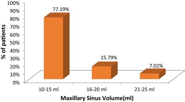
Distribution of patients according to maxillary sinus volume
Maxillary Sinus Bony Walls Thickness in Chronic Rhinosinusitis
In the present study, we found that the mean thickness of superior wall is 1.7 mm (SD: 0.3 mm) whereas the anterolateral, medial and posterolateral walls has a mean thickness of 1.6 mm (SD: 0.3 mm). The maximum bony wall thickness for anterolateral wall is 2.2 mm whereas the minimum thickness is 1.0 mm for the same. Posterolateral bony wall has maximum thickness of 2.1 mm while minimum thickness is 0.6 mm. Both superior and medial bony walls has the minimum bony thickness of 0.8 mm while the maximum bony wall thickness is 2.2 and 2.3 mm respectively (Table 2, Fig. 4).
Table 2.
Descriptive statistics for thickness of wall in CRS
| Maxillary sinus wall | N | Minimum | Maximum | Mean | Std. Deviation |
|---|---|---|---|---|---|
| Anterolateral | 57 | 1.00 | 2.2 | 1.6 | 0.30 |
| Posterolateral | 57 | 0.60 | 2.1 | 1.6 | 0.30 |
| Medial | 57 | 0.80 | 2.2 | 1.7 | 0.30 |
| Superior | 57 | 0.80 | 2.3 | 1.6 | 0.30 |
Fig. 4.
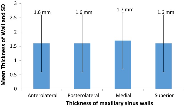
Descriptive statistics for thickness of wall in CRS
Discussion
Computed tomographic imaging of sinonasal region has become the gold standard in the evaluation of patients with chronic sinusitis. Its ability to accurately map out the bony and soft tissue anatomy of the paranasal sinuses has proven invaluable to the endoscopic surgeon in the diagnostic workup [3].
Maxillary Sinus Volume in Chronic Rhinosinusitis
In the present study, 44 (77.19%) patients of chronic rhinosinusitis has the maxillary sinus volume between 10 and 15 ml followed by 9 (15.79%) patients who have maxillary sinus volume between 16 and 20 ml and 4 (7.02%) patients has maxillary sinus volume between 21 and 25 ml. The mean value of maxillary sinus volume in present study is 13.14 ml.
The present study correlates with the study conducted by Cho et al. [2] which shows that CRS group had significantly smaller maxillary sinus volume than did controls (P = 0.001). In this study author also states that hyperostosis of maxillary sinus tended to increase maxillary walls thickness which ultimately results into decrease in maxillary sinus volume. Study conducted by Kim et al. [4] demonstrates that the volume of the maxillary sinus in the controls was much higher than that in the CRS group. The difference between the two groups was statistically significant (P = 0.04).
Maxillary Sinus Bony Walls Thickness in Chronic Rhinosinusitis
In the present study, we found that the mean thickness of superior wall is 1.7 mm (SD: 0.3 mm) whereas the anterolateral, medial and posterolateral walls has a mean thickness of 1.6 mm (SD: 0.3 mm). The maximum bony wall thickness for anterolateral wall is 2.2 mm whereas the minimum thickness is 1.0 mm for the same. Posterolateral bony wall has maximum thickness of 2.1 mm while minimum thickness is 0.6 mm. Both superior and medial bony walls has the minimum bony thickness of 0.8 mm while the maximum bony wall thickness is 2.2 and 2.3 mm respectively.
In the comparative study conducted by Cho et al. [2] 99 adult patients were included: 52 controls (having septal deviation without CRS) and 47 patients with CRS. The mean anterolateral bony wall thickness in control group is 1.10 mm whereas in CRS group is 1.46 mm. The mean posterolateral bony wall thickness in control group is 1.03 mm whereas in CRS group it is 1.42 mm.
Present study correlates with findings of study conducted by Cho et al. [2] who concluded that in chronic rhinosinusitis due to hyperostosis there is increase in bony wall thickness in chronic rhinosinusitis.
The above findings are seen in patients with CRS i.e. infection of paranasal sinuses for more than 12 weeks. The patients which has longer duration of disease have more thicker walls.
Clinical Implication
Present study shows that CRS causes maxillary sinus wall thickening and decreasing in maxillary sinus volume and it also shows that more the wall thickness and lesser the maxillary sinus volume, severe is the disease which necessitates the Functional Endoscopic Sinus Surgery as a treatment modality in order to treat and to prevent the further complications of CRS.
Conclusion
Hyperostosis of maxillary sinus tended to increase maxillary sinus walls thickness which ultimately results into decrease in maxillary sinus volume in chronic rhinosinusitis patients.
Footnotes
Publisher's Note
Springer Nature remains neutral with regard to jurisdictional claims in published maps and institutional affiliations.
References
- 1.Benninger MS, Ferguson B, Hadley J, et al. Adult chronic rhinosinusitis: definitions diagnosis, epidemiology, and pathophysiology. Otolaryngol Head Neck Surg. 2003;129(3(suppl)):S1–S32. doi: 10.1053/hn.2003.v128.amhn0312811. [DOI] [PubMed] [Google Scholar]
- 2.Cho SH, et al. Factors for maxillary sinus volume and craniofacial anatomical features in adults with chronic rhinosinusitis. Arch Otolaryngol Head Neck Surg. 2010;136(6):610–615. doi: 10.1001/archoto.2010.75. [DOI] [PubMed] [Google Scholar]
- 3.Tiwari R, Goyal R. Study of anatomical variations on CT in chronic sinusitis. Indian J Otolaryngol Head Neck Surg. 2015;67(1):18–20. doi: 10.1007/s12070-014-0734-2. [DOI] [PMC free article] [PubMed] [Google Scholar]
- 4.Kim HY, Kim MB, Dhong HJ, et al. Changes of maxillary sinus volume and bony thickness of the paranasal sinuses in longstanding pediatric chronic rhinosinusitis. Int J Pediatr Otorhinolaryngol. 2008;72(1):103–108. doi: 10.1016/j.ijporl.2007.09.018. [DOI] [PubMed] [Google Scholar]