



Abstract
Complications following the total laryngectomy with or without partial pharyngectomy with neck dissection for laryngeal and pyriform fossa malignancies like aspiration, pharyngocutaneous fistula wound infection, flap necrosis, haematoma, chyle fistula and carotid blowout can cause serious implication on the final outcome of the treatment, which leads to increased postoperative morbidity, hospital stay and hospital cost. A prospective study in the Department of Otolaryngology and Head–Neck Surgery, JSS Hospital, Mysore, from November 2014 to July 2016. 30 patients undergoing Total laryngectomy with or without partial pharyngectomy for laryngeal and pyriform fossa were included in this study. The presentation, diagnosis, and management of the complications that were occurred, were discussed. The age of the patients vary between 32 and 76. Also, male preponderance was seen with approximately M:F ratio 3:1. Out of these 30 patients, 6 patients developed complications. The most common complication was pharyngocutaneous fistula (2 patients, 6%), which was developed after the 7th day. It was managed conservatively in both patients, wound infection was a second complication (2, 6%). Other complications were drain failure (1, 3%) and chylous fistula (1, 3%). The Most common complications after total laryngectomy with or without partial pharyngectomy with neck dissection in our study were wound infection and pharyngocutaneous fistula. Assessment of risk factors, early recognition of complications per operative protocols with improvised techniques are necessary to reduce incidence of complication after total laryngectomy with or without partial pharyngectomy with neck dissection.
Keywords: Laryngeal cancer, Laryngectomy, Partial pharyngectomy, Pharyngocutaneous fistula
Introduction
Carcinoma of the larynx and pyriform sinus (1:100,000 of the population) which is the most common presentation of hypopharyngeal cancer constitutes about 25% of all the head and neck malignancies. There are many therapeutic options available in the treatment of laryngeal and pyriform fossa cancer, which include Surgical (total laryngectomy with or without partial pharyngectomy) organ preservation modalities (chemoradiation and combined therapy) [1]. Total Laryngectomy is a radical procedure which involves removal of the total larynx. These procedures are useful in the treatment of advanced laryngeal and pyriform fossa cancer like T3, T4a, and T4b. Laryngectomy is one of the most frequently done oncological surgeries in this part of the world, with a high incidence of morbidity as the patient present at a late stage of the disease. Complications following the total laryngectomy with or without partial pharyngectomy with neck dissection for laryngeal and pyriform fossa malignancies like aspiration, pharyngocutaneous fistula wound infection, flap necrosis, haematoma, chyle fistula and carotid blowout can cause serious implication on the final outcome of the treatment, which leads to increased postoperative morbidity, hospital stay and hospital cost. Through improvised techniques and pre and post operative protocols the incidence of complications have reduced, none the less they occur.
So this study was carried on to know the various factors causing Complications following the total laryngectomy with or without partial pharyngectomy with neck dissection and to draw the pre operative, intra operative and post operative protocols with improvised techniques to decrease the incidence of complications.
Materials and Methods
Study Subjects
Patients who underwent Total laryngectomy with or without partial pharyngectomy for laryngeal and pyriform fossa malignancies in the Department of ENT, JSS Hospital Mysore.
Study Size
30 cases of total laryngectomy with or without partial pharyngectomy for laryngeal and pyriform fossa malignancies.
Sampling Technique
Non-probability purposive sampling technique was used in this study.
Inclusion Criteria
All those patients who underwent total laryngectomy with or without partial pharyngectomy for laryngeal and pyriform fossa malignancies with/without additional surgical procedure like radical or modified radical or selective neck dissection for histologically proven carcinoma larynx and pyriform fossa with/without neck metastasis.
Exclusion Criteria
Who opt for organ preservation modality (chemo and radiotherapy) and
Patients who did not give valid consent.
Methods of Collection of Data
Patients selected based on standard clinical proforma.
An informed consent was taken from the patient.
Patients who underwent total laryngectomy with or without partial pharyngectomy for histopathologically proven carcinoma larynx and pyriform fossa to be included. Followed a minimum for 3 months for postoperative period complications.
All patients were observed for post-operative complications during their 5 days stay in the hospital and after discharge from hospital a regular follow-up visit record was maintained.
Weekly follow up for the first month, fortnightly during next 2 months and monthly follow-up for 6 months was advised. Later on patients were called for follow up after every 6 months for 1 year. During each follow-up visit a thorough clinical examination was done in all patients and appropriate investigations were carried out where indicated. A complete record of complications, their diagnosis and treatment was maintained during this period.
Statistical Methods Employed
Following methods were employed in the present study:
Descriptive statistics
Chi square test
Kappa
McNemar test
Results
There were 30 patients included in the study, overall if we see, the age of the patients vary between 32 and 76 and peak incidence is noted between 51 and 70 (60%) age group. Also, male preponderance was seen with approximately M: F ratio 3:1 i.e. out of a sample of 30 patients 23 were males (76.7%) and 7 were females. Out of these 30 patients, 5 patients developed complications. The most common complication was pharyngocutaneous fistula (2 patients, 6%), which was developed after the 7th day. It was managed conservatively in both patients, wound infection was a second complication (2, 6%). Other complications were drain failure (1, 3%) and chylous fistula (1, 3%).
Discussion
Complications following total laryngectomy (Fig. 1) with or without partial pharyngectomy with neck dissection can cause serious implications on the final outcome of the treatment. For example Pharyngocutaneous fistula can prolong hospitalization, delays the initiation of radiotherapy, there by increases the morbidity and mortality, Severe infection with flap necrosis resulting in a carotid blowout can be life threatening. Similarly, late complications like pharyngeal and tracheal stomal stenosis can result in swallowing and breathing difficulty respectively. It is, therefore, important to diagnose these complications early so that timely intervention can be done.
Fig. 1.
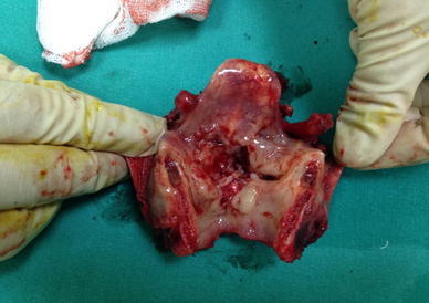
Total laryngectomy specimen
Out of the 30 patients, 8 (26%) patients treated with surgery alone, whereas 22 (74%) patients underwent both surgery and radiotherapy. Total laryngectomy in 30 patients (100%), partial pharyngectomy (40%), various types of neck dissection (100%) and Tracheoesophageal prosthesis inserted in 22 patients at the same sitting.
30 patients who underwent Total laryngectomy with or without Partial pharyngectomy with neck dissection were evaluated for various types of complications intraoperatively and post-operatively and followed for minimum of 3 months. In our study, the minor complications were seen in five patients, among them most common early complication is pharyngocutaneous fistula in 2 patients (6%) followed by wound sepsis in 2 (6%) patients and drain failure in 1 (3%) patients (Fig. 2).
Fig. 2.
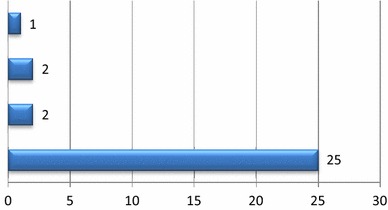
Early complications of laryngectomy
In our study, although the age of the patients vary between 32 and 76 years with male preponderance (M:F—3:1). There is no correlation of complications neither with gender nor with age, However a study done by Lagier et al. [2], highlighted the importance of preoperative oncogeriatric evaluation from the age of 65 years to optimize surgical management.
Eventhough incidence of malignancy is more in smokers and alcoholic [3] (80%), there is no effect of the same on the incidence of post operative complications in our study. But there was a study conducted on Tobacco Exposure and Complications in Laryngeal Surgery by Fiorini [4], where Smoking status, and alcohol abuse were statistically associated with a major incidence of local complications and their study concluded that Tobacco exposure is implicated not only in the pathogenesis of head and neck cancer but also play a key role in the development of local complications by delaying wound healing in patients who undergo laryngeal cancer surgery.
The socio-economic distribution of 30 patients according to Kuppuswamy classification, most of them are belongs to the lower class i.e. class 4 around 77% (23) and class 3 23% (7). Which shows a strong correlation between the poor socio-economic group and carcinoma of laryngeal and pyriform fossa cancer, which is similar to study conducted by Arsenijevic et al. [5], in his study, he concluded that People of the lower educational level and poor socioeconomic status had increased the risk of getting laryngeal cancer. But almost all the patients in our study group belongs to the low socio economic group, we couldn’t draw any relation between socio economic status and complications.
On reviewing literature, it has been found out that, only few studies have been carried out on the relationship between nutritional parameters and the incidence of postoperative complications. Unintentional weight loss, and a lesser degree albumin, were predictive for postoperative complications. Weight loss seems to be the most important parameter for predicting major postoperative complications; patients with > 10% weight loss during the 6 months before surgery are at greater risk for the occurrence of major postoperative complications and If there is good preoperative nutritional status, the onset of postoperative malnutrition is thus avoided.
In all of our patients
No weight loss > 10–15% within 6 months before surgery,
BMI > 18.5%,
serum albumin > 3 g/dl.
So none of our patients were malnourished pre operatively even though all patient were well nourished in our study. Care was taken to prevent post operative nutritional impairment by following few protocols:
Tube feeding for patients starts within 24–48 h after surgery
Each patient was received 1.2–1.4 g/kg/day of proteins
serum hemoglobin was with in normal limit for all the patients (an average all the patients were given 1 pint PRBC to maintain Hemoglobin level between 12.5 and 14.5)
According to the results of this study, supportive treatments and supplementary nutritional supports is highly suggested in all patients undergoing laryngectomy, even with acceptable preoperative nutritional indices. These supportive approaches can be also advisable in patients leaving hospital without any serious problem. In regards to the high catabolism waste in cancer patients due to underlying diseases or surgery, special attention to the patients’ nutritional dietary regimens is recommended.
In this study 2 patients (6%) developed the wound infection immediate postoperatively, In both cases, the specimen for culture and sensitivity revealed staph aureus species and were found sensitive to aminoglycosides who were successfully treated with appropriate antibiotics and adequate aseptic dressing Wound healed within 2 weeks. Other patient having the fever only for 1 day i.e. probably due to stress induced as there is no reason identified. This less incidence of wound infection achieved through
Prophylactic antibiotic therapy (ceftriaxone 1 g with sulbactam 500 mg to cover aerobic organism and Metrogyl 500 mg to cover anaerobic organism was given 12th hourly) in the preceding 36 h to surgery and continuation of the same postoperatively for 5 days.
Hematomas were avoided by careful hemostasis and continuous negative suction drainage post operatively.
The Maintenance of uncontaminated nature of the neck surgical field explains this low rate of infection in this study.
This is less when compare to many other studies like a study done Maharajan et al. [10] where the wound infection is 20%, which suggests that pre-operative improvement in general condition, selection of the patient and planned surgery improves outcome, similarly Seth et al. [6] concluded that preoperative radiation, prolonged operative time, low albumin and diabetes were independently associated with postoperative wound infections, these results will help to identify the patients at risk for wound complications thus allowing for heightened surveillance and preventive measures where possible. Similarly, Arriaga et al. observed that medical complications were a significant cause of morbidity in total laryngectomy, a focused pre-anesthetic risk factor analysis by a medical team familiar with head and neck cancer patients assures prompt identification and management of these complications.
And only 1 patient have the drain failure and none of the patients developed the hematoma, the less incidence of drain failure was because of following protocols.
The steps followed was,
The drain is placed most dependent part of the wound.
The drain is passed out of the skin flap in the usual way, leaving the perforated end in the wound completely intact. After the trocar has been cut.
A bite was taken through the drain site with a length of non-absorbable suture and was secured to the skin after the black mark.
This was when comparable to a study done by Reddy et al. [7] is less, where Hematoma due to drain block was seen in two patients. One developed abscess after 3 weeks which was treated by incision and drainage and daily dressings.
In our study there is no delay in the wound healing in any of the patient. This was achieved mainly because of protocols what we followed i.e. Sterile operative field, Careful drain placement (as explained above), Gentle tissue handling, Precise use of cautery and keeping the flaps moist, Gentle curved skin crease incisions and including platysma in skin flap (subplatysmal flap) helps to achieve good cosmesis and flap viability, Measures taken to prevent wound infection (as explained above), Good nutritional status of the patient (as explained above).
In our study none of the patient developed wound dehiscence (0%). But Pellini et al. [8] observed, wound dehiscence was the most frequent major complication and 3 out of 8 cases required a pedicled flap for repair in their in their study.
This was achieved mainly because of following protocols
A tension free skin closure.
Prevention of wound infection (as explained below).
Preventing Fistula formation by following proper surgical protocols while closing the pharynx (explained below).
Well designed non ischemic neck skin flaps (subplatysmal flaps).
Post operative local wound care.
In this study the incidence pharyngocutaneous fistula is less, when compare to many other studies like A study conducted by Maroku et al. [9], showed as pharyngocutaneous fistula is the most common complication in the immediate postoperative period following total laryngectomy, the presence of early postoperative fistula was established in 4 of the 37 patients (13%). and in one more study done by Maharjan et al. [10] included 54 patients, with an average age of 58 years, 20 patients developed complications postoperatively. The most common complication was pharyngocutaneous fistula (6 patients, i.e. 11%). In our study, the incidence of pharyngeal cutaneous fistula is 6%, was achieved because the pharyngeal defect was closed with a T-type closure (Fig. 3).
Fig. 3.
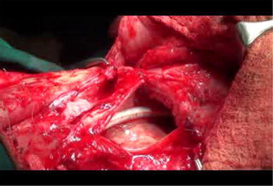
Pharyngeal T shape closure
A 2-layered pharyngeal closure was followed i.e.
1 st layer 3-0 vicryl running modified Connell or true Connell technique (care was taken to invert the mucosa after each pass. This Inversion was achieved with an absorbable stich that runs horizontally through the submucosa, care was taken to avoid overtightening to avoid ischemia).
2nd layer Approximate inferior constrictors and suture constrictors to suprahyoid muscles with interrupted 3-0 vicryl (care was taken during this layer of closure to avoid overtightening to avoid dysphagia. Consequently, it decreased the average time of patient hospitalization and the costs).
These two pharyngocutaneous fistulae were developed around the 7th day. The aim of conservative treatment is to preserve and restore the injured region, It was constituted by the continuation of the nasogastric tube for 3 weeks, compressive bandage around the neck, draining the exudate liquid retained under the patch, maintenance of hemoglobin levels above 12.5 g/dl, adequate nutrition, high doses of intravenous antibiotics (ceftriaxone 1 g, sulbactam 500 mg and Metrogyl 500 mg) and Intramuscular Glycopyrrolate 0.1 mg IM for every 8 h daily to reduce the secretions; With these conservative management fistula healed completely within 3 weeks. These two pharyngocutaneous fistulae were developed in pyriform fossa malignancies, that shows the more chance of pharyngocutaneous fistula when patient is undergoing total laryngectomy with partial pharyngectomy than laryngectomy alone.
Steps followed while making the stoma:
The trachea is entered transversely between the second and third tracheal cartilages unless a subglottic extension of the tumor exists. An angled cut is then carried out superiorly and posteriorly using Mayo scissors beveling the tracheotomy incision.
A single 2-0 Prolene half mattress suture is placed to stabilize the trachea to inferior skin flap and begin maturation of the stoma.
The posterior wall of the trachea is then sharply transected into the space between the trachea and esophagus by keeping the dissection plan in the midline (as lateral part contain blood vessel supplying the trachea).
The distal tracheal stump, which had been previously secured to the inferior skin flap using a Prolene stitch, is then matured into a tracheal stoma. 2-0 Prolene sutures are placed in a half-mattress (SKIN–TRACHEA–SKIN) fashion to mature the stoma. While fashioning tracheotomy, bare cartilage was covered with skin. Stoma care was taken by following these 3 things.
Neck dissection, mostly on the left side, carries a risk of chylous fistulae. In our study only one patient (3.3%) had chylous leak i.e. on let side and was treated conservatively by
Nil per orally with low protein diet
Fat-restricted diet, and
Compressive dressings
Close monitoring of volume, electrolytes and nutritional status is of vital importance in managing these cases.
This is similar to the study done by Rogério Aparecido Dedivitis in 2011, there were three cases (6%) following radical neck dissection one was treated conservatively (nil per orally and compressive dressings), and two were re-operated.
If we go through the literature, Thoracic duct opens into IJV, subclavian vein or the angle formed by their junction, So precise knowledge of these anatomic relationships is important to avoid injuring the duct during the time of neck dissection. The critical protocols what we followed during neck dissection to prevent the chylous fistula were.
If the chylous fistula is suspected (clear fluid or milky appearance), every attempt should be made to seal it at the time of surgery by
Identifying it by head down positions—Trendelenburg position
And performing modified Valsalva maneuver.
Because the vessel is quite fragile and surrounded by fatty tissue, which makes it prone to tearing, therefore, direct clamping and ligating may be counter productive.
So the duct is ligated without going through the vessel wall, along with surrounding tissue with a non-absorbable suture or vascular clips.
If that fails, fibrin sealant and collagen felt or vicryl mesh may be used.
Diathermy does not seal the fragile lymphatic vessels.
There is no internal jugular vein bleed or rupture (0%) (Fig. 4) in our study and we routinely attempted to preserve at least one of the internal jugular vein to prevent post operative lymphedema. And vital steps we followed while mobilizing the IJV were
It was good practice to tie off tributaries away from IJV wall to avoid eddy currents and subsequent thrombosis formation
It should be mobilized circumferentially using a spreading motion perpendicular to the vessel wall
In event of large accidental IJV rent, this can be repaired using a running 6-0 vascular suture.
Fig. 4.
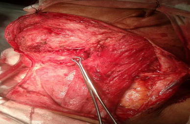
Neck dissection showing left level II lymph node adherent to IJV
No Carotid rupture was reported in our study (0%). As we go through literature, The reasons quoted behind Carotid artery rupture are, wound break down because of previous irradiation, secondary infection and the poor metabolic condition of the patient. It is a fatal complication resulting in immediate mortality if not intervened immediately.
To avoid the same, following prophylactic measures were taken in our study.
Avoided flap necrosis
Avoided salivary fistula (explained above)
Avoided wound infection
And along with these measures, the carotid artery was with muscles before closure.
None of our the patients developed tracheal stomal stenosis (0%), which was less compare to many studies, a study done by Kazi Atikuzzaman et al. [11]. Here, one patient (6.6%) developed tracheostomal stenosis (Fig. 5).
Fig. 5.
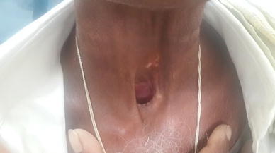
Post laryngectomy tracheal stoma
The reason behind it was
The technique which we were used while we making the stoma and the stoma care after that (explained above).
- Stoma care
- Check your stoma: Check around the stoma and inside the trachea for mucus and crusts. You may need to use a flashlight to check your stoma
- Clean your stoma: The skin around your stoma needs to be clean and free from secretions to avoid irritation and problems. Humidification and saline spray
- Cover your stoma: Covering your stoma helps keep it clean and healthy by preventing dust particles from entering your airway. It also retains the warmth in the air you breathe
- Tracheostomy tube care.
In our study, none of the patients (0%) developed deep vein thrombosis, After going through literature, DVT is mostly observed in elderly age group patients, where surgeries are lasting for more duration, prolong bedridden patients, and patients with the previous history of deep vein thrombosis, pulmonary embolism, myocardial infarction, and thrombophilia.
The most probable reasons behind this being
Most of our patients were below 65 years
In our study, the average time taken for total laryngectomy with selective neck dissection was around is around 210–260 min and total laryngectomy with partial pharyngectomy with RND is around 310–340 min
After immediate post operative day patients were provided with passive leg exercises
Patients were mobilized immediately from 2nd post operative day.
None of our patients (0%) developed late complication like lymphedema, recurrence. As like literature tells, the reason for lymphedema, When both the internal jugular veins are ligated, lymphedema often follows owing to interruption of the lymphatic drainage channels from the head. Lymphedema generally occurs when venous return is compromised after bilateral radical ND; rarely, it is massive and accompanied by cerebral edema and even death.
So we routinely attempted to preserve
At least one of the internal jugular veins and
External jugular vein; so there is no complication even after bilateral Radical Neck dissection, and there were no cerebral complications.
The late complications in this study was hypothyroidism (25 patients) (Fig. 6), Among 25 Hypothyroidism patients majority were total thyroidectomy with radiotherapy i.e. 13 patients (52%) followed by hemithyroidectomy with radiotherapy 8 patients (32%) and total thyroidectomy alone in 4 patients (16%). This suggests the chance of hypothyroidism was more in those who underwent radiotherapy with total thyroidectomy, which was addressed with regular thyroid function tests and kept on appropriate thyroxine dose.
Fig. 6.
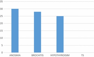
Sequelae of laryngectomy
So Attention was given
To Preserving intact the vasculature of the contralateral thyroid lobe whenever it is necessary.
To remove the ipsilateral thyroid lobe during a laryngectomy.
Proper postoperative assessment of thyroid gland function is desirable in all these patients especially those who undergoing total thyroidectomy with post op radiotherapy, to identity those at risk of hypothyroidism and to avoid unnecessary morbidity.
Tracheobronchitis is the late sequelae of laryngectomy, only few patients developed Tracheobronchitis in our study. Tracheobronchitis is common in patients with an altered airway due to the lack of normal humidification by the mouth and nose. Mucous membranes lining the trachea become dry from exposure and secretions become crusty. The Methods we followed to increase stomal and tracheal humidity include
Applying humidified oxygen via a tracheostomy collar instilling 3–5 ml of normal saline into the laryngectomy stoma as needed.
Applying a moistened cover over the laryngectomy stoma.
Conclusion
Laryngectomy is a procedure which is associated with many complications in the earlier days, the incidence of complications has come down with growing expertise among otolaryngologists. Even though we encountered some minor complications in the postoperative period and were successful in managing all of them conservatively. The most common complications after total laryngectomy with or without partial pharyngectomy with neck dissection in our study were wound infection and pharyngocutaneous fistula.
Assessment of risk factors, early recognition of complications with good pre operative, intra operative and post operative protocols along with improvised techniques are necessary to reduce the complication after total laryngectomy with or without partial pharyngectomy.
None the less, many more prospective studies and medical audit of the cases should be done to identify the incidence and reasons for the complications to improve the outcome of the surgery which helps in reducing the mortality and morbidity associated with the procedure.
References
- 1.Gollo A, Moi R, Simonelli M, et al. Salvage resection after previous laryngeal surgery: total laryngectomy with en bloc resection of the overlying cervical skin. Arch Otolaryngol Head Neck Surg. 2001;127:786–789. [PubMed] [Google Scholar]
- 2.Lagier A, et al. The influence of age on postoperative complications after total laryngectomy or pharyngolaryngectomy. Eur J Surg Oncol. 2014;40(2):202–207. doi: 10.1016/j.ejso.2013.09.010. [DOI] [PubMed] [Google Scholar]
- 3.Muscat JE, Wynder EL. Tobacco, alcohol, asbestos, and occupational risk factors for laryngeal cancer. Cancer. 1992;69:2244–2251. doi: 10.1002/1097-0142(19920501)69:9<2244::AID-CNCR2820690906>3.0.CO;2-O. [DOI] [PubMed] [Google Scholar]
- 4.Fiorini FR, et al. Tobacco exposure and complications in conservative laryngeal surgery. Cancers (Basel) 2014;6(3):1727–1735. doi: 10.3390/cancers6031727. [DOI] [PMC free article] [PubMed] [Google Scholar]
- 5.Arsenijevic S, et al. Demographic characteristics of patients with laryngeal cancer and their socio-economic status. J BUON. 2010;15(1):131–135. [PubMed] [Google Scholar]
- 6.Schwartz SR, Yueh B, Maynard C, Daley J, Henderson W, Khuri SF. Predictors of wound complications after laryngectomy: a study of over 2000 patients. Otolaryngol Head Neck Surg. 2004;131(1):61–68. doi: 10.1016/j.otohns.2003.08.028. [DOI] [PubMed] [Google Scholar]
- 7.Reddy SL, Reddy SD, Prasad AVVSL. A prospective study of total laryngectomy and associated complications. Int J Phonosurg Laryngol. 2012;2(1):20–22. doi: 10.5005/jp-journals-10023-1029. [DOI] [Google Scholar]
- 8.Pellini R, et al. Predictive factors for postoperative wound complications after neck dissection. Acta Otorhinolaryngol Ital. 2013;33(1):16–22. [PMC free article] [PubMed] [Google Scholar]
- 9.Markou KD, Vlachtsis KC, Nikoolau AC, Petridids DG, Koulouas AI, Danilidis IC. Incidence and predisposing factors of pharyngocutaneous fistula formation of total laryngectomy. Eur Arch Otorhinolaryngol. 2004;261(2):61–67. doi: 10.1007/s00405-003-0643-6. [DOI] [PubMed] [Google Scholar]
- 10.Maharajan R, et al. Early complications of total laryngectomy: a retrospective study. Nepal J ENT Head Neck Surg. 2010;1(2):17–18. doi: 10.3126/njenthns.v1i2.4759. [DOI] [Google Scholar]
- 11.Atikuzzaman K, et al. Postoperative complications and its management after total laryngectomy. Bangladesh J Otorhinolaryngol. 2013;19(2):82–86. doi: 10.3329/bjo.v19i2.16366. [DOI] [Google Scholar]