1.
Dear Editors,
Mucinous adenocarcinoma of the abdomen is a rare disease, usually caused by the abdominal metastasis of tumours. Here, we report a case of mucinous adenocarcinoma of the abdomen due to appendiceal mucinous adenocarcinoma (MA).
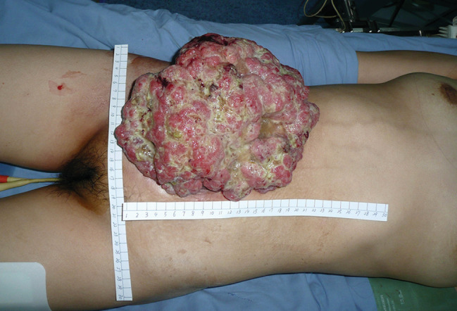
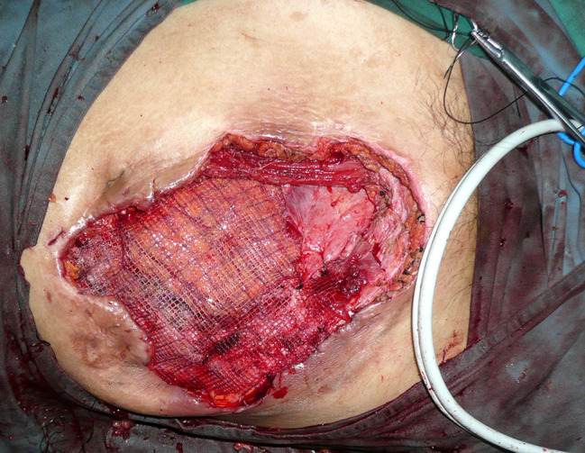
A 36‐year‐old woman presented to the hospital with a large tumour in the right lower quadrant of the abdomen. The patient had undergone an ileocecectomy for an MA 3 years previously. However, 2 years later, a tumour, which was gradually increasing in size, appeared along the incision line. Physical examination revealed a 20 × 23 cm cauliflower‐shaped mucinous neoplasm with tenderness in the right lower quadrant of the abdomen (Figure 1). Laboratory examination revealed a low haemoglobin concentration of 82 g/L. Computed tomography of the abdomen uncovered a mass in the right lower quadrant invading the abdominal wall and part of the intestine. After transfusion of two units of red blood cells, the haemoglobin increased to 95 g/L. Surgical excision of the abdominal wall and part of the intestine where tumour infiltration had occurred was performed. There was a large coloboma in the right lower abdomen; therefore, a patch graft procedure was paved (Figure 2) and the abdominal wall was closed. A diagnosis of MA of the abdomen was made on the basis of the histopathological report. The patient was followed‐up and succumbed to metastases due to MA of the abdomen during the 11th month of follow‐up (informed consent was obtained from the patient to publish these images).
Figure 1.

A 20 × 23 cm cauliflower‐shaped mucinous neoplasm in the right lower quadrant of the abdomen
Figure 2.

A patch graft procedure was paved in the right lower quadrant of the abdomen
MA is an extraordinarily rare disease in general surgery, which has an insidious onset and there is no obvious symptoms at the early stage. The disease usually presents as acute appendicitis. The treatment strategy includes completely surgical resection intraoperatively and adjuvant chemotherapy postoperatively.1 How to select the type of operation? When carcinoma is localised in the mucosa, a simple appendectomy is enough; while the Carcinoma infiltrated to whole cecum wall or regional lymph nodes, a right hemicolectomy including omentectomy is needed.2 Spontaneous rupture or iatrogenic rupture of MA could lead to serious outcome such as implanted metastases to peritoneal cavity and abdominal wall. The present case most probably suffered from a rupture. Therefore, faced to the MA, doctor should pay attention to the integrity of carcinoma.
CONFLICT OF INTEREST
The authors declare that they have no competing interests.
FINANCIAL DISCLOSURES
National Natural Science Foundation of China, Grant/Award Numbers: 81701965, 81872255; Natural Science Foundation of Liaoning Province, Grant/Award Number: 201800199; Dalian Medical Science Research Project Grant/Award Number: 1711038
REFERENCES
- 1. Hajiran A, Baker K, Jain P, Hashmi M. Case of an appendiceal mucinous adenocarcinoma presenting as a left adnexal mass. Int J Surg Case Rep. 2014;5:172‐174. [DOI] [PMC free article] [PubMed] [Google Scholar]
- 2. Sugarbaker PH. New standard of care for appendiceal epithelial neoplasms and pseudomyxoma peritonei syndrome? Lancet Oncol. 2006;7:69‐76. [DOI] [PubMed] [Google Scholar]


