Abstract
The purpose of this study was to investigate the relation between external and internal load and the response of the patellar tendon structure assessed with ultrasound tissue characterization (UTC) in elite male volleyball players during preseason. Eighteen players were followed over 7 weeks, measuring four load parameters during every training and match: volume (minutes played), rating of perceived exertion (RPE) (ranging from 6 to 20), weekly load (RPE*volume), and jump frequency (number of jumps). Patellar tendon structure was measured biweekly using UTC, which quantifies tendon matrix stability resulting in four different echo types (I‐IV). On average, players spent 615 min/wk on training and matches with an RPE of 13.9 and a jump frequency of 269. Load evaluation shows significant changes over the 7 weeks: Volume and weekly load parameters were significantly higher in week 3 than week 7 and in week 4 than week 2. Weekly load performed in week 4 was significantly higher than week 7. No significant changes were observed in tendon structure. On the non‐dominant side, no significant correlations were found between changes in load parameters and echo types. At the dominant side, a higher weekly volume and weekly load resulted in a decrease of echo type I and a higher mean RPE in an increase of echo type II. The results of this study show that both external and internal load influence changes in patellar tendon structure of elite male volleyball players. Monitoring load and the effect on patellar tendon structure may play an important role in injury prevention.
Keywords: athletes, imaging, jumper’s knee, periodization, tendinopathy
1. INTRODUCTION
Volleyball players are frequently affected by knee problems, including overload injuries such as patellar tendinopathy (PT).1, 2 PT has a high prevalence among both non‐elite and elite volleyball athletes (14%, 45%).3, 4 The rates are almost twice as high for male players than for female players.4 The fact that this injury can affect athletes’ ability to perform optimally4 and the poor clinical improvement after treatment5 stresses the importance of prevention.
Literature has identified some evidence supporting intrinsic factors such as gender, weight, and body mass index (BMI) as risk factors for developing PT.6, 7 Extrinsic risk factors, including player position2 and training load,8 are also risk factors for developing PT. Of all risk factors, training load (such as training/competition volume) seems to be the most important.8 Visnes et al8 showed that one extra training hour or one extra set per week significantly increased the risk for patellar tendinopathy in young players. This was likely due to the increased exposure to jumping loads. Additionally, the ratio of acute training load (load of the last week) and chronic load (4‐week rolling average of load), called acute:chronic load ratio (ACWR), is reported to have an effect on the risk of injury.9 In team sports, assessment of internal training load (the psycho‐physiological response to this load) is also relevant as player's exposure to the load dosage (external load) may be consistent across players of the same team.10, 11, 12 Hence, to monitor and control training load, it is necessary to have a measure of both external and internal load.12
The effect of load on tendons structure can either be positive (tendon adaptation) or be negative (tendon maladaptation).13 According to the tendon continuum model, proposed by Cook and Purdam,14 there will be tendon adaptation if an optimized load is applied to the tendon. Too much training load without an appropriate period of rest between sessions can lead to changes in tendon structure, resulting in reactive tendinopathy, tendon disrepair, and even degenerative tendon abnormalities.14 Unfortunately, the exact turning point for the amount of tendon loading, where physiological adaptation changes into pathological adaptation, remains unclear.14 This fact highlights the necessity of investigating changes on tendon structure with new imaging techniques of subjects who are exposed to high amount of load.
Ultrasound tissue characterization is one of the several ultrasound‐based imaging techniques used to detect load‐related changes in tendon structure.15, 16 This equipment quantifies the structure of the tendon by calculating percentages of four echo types from echo type I, which represents highly stable structures, to echo type IV, representing amorphous structures. The quantification of tendon structure offers the possibility to monitor subtle changes and thereby address the limitations of conventional imaging techniques.17
Previous studies investigating the relation between load and changes in patellar tendon structure did not measure load accurately.18 To the best of our knowledge, there is only one study that investigated changes in patellar tendon structure in relation to the amount of load performed.19 These authors followed Australian football players during the preseason and found that only two load parameters (monotony and total distance) may have had small positive effects on the changes in structure of the non‐dominant patellar tendon. More research is therefore needed to understand the relation between load and changes in patellar tendon structure.
The aim of this study was to investigate the relation between external and internal load and the response of the patellar tendon structure as assessed with UTC in male volleyball players during preseason.
2. METHODS
2.1. Study design and participants
A total of 18 male volleyball players from two teams of the top‐two Dutch volleyball leagues were recruited for this study. Before the start of preseason, all players completed a questionnaire about age, playing position, and present and past injuries; height, weight, and body fat percentage were measured. During 7 weeks, the load was monitored and UTC scans were performed every 2 weeks. Load and UTC measurements are shown in detail in Figure 1.
Figure 1.

Load and UTC measurements over the 7‐week preseason
Permission from the coaches was obtained, and all participants gave informed consent after being fully informed about the study. This study was approved by the ethical committee of the Center for Human Movement Sciences, University of Groningen, University Medical Center, Groningen (UMCG), The Netherlands.
2.2. Load monitoring
Both external and internal loads were monitored over the 7 weeks during training (gym and volleyball practice) and matches. The total of 7 weeks was chosen based on the preseason duration of the two volleyball teams included in this study, which is similar to previous studies.20, 21 While external load measures an athlete's training or competition load, internal load assesses the internal physiological and psychological response to the external load.10, 11, 12
2.3. External load
Two different external load measurements were performed: training volume and jump frequency (number of jumps). Training volume was defined as the total duration (minutes) of each training and match session. At the end of each session, players were asked to fill out a training log, which includes the duration of the session.
To count the jump frequency, authors applied two different methods. Firstly, jump frequency was measured using the Zephyr BioHarness 3TM (Zephyr Technology), which contains a triaxial accelerometer (100 Hz). Jumps were counted based on pattern recognition in the vertical acceleration data. Acceleration characteristics were linked to the three stages of a jump (take‐off, flight, and landing). The flight stage was identified as the area between the peaks in the vertical acceleration, which occurs shortly before take‐off and shortly after landing (acceleration between these peaks is close to zero). These phases were detected using a customized algorithm created in MATLAB (MathWorks) to identify jumps. Spike jumps, block jumps, setting jumps, and serve jumps were included. Low‐intensity hop jumps were excluded.
Secondly, all training sessions and matches were videotaped in case of missing data from the Zephyr Bioharness 3 and during tournaments abroad. In both cases, the videos were reviewed and the jumps were manually counted. Percentage agreement between estimated jumps from the accelerometer and data obtained by video analysis was calculated. The agreement ranged from 88.2% to 100%. Weekly volume and weekly jump frequency were defined as the sum of the load and jump frequency, respectively, for all training and match sessions.
2.4. Internal load
Internal load was measured using the rating of perceived exertion (RPE), defined as the perceived intensity level of a physical activity.22 The session RPE was taken 30 minutes after each training or match to ensure that the perceived exertion captured the entire session and did not refer to the last part only.22 Load was calculated by the product of the session RPE and volume of the training or match.23, 24 The sum of all training and match loads for each week determined the weekly load. This method has been shown to be valid for quantifying training and match load.22, 23
2.5. Ultrasound tissue characterization (UTC)
Structure of the patellar tendon was quantified using UTC. A standardized protocol was used to take the UTC scans over the 7‐week period. Participants lay on a treatment bench with their knee angled in approximately 100° flexion. An ultrasound probe (SmartProbe 12L5‐V; Terason 2000+; TeraTech) was affixed to a tracking device (UTC Tracker; UTC Imaging) to standardize transducer tilt angle. The tracker device moves the probe automatically over the length of the tendon, capturing transverse images every 0.2 mm. The ultrasound probe in the tracking device was placed perpendicular to the long axis of the tendon, moving from proximal to distal. All scans were performed at the beginning of the week and before training, to avoid bias of short‐term adaptations of training. Both patellar tendons were scanned for each participant. All scans were taken by a single examiner who was experienced with UTC (LMR).
The consecutive transversal images were used to create 3D reconstructions. Consistency of intensity and distribution of gray images were calculated over a distance of 4.8 mm using UTC algorithms. Four echo types can be discriminated based on consistency, with echo type I representing the most stable pattern and echo type IV the least stable.17 Tendon structure was quantified by calculating the percentages of these four echo types in multiple regions of interest (ROIs) placed around the border of the tendon in the transverse view. ROIs were selected at intervals no >5 mm from the apex of the patella to 20 mm distally.25, 26 Contours were drawn every 5 mm, and window size 17 was used for imaging analysis. All tendons contours were marked by one investigator (LMR). Poor‐quality scans were excluded.
2.6. Statistical analysis
SPSS (version 23) was used for all statistical analyses. Descriptive statistics (mean and SD) were calculated for the load parameters (weekly training volume, training intensity, load, and jump frequency) and echo type percentages. ACWR was calculated over preseason weeks 4‐7 by dividing the acute workload (load of week 7) by the chronic load (mean of weeks 4‐7).9 To determine the dominant side, athletes were asked: “If you would shoot a ball on a target, which leg would you use to shoot the ball?” This question is appropriate to determine leg dominance in bilateral mobilizing task.27
Completion of training logs was implemented with 99% compliance, and 98% of jump frequency data were available. Missing data were caused by players who forgot to fill in their RPE score and a technical failure of the video camera during a friendly match. In case of missing values in the training log, data were replaced with the weekly average volume and RPE score if at least 75% data within that week were available.28 Missing jumps were replaced with the mean jump frequency per set (calculated from the previously played match), multiplied by the number of played sets of that match for each player separately.29 Sixteen participants completed all five scans, and two participants were unable to be present at one of the scan sessions for personal reasons. Six UTC scans were excluded because of poor quality. A total of 77 scans were included.
A simple t test was used to compare the non‐dominant and dominant sides for tendon structure expressed in the four echo type percentages. To determine the changes in load parameters and echo types over preseason, a general linear model (GLM) analysis was performed, with the Bonferroni correction (P ≤ 0.01). Linear regression analysis was used to investigate the association between the sum of the load (independent) during the 7‐week preseason with the changes between the final (week 7) and the second (week 2) UTC measurement (dependent), adjusting for UTC baseline (week 1) and for load variation over the weeks (Figure 2). Additionally, linear regression analysis was used to investigate the association between the ACWR of week 7 and the changes in UTC.9
Figure 2.

Statistical model used in the manuscript to investigate the relation between load (over 7‐wk) and changes on tendon structure
3. RESULTS
During the 7 weeks, a total of 88 training sessions and matches were monitored. Athletes practiced six times a week on average, including weight training, volleyball training, and combined training sessions. Players spent a mean of 615 (SD 134) minutes on training and matches per week with a mean jump frequency of 269 (SD 90) per week. Mean RPE was 13.9 (SD 0.7), indicating “somewhat hard” intensity.
3.1. Players’ characteristics
The anthropometric characteristics of the players are presented in Table 1. Players of five different field positions were included in this study: setter (6), outside hitter (4), opposite (3), middle hitter (3), and libero (2). Of the 18 players included, six had been previously diagnosed with PT (4 dominant side and 2 bilateral symptoms). Three of them had actual symptoms at the beginning of preseason (2 dominant side and 1 bilaterally).
Table 1.
Participant characteristics at baseline
| Measure | Mean (N = 18) |
|---|---|
| Anthropometrics characteristics | |
| Age (y) | 23.0 (4.3) |
| Height (m) | 1.9 (0.0) |
| Weight (kg) | 89.8 (9.6) |
| BMI (kg/m2) | 23.1 (2.1) |
| Ultrasound tissue characterization | |
| Echo type I (%) | |
| Non‐dominant | 65.1 (9.5) |
| Dominant | 65.7 (9.1) |
| Echo type II (%) | |
| Non‐dominant | 31.4 (6.8) |
| Dominant | 30.7 (5.8) |
| Echo type III (%) | |
| Non‐dominant | 2.5 (2.9) |
| Dominant | 2.5 (3.4) |
| Echo type IV (%) | |
| Non‐dominant | 1.0 (1.3) |
| Dominant | 1.0 (1.5) |
Displayed values are means (SD).
Abbreviations: kg, kilograms; m, meters; m2, square meters; VISA‐P, Victorian Institute of Sport Assessment‐Patella.
3.2. Load parameters
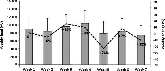
Means and SD of all load parameters for each week are shown in Table 2. Weekly volume and weekly load parameters were significantly higher in week 3 than week 7 and in week 4 than week 2. Moreover, the weekly load performed in week 4 was significantly higher than that of week 7. No significant changes over the weeks were observed for RPE or weekly jumps frequency parameters. The ACWR in week 7 for measures of athletic load (mean RPE, weekly volume, weekly load, and jumps) ranged from 0.87 to 1.00. Weekly load and relative weekly changes, in percentages, are presented in Figure 3.
Table 2.
Mean and SD of the load parameters
| Weekly volume (min) | Mean RPE score (6‐20) | Weekly load (AU) | Weekly jump frequency (jumps) | |
|---|---|---|---|---|
| Week 1 | 638 (193) | 14.1 (1.0) | 8976 (2741) | 262 (84) |
| Week 2 | 599 (211)b | 13.8 (1.1) | 8398 (3211)b | 265 (93) |
| Week 3 | 687 (207)a | 13.8 (1.1) | 9587 (3010)a | 293 (131) |
| Week 4 | 717 (208) | 14.2 (1.0) | 10 437 (3257)a | 320 (132) |
| Week 5 | 559 (199) | 13.6 (1.1) | 7897 (3066) | 229 (132) |
| Week 6 | 590 (207) | 13.7 (1.4) | 8419 (3180) | 252 (125) |
| Week 7 | 522 (162) | 14.0 (1.1) | 7433 (2467) | 266 (138) |
| SUM | 4312 (940) | 13.9 (0.7) | 61 148 (13 945) | 18 086 (634) |
Abbreviations: AU, arbitrary units; RPE, rate of perceived exertion.
Significant difference compared to week 7.
Significant difference compared to week 4.
Figure 3.

Means and standard deviations and weekly change (%) of weekly load
3.3. Tendon structure
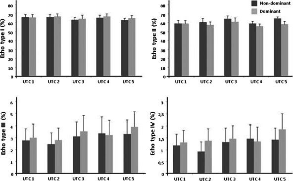
No significant differences in echo type percentages between the non‐dominant and dominant sides were observed at the beginning of the preseason period (echo type I, P = 0.628; echo type II, P = 0.705; echo type III, P = 0.389; and echo type IV, P = 0.455) or at the end (echo type I, P = 0.735; echo type II, P = 0.136; echo type III, P = 0.529; and echo type IV, P = 0.648). For the changes in percentages of the four different echo types over the 7 weeks, no significant changes were observed for the dominant or non‐dominant side (Figure 4).
Figure 4.

Echo type percentages over the 7‐week preseason
3.4. Relation between load parameters and changes in patellar tendon structure
No significant correlations were found between load and changes in echo types (I‐IV) for the non‐dominant side. For the dominant side, higher cumulative weekly volumes and loads resulted in a significant decrease of echo type I (weekly volume: standardized coefficient = –0.588, P = 0.020; weekly load: standardized coefficient t = –0.0.586, P = 0.022). A higher mean RPE resulted in a significant increase of echo type II (mean RPE: standardized coefficient = 0.681, P = 0.031). No significant correlations between load parameters and changes in echo types III and IV were found. No significant correlations between ACWR parameters of week 7 (mean RPE, weekly volume, weekly load, and jumps) and changes in echo types (I‐IV) were observed.
4. DISCUSSION
The present study investigated the relation between load performed over seven weeks of preseason and changes in patellar tendon structure of male elite volleyball players. This is the first study to compare amount of load (measured during every training and match) and changes in patellar tendon structure (measured biweekly). At the non‐dominant side, there was no significant relation between all load parameters investigated and percentage of echo types (I‐IV). At the dominant side, higher cumulative weekly volume and weekly load were related to a significant decrease of echo type I percentage. A higher mean RPE was related to an increase of echo type II percentage. No significant relations were observed between load and percentages of echo types III and IV at the dominant side.
The patellar tendon structure at the dominant side showed a relation with a higher amount of load, decreasing echo type I, and increasing echo type II percentages, findings that might suggest a downward shift in the continuum model.14 This is the first study to observe such changes in the patellar tendon structure of volleyball players. Our findings are in line with previous results among Australian football players demonstrating that load (monotony and high‐intensity running) is related to positive effects in the patellar tendon structure.19 It should be noted, however, that Australian football players are a population with different jump characteristics30 and a lower risk of patellar tendinopathy31 than volleyball players. By contrast, van Ark et al (2015) observed no significant changes in the tendon structure of young volleyball players during a 5‐day tournament. Possible explanations might be that the authors did not measure the load accurately and that the tournament only lasted 5 days, which was likely not long enough to cause tendon adaptation/maladaptation. We speculate that the changes observed in the present study occur as a short‐term adaptation to overload. The tendon still has the potential to revert to normal if sufficient recovery time is given and further training load is balanced.32
It is notable that the players were exposed to only subtle changes in load during preseason, and they performed a low amount of load over those 7 weeks. The athletes jumped much less (weekly jump frequency range 228‐319 jumps) compared to a previous study observing that athletes jumped up to 300 times in one single match.33 The first explanation for the low load performed would be the fact that this study was conducted during the preseason, when athletes are returning from a low level of activities (holiday). Another explanation could be that because of this study, coaches and medical staff were more aware of the risks of injuries such as patellar tendinopathy. As we demonstrated, no spikes in load were observed during preseason.
Regarding the results of the internal and external load measurements, we observed that both load measurements were related to changes in patellar tendon structure. Although both load measurements were related to the changes in echo type I, only internal load was related to changes in echo type II. These results are in line with previous findings (in a population of cricket athletes) that internal load is twice as predictive for risk of injury as external loads.34 However, evidence exists that both internal and external load indicators are related to overuse injuries.29 It is important to point out that external load does not include individual characteristics of players and is thereby limited in its quantification of the actual training.13 Hence, our results support that internal and external load should be monitored to better understand changes in patellar tendon structure, based on the fact that different load measurements were able to detect different changes in echo types.
The low amount of load performed during the 7‐week preseason might explain the non‐significant changes in patellar tendon structure (echo types I‐IV). One explanation for these results is that coaches and medical staff allowed sufficient rest to the tendon. In this way, the tendon was not heavily overloaded and no structural changes occurred. Another explanation for our findings may be that 7 weeks is too short to observe significant changes in tendon structure. A previous study that followed Australian football players for a longer period of time (16‐week preseason) observed an improvement of the patellar tendon structure at the end of the preseason.19 However, as discussed previously, the differences between sport modalities should be taken in consideration.
Another interesting finding in this study is that the structure of the patellar tendon in the dominant side behaved differently than the non‐dominant side—possibly because tendon structure may be influenced not only by jump frequency but also by the biomechanics involved. For example, during landing the knee is in external rotation on the non‐dominant side and in internal rotation on the dominant side.35 This is in line with our results that show a significant relation between load and changes in tendon structure on the dominant side—which as a result of these different biomechanical features of landing may explain the dominant/non‐dominant variation.
This is the first study to investigate the relation between load (weekly) and changes in patellar tendon structure (biweekly) of male elite volleyball players. In contrast to previous studies, we examined the relationship between changes in load and tendon structure considering the variation in load over the study period and the baseline UTC measurement. Taking load variation into consideration during the analysis is important as changes in load over the period investigated may influence the tendon structure. Load changes that occur during the period investigated might increase the risk for developing overuse injuries, such as patellar tendinopathy.9, 36 Moreover, the UTC baseline measurement is important to consider the tendon structure prior to preseason loading. This study also presents some limitations that should be considered when interpreting the results. Firstly, athletes with and without previous or current symptoms were included. To our knowledge, there is no study using UTC to investigate the different responses to load of subjects with and without symptoms. One study that investigated changes in tendon structure with grayscale US observed that tendons without hypoechoic areas (only 24% were painful) showed similar response to load compared to tendons with hypoechoic areas (59% were painful), developing diffuse thickening.37 However, this should be interpreted with caution since the US might be not sensitive to subtle changes in tendon structure as UTC. Secondly, the load performed on the day previous to the UTC measurement could have influenced the results.38 In Australian football players, the Achilles tendon structure changed significantly 2 days post‐game (reduction in echo type I) and returned to baseline 4 days post‐game.38 However, other studies involving novice runners and much lower running volumes did not observe this Achilles tendon response at 2 and 7 days post‐activity.18, 39 The acute tendon response to load might depend on different factors, including the amount of load players are exposed to and the tendon load capacity. To minimize this effect, the UTC measurements were performed at the beginning of the week and players refrained from physical activity on the day of measurement. Thirdly, no distinction between jump height or between single‐ and double‐leg jumps was made in this study. It is known that players who jump higher expose their tendons to a higher load.40 Further research could explore the link between jump height and changes in tendon structure.41
5. PERSPECTIVE
This study emphasizes the importance of monitoring load, which could play an important role in preventing overload injuries. Given its relevance for elite volleyball players, research into the relation between load and changes in tendon structure is of great interest for athletes, medical staff, and coaches. Using imaging tests in combination with load measurement might provide additional information about the risk for developing patellar tendinopathy. This study also showed that measuring internal load in addition to external load may provide valuable insights into the relationship of load to tendon in this population. More research is needed to better understand whether the transient changes between echo types I and II are a positive adaptation or a maladaptation of the tendon to the load, and to investigate the use of UTC in a larger sample to differentiate the response to load of different subgroups (with and without symptoms, for example) plus include jump height. Moreover, future research should focus on monitoring players during the season using UTC to gain more information about how this affects tendon structure.
ACKNOWLEDGEMENTS
This work was supported by CNPq, National Council for Scientific and Technological Development—Brazil. The authors would like to thank the athletes and coaches for their assistance in the execution of this study.
Rabello LM, Zwerver J, Stewart RE, van den Akker‐Scheek I, Brink MS. Patellar tendon structure responds to load over a 7‐week preseason in elite male volleyball players. Scand J Med Sci Sports. 2019;29:992–999. 10.1111/sms.13428
REFERENCES
- 1. Ferretti A, Papandrea P, Conteduca F. Knee injuries in volleyball. Sport Med. 1990;10(2):132‐218. [DOI] [PubMed] [Google Scholar]
- 2. Bere T, Kruczynski J, Veintimilla N, Hamu Y, Bahr R. Injury risk is low among world‐class volleyball players: 4‐year data from the FIVB injury surveillance system. Br J Sports Med. 2015;49(17):1132‐1137. [DOI] [PMC free article] [PubMed] [Google Scholar]
- 3. Zwerver J, Bredeweg SW, van den Akker‐Scheek I. Prevalence of jumper’s knee among nonelite athletes from different sports. Am J Sports Med. 2011;39(9):1984‐1988. [DOI] [PubMed] [Google Scholar]
- 4. Lian Ø, Refsnes P‐E, Engebretsen L, Bahr R. Prevalence of jumper’s knee among elite athletes from different sports. Am J Sports Med. 2005;31(3):408‐413. [DOI] [PubMed] [Google Scholar]
- 5. van Rijn D, van den Akker‐Scheek I, Steunebrink M, Diercks RL, Zwerver J, van der Worp H. Comparison of the effect of 5 different treatment options for managing patellar tendinopathy. Clin J Sport Med. 2017. [Epub Ahead of print]. [DOI] [PubMed] [Google Scholar]
- 6. de Vries AJ, van der Worp H, Diercks RL, van den Akker‐Scheek I, Zwerver J. Risk factors for patellar tendinopathy in volleyball and basketball players: a survey‐based prospective cohort study. Scand J Med Sci Sport. 2015;25(5):678‐684. [DOI] [PubMed] [Google Scholar]
- 7. van der Worp H, van Ark M, Roerink S, Pepping G‐J, van den Akker‐Scheek I, Zwerver J. Risk factors for patellar tendinopathy: a systematic review of the literature. Br J Sports Med. 2011;45(5):446‐452. [DOI] [PubMed] [Google Scholar]
- 8. Visnes H, Bahr R. Training volume and body composition as risk factors for developing jumper’s knee among young elite volleyball players. Scand J Med Sci Sport. 2013;23(5):607‐613. [DOI] [PubMed] [Google Scholar]
- 9. Gabbett TJ. The training‐injury prevention paradox: should athletes be training smarter and harder? Br J Sports Med. 2016;50(5):273‐280. [DOI] [PMC free article] [PubMed] [Google Scholar]
- 10. Bangsbo J. The physiology of soccer—with special reference to intense intermittent exercise. Acta Physiol Scand Suppl. 1994;619:1‐155. [PubMed] [Google Scholar]
- 11. Vanrenterghem J, Nedergaard NJ, Robinson MA, Drust B. Training load monitoring in team sports: a novel framework separating physiological and biomechanical load‐adaptation pathways. Sport Med. 2017;47(11):2135‐2142. [DOI] [PubMed] [Google Scholar]
- 12. Impellizzeri FM, Rampinini E, Marcora SM. Physiological assessment of aerobic training in soccer. J Sports Sci. 2005;23(6):583‐592. [DOI] [PubMed] [Google Scholar]
- 13. Drew MK, Finch CF. The relationship between training load and injury, illness and soreness: a systematic and literature review. Sport Med. 2016;46(6):861‐883. [DOI] [PubMed] [Google Scholar]
- 14. Cook JL, Purdam CR. Is tendon pathology a continuum? A pathology model to explain the clinical presentation of load‐induced tendinopathy. Br J Sports Med. 2009;43:409‐416. [DOI] [PubMed] [Google Scholar]
- 15. Malliaras P, Cook J. Changes in anteroposterior patellar tendon diameter support a continuum of pathological changes. Br J Sports Med. 2011;45(13):1048‐1051. [DOI] [PubMed] [Google Scholar]
- 16. Malliaras P, Cook J. Patellar tendons with normal imaging and pain: change in imaging and pain status over a volleyball season. Clin J Sport Med. 2006;16(5):388‐391. [DOI] [PubMed] [Google Scholar]
- 17. van Schie H, de Vos RJ, de Jonge S, et al. Ultrasonographic tissue characterisation of human Achilles tendons: quantification of tendon structure through a novel non‐invasive approach. Br J Sports Med. 2010;44(16):1153‐1159. [DOI] [PubMed] [Google Scholar]
- 18. van Ark M, Docking SI, van den Akker‐Scheek I, et al. Does the adolescent patellar tendon respond to 5 days of cumulative load during a volleyball tournament? Scand J Med Sci Sport. 2016;26(2):189‐196. [DOI] [PubMed] [Google Scholar]
- 19. Esmaeili A, Stewart AM, Hopkins WG, Elias GP, Aughey RJ. Effects of training load and leg dominance on achilles and patellar tendon structure. Int J Sports Physiol Perform. 2017;12:122‐126. [DOI] [PubMed] [Google Scholar]
- 20. Trajković N, Milanović Z, Sporis G, Milić V, Stanković R. The effects of 6 weeks of preseason skill‐based conditioning on physical performance in male volleyball players. J Strength Cond Res. 2012;26(6):1475‐1480. [DOI] [PubMed] [Google Scholar]
- 21. Newton RU, Kraemer WJ, Häkkinen K. Effects of ballistic training on preseason preparation of elite volleyball players. Med Sci Sports Exerc. 1999;31(2):323‐330. [DOI] [PubMed] [Google Scholar]
- 22. Impellizzeri FM, Rampinini E, Coutts AJ, Sassi A, Marcora SM. Use of RPE‐based training load in soccer. Med Sci Sports Exerc. 2004;36(6):1042‐1047. [DOI] [PubMed] [Google Scholar]
- 23. Foster C. Monitoring training in athletes with reference to overtraining syndrome. Med Sci Sports Exerc. 1998;30(7):1164‐1168. [DOI] [PubMed] [Google Scholar]
- 24. Kenttä G, Hassmén P. Overtraining and recovery. A conceptual model. Sport Med. 1998;26(1):1‐16. [DOI] [PubMed] [Google Scholar]
- 25. Rudavsky A, Cook J, Magnusson SP, Kjaer M, Docking S. Characterising the proximal patellar tendon attachment and its relationship to skeletal maturity in adolescent ballet dancers. Muscles Ligaments Tendons J. 2017;7(2):306‐314. [DOI] [PMC free article] [PubMed] [Google Scholar]
- 26. van Ark M, Cook JL, Docking SI, et al. Do isometric and isotonic exercise programs reduce pain in athletes with patellar tendinopathy in‐season? A randomised clinical trial. J Sci Med Sport. 2016;19(9):702‐706. [DOI] [PubMed] [Google Scholar]
- 27. Van MN, Meddeler BM, Hoogeboom TJ, Nijhuis‐van M, Sanden D, Van Cingel R. How to determine leg dominance: the agreement between self‐reported and observed performance in healthy adults. PLoS ONE. 2017;12(12):e0189876. [DOI] [PMC free article] [PubMed] [Google Scholar]
- 28. Brink M, Nederhof E, Visscher C, Schmikli S, Lemmink K. Monitoring load, recovery, and performance in young elite soccer players. J Strength Cond Res. 2010;24(3):597‐603. [DOI] [PubMed] [Google Scholar]
- 29. Jaspers A, Kuyvenhoven JP, Staes F, Frencken W, Helsen WF, Brink MS. Examination of the external and internal load indicators’ association with overuse injuries in professional soccer players. J Sci Med Sport. 2018;21(6):579‐585. [DOI] [PubMed] [Google Scholar]
- 30. Laffaye G, Wagner PP, Tombleson T. Countermovement jump height: gender and sport‐specific differences in the force‐time variables. J Strength Cond Res. 2014;28(4):1096‐1105. [DOI] [PubMed] [Google Scholar]
- 31. Morton S, Williams S, Valle X, Diaz‐Cueli D, Malliaras P, Morrissey D. Patellar tendinopathy and potential risk factors: an international database of cases and controls. Clin J Sport Med. 2017;27(5):468‐474. [DOI] [PubMed] [Google Scholar]
- 32. Cook JL, Rio E, Purdam CR, Docking SI. Revisiting the continuum model of tendon pathology: what is its merit in clinical practice and research? Br J Sports Med. 2016;50(19):1187‐1191. [DOI] [PMC free article] [PubMed] [Google Scholar]
- 33. Bahr MA, Bahr R. Jump frequency may contribute to risk of jumper’s knee: a study of interindividual and sex differences in a total of 11,943 jumps video recorded during training and matches in young elite volleyball players. Br J Sports Med. 2014;48(17):1322‐1326. [DOI] [PubMed] [Google Scholar]
- 34. Hulin BT, Gabbett TJ, Blanch P, Chapman P, Bailey D, Orchard JW. Spikes in acute workload are associated with increased injury risk in elite cricket fast bowlers. Br J Sports Med. 2014;48(8):708‐712. [DOI] [PubMed] [Google Scholar]
- 35. Sinsurin K, Srisangboriboon S, Vachalathiti R. Side‐to‐side differences in lower extremity biomechanics during multi‐directional jump landing in volleyball athletes. Eur J Sport Sci. 2017;17(6):699‐709. [DOI] [PubMed] [Google Scholar]
- 36. Timoteo TF, Debien PB, Miloski B, Werneck FZ, Gabbett T, Filho M. Influence of workload and recovery on injuries in elite male volleyball players. J Strength Cond Res. 2018:1‐6. [Epub ahead of print] [DOI] [PubMed] [Google Scholar]
- 37. Malliaras P, Purdam C, Maffulli N, Cook J. Temporal sequence of greyscale ultrasound changes and their relationship with neovascularity and pain in the patellar tendon. Br J Sports Med. 2010;44(13):944‐947. [DOI] [PubMed] [Google Scholar]
- 38. Rosengarten SD, Cook JL, Bryant AL, Cordy JT, Daffy J, Docking SI. Australian football players’ Achilles tendons respond to game loads within 2 days: an ultrasound tissue characterisation (UTC) study. Br J Sports Med. 2015;49(3):183‐187. [DOI] [PubMed] [Google Scholar]
- 39. Heyward OW, Rabello LM, van der Woude L, et al. The effect of load on Achilles tendon structure in novice runners. J Sci Med Sport. 2018;21:661‐665. [DOI] [PubMed] [Google Scholar]
- 40. Visnes H, Aandahl HÅ, Bahr R. Jumper’s knee paradox—jumping ability is a risk factor for developing jumper’s knee: a 5‐year prospective study. Br J Sports Med. 2013;47:503‐507. [DOI] [PubMed] [Google Scholar]
- 41. Skazalski C, Whiteley R, Hansen C, Bahr R. A valid and reliable method to measure jump‐specific training and competition load in elite volleyball players. Scand J Med Sci Sport. 2018;28:1578‐1585. [DOI] [PubMed] [Google Scholar]


