Summary
Fluorescent proteins (FPs) are of vital importance to biomedical research. Many of the currently available fluorescent proteins do not fluoresce when expressed in non‐native environments, such as the bacterial periplasm. This strongly limits the options for applications that employ multiple FPs, such as multiplex imaging and Förster resonance energy transfer (FRET). To address this issue, we have engineered a new cyan fluorescent protein based on mTurquoise2 (mTq2). The new variant is dubbed superfolder turquoise2ox (sfTq2ox) and is able to withstand challenging, oxidizing environments. sfTq2ox has improved folding capabilities and can be expressed in the periplasm at higher concentrations without toxicity. This was tied to the replacement of native cysteines that may otherwise form promiscuous disulfide bonds. The improved sfTq2ox has the same spectroscopic properties as mTq2, that is, high fluorescence lifetime and quantum yield. The sfTq2ox‐mNeongreen FRET pair allows the detection of periplasmic protein‐protein interactions with energy transfer rates exceeding 40%. Employing the new FRET pair, we show the direct interaction of two essential periplasmic cell division proteins FtsL and FtsB and disrupt it by mutations, paving the way for in vivo antibiotic screening.
Significance
The periplasmic space of Gram‐negative bacteria contains many regulatory, transport and cell wall‐maintaining proteins. A preferred method to investigate these proteins in vivo is by the detection of fluorescent protein fusions. This is challenging since most fluorescent proteins do not fluoresce in the oxidative environment of the periplasm. We assayed popular fluorescent proteins for periplasmic functionality and describe key factors responsible for periplasmic fluorescence. Using this knowledge, we engineered superfolder mTurquoise2ox (sfTq2ox), a new cyan fluorescent protein, capable of bright fluorescence in the periplasm. We show that our improvements come without a trade‐off from its parent mTurquoise2. Employing sfTq2ox as FRET donor, we show the direct in vivo interaction and disruption of unique periplasmic antibiotic targets FtsB and FtsL.
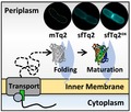
The periplasm of Gram‐negative bacteria is challenging to research as most fluorescent proteins do not function in its environment. We developed a new fluorescent protein, sfTq2ox, that is able to efficiently transport to, fold and mature in the periplasm. The beneficial properties of sfTq2ox come without a trade‐off allowing the development of a highly efficient FRET assay for the periplasm.
Introduction
In Gram‐negative bacteria, the cytoplasm is enveloped by an inner membrane (IM) and an asymmetric outer membrane (OM). The space between the IM and OM is called the periplasm and contains the protective peptidoglycan layer. As much as 30% of Escherichia coli’s proteins are predicted to localize to the envelope and many essential processes function fully or partly within the periplasm (Weiner and Li, 2008). The protein interactions in the periplasm are of great interest for biotechnological and medical purposes like synthesis of exogenous proteins and antibiotic development (Harvey et al., 2004). The most direct way to observe these proteins in living cells is by fluorescence microscopy of genetically encoded fusions to fluorescent proteins (FPs). Fluorescence also provides a means of detecting protein‐protein interactions by Förster resonance energy transfer (FRET). The stringent distance dependence for FRET is ideal to detect direct protein‐protein interactions as these also occur in the nanometre range, whereas indirect protein interactions usually occur on a larger distance scale and are not detectable by FRET (Piston and Kremers, 2007).
In vivo studies of proteins in the periplasm are challenging because of its oxidizing environment and toxicity associated with protein over‐expression (Meiresonne et al., 2017). Expression of FPs in the periplasm does not always result in fluorescence and only a limited number of FPs have been shown to fold and mature under periplasmic conditions. Recently, we observed good expression of mNeongreen (mNG) and used it as a donor to the mCherry (mCh) acceptor FP in an in vivo periplasmic FRET assay (Meiresonne et al., 2017). The mNG‐mCh FRET pair has an R 0 of 5.5 nm (the distance at which 50% FRET occurs) and allowed the detection of periplasmic protein‐protein interactions with a dynamic range of up to 16% energy transfer efficiency. However, this is only half the range of what can be achieved for cytoplasmic FRET pairs.
To improve our periplasmic FRET assay, popular existing FPs were screened for periplasmic fluorescence but none was adequate. Therefore, we rationally designed a novel FP (dubbed sfTq2) and further optimized it for periplasmic functionality creating sfTq2ox. This process revealed the factors important for periplasmic fluorescence. sfTq2ox has biophysical properties equal to its parent FP mTq2. Expressing it in the periplasm comes at greatly reduced toxicity, resulting in bright cyan fluorescence. sfTq2ox forms a FRET pair with mNG with an R 0 of 6.0 nm and allows exceptionally high rates of energy transfer in the cytoplasm and periplasm of E. coli. Employing our new assay, we show the periplasmic interaction of the essential cell division proteins FtsB and FtsL. This work breaks ground for new research and provides microbiologists with new tools to use fluorescent techniques in the periplasm.
Results
Robust folding is the first prerequisite for periplasmic FP fluorescence
To optimize our periplasmic mNG‐mCh FRET assay (Meiresonne et al., 2017), a higher R 0 value FRET pair was sought for. This could be achieved by finding an acceptor FP with high extinction coefficient and/or high spectral overlap with the mNG donor to replace mCherry. However, most FPs do not fluoresce in the Gram‐negative periplasm due to hampered folding of their ß‐barrel or maturation defects. These challenges are still insufficiently understood. FPs evolved to function in the eukaryotic cytoplasm and are not adapted to foreign environments. Especially challenging are compartments where protein folding occurs after translocation across a membrane as is the case for the eukaryotic endoplasmic reticulum (ER) and the bacterial periplasm. These are oxidative environments and cysteines may form promiscuous disulfide bridges resulting in non‐fluorescent oligomers (Costantini et al., 2015). The Anthozoa‐derived mFruits and mScarlets were thought to be good candidate acceptor FPs because of their lack of native cysteines and favourable spectroscopic properties to form a FRET pair with the established periplasmic FP mNG instead of mCherry (Table S1). The FPs were expressed in the periplasm of E. coli by co‐translational translocation through the sec‐translocase (Fig. 1).
Figure 1.
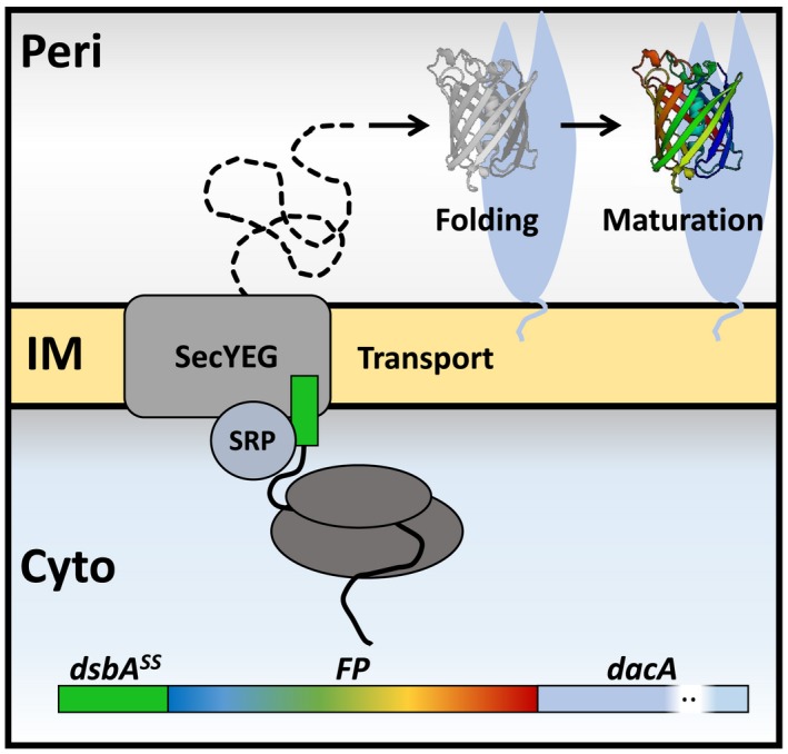
Co‐translational expression of FPs in the periplasm. FP fusions were expressed in the periplasm and attached to the periplasmic (Peri) side of the inner membrane (IM) through PBP5 (encoded by dacA). The DsbA (dsbass) signal sequence is a substrate for the signal recognition particle (SRP) that directs co‐translational transport through the SecYEG translocase of the N‐terminal fused gene product translated in the cytoplasm (Cyto). The FP fusion is translocated to the periplasm and the signal sequence is cleaved off. Here the FP is exposed to periplasmic conditions under which it needs to efficiently fold its ß‐barrel and subsequently mature its chromophore to become fluorescent. FPs that are unable to fluoresce in the periplasm thus have problems with either folding or chromophore maturation.
Periplasmic FP fusions were tested for expression‐related toxicity by growing E. coli in a plate reader in rich medium at 37°C and inducing expression at a concentration range of isopropyl‐ß‐D‐thiogalactopyranoside (IPTG) while monitoring growth and fluorescence. All periplasmic FP constructs resulted in toxicity‐associated growth curves correlating with the level of induction (Fig. S1a). Expression of cysteine‐less FPs seemed less toxic compared to sfGFP and mNG. Cultures that grew relatively unimpaired produced fluorescent signals over time, which were confirmed to be periplasmic by fluorescence microscopy (Fig. S1B and C). However, expressing the same constructs for two mass doubling times at non‐toxic concentrations did not result in all‐round periplasmic fluorescence for living, fixed or fixed and matured samples. Of the tested mFruits, only mCherry was able to fluoresce in the periplasm where the others only showed faint and grainy fluorescence of near background intensity (Fig. S2). The mScarlets performed better and mScarlet‐I showed fluorescence in living, fixed or fixed and matured cells. mScarlet and mScarlet‐H resulted in clearer periplasmic signals only after maturation. Yet, mCherry outperformed the mScarlets with brighter initial periplasmic fluorescence (Fig. 2).
Figure 2.
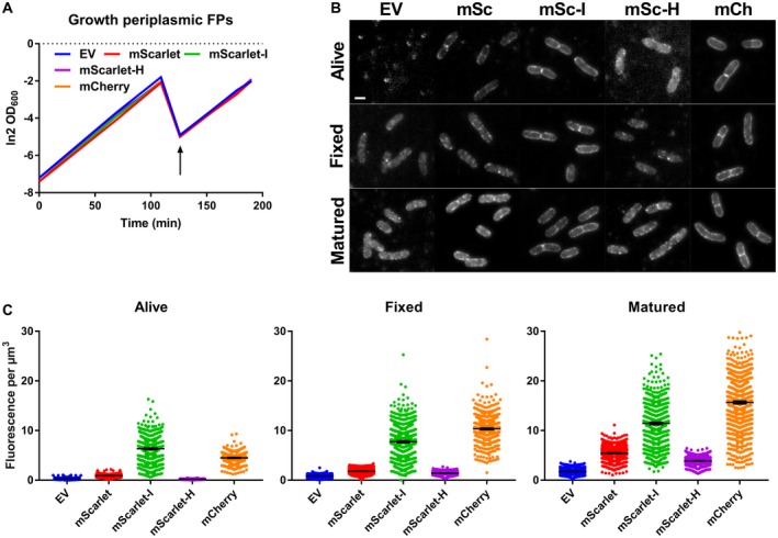
mCherry is the preferred red FP in the periplasm. A. LMC500 carrying DsbAss‐FP‐PBP5 plasmids for the periplasmic expression of the mScarlet FPs and mCh were grown as flask cultures in TY at 37°C and induced with 15 µM IPTG (arrow). Induction did not alter growth rates compared to a control carrying an empty plasmid (EV). B. Fluorescence images of living, fixed and fixed and matured cells showed varying levels of periplasmic fluorescence. Note that the greyscales were set for automatically visualizing any signal. The scale bar represents 2 µm. C. Quantification of fluorescence signals from all samples revealing strong periplasmic signals for mSc‐I and mCherry. The respective number of cells analysed for the living, fixed or fixed and matured samples were: EV 415, 928 and 689, mSc 623, 868 and 1165, mSc‐I 672, 1009 and 952, mSc‐H 618, 569 and 758 and mCh 477, 792 and 1144. The error bars at the mean indicate the 95% confidence interval.
Given the modest brightness of mCherry compared to the other mFruits and mScarlets, it is likely that these still suffer from adverse effects in the periplasm. None of them contain cysteines excluding promiscuous disulfide bond formation as a cause for their ineffective fluorescence. Chromophore maturation continues after fixation while protein folding stops. It is thus likely that the non‐fluorescing FP fraction had not folded properly under periplasmic conditions. This is supported by the strong fluorescence signals from the periplasm of stationary living cells where protein folding could continue over time (Fig. S1).
Together, these results show that periplasmic conditions can be inhospitable for FPs regardless of their lack of cysteines and suggest that proper folding is the first prerequisite for periplasmic fluorescence. The use of co‐translational transport for the periplasmic expression of FPs is a good method to select for well‐folding variants and low‐expression toxicity. mCherry is still the preferred periplasmic FP and FRET acceptor to mNG. Although no superior acceptor was found, the same tools could be employed to select for a high quantum yield donor FP to mNG as an acceptor. Indeed, mNG was shown to be an efficient acceptor to the cyan FP mTq2 with an R 0 value of 6.0 nm with FRET efficiencies in eukaryotes of 50–60% (Shaner et al., 2013; Mastop et al., 2017), making mTq2 a logical candidate to optimize for expression in the periplasm.
Superfolder mTq2 folds and matures in the periplasm
Aequorea victoria‐derived FPs contain two cysteines, C48 and C70, and were therefore initially not tested. However, sfGFP folds and fluoresces in the periplasm of E. coli (Aronson et al., 2011) despite the presence of cysteines (Pédelacq et al., 2006). This observation further suggests that the folding rate may be more important than the presence of cysteines for optimal periplasmic chromophore development.
Bright cyan FPs are great FRET donors because of their high quantum yield (QY). mTurquoise2 (mTq2) has a QY of 93% and was shown to allow high FRET efficiencies with mNG (Goedhart et al., 2012; Shaner et al., 2013; Mastop et al., 2017). It can be paired with green, yellow or orange acceptors that have a high molar extinction coefficient (ɛ) resulting in relatively high R 0 values and strong FRET pairs. mNG is the preferred FRET acceptor of mTq2 in eukaryotic cells with an R 0 of 6.0 nm and FRET efficiencies ranging up to 50–60% (Shaner et al., 2013; Mastop et al., 2017).
mTq2 was tested in the periplasm of E. coli but it did not fluoresce (Fig. 3A). Since mTq2 is also derived from Aequorea victoria, superfolding mutations S30R, Y39N, N105T F99S, and I171V (Fukuda et al., 2000; Pédelacq et al., 2006) were introduced based on sfGFP to create superfolder mTq2 (sfTq2). sfGFP mutations M153T and V163A were already in mTq2 and Y145F was not included since it is close to the chromophore and could affect maturation and indeed negatively impact periplasmic fluorescence (Fig. S3). A206V was not introduced since it may increase dimerization tendency (Cranfill et al., 2016).
Figure 3.
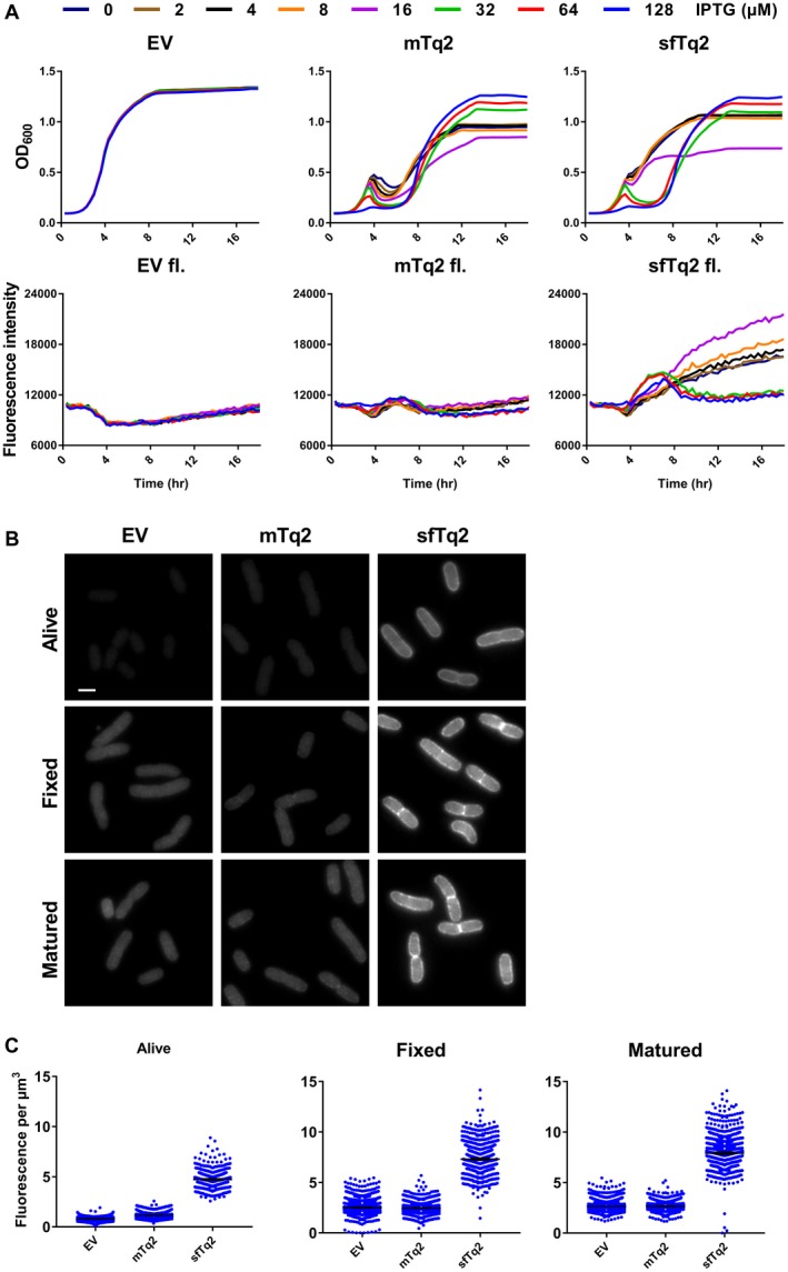
Superfolder mTurquoise2 fluoresces in the periplasm. A. Plate reader growth data in rich medium at 37°C show that the periplasmic sfTq2 expression is less toxic at high induction than the original mTq2. Plate reader fluorescence measurements confirm that periplasmic sfTq2 gives fluorescent signals while mTq2 does not. B. Fluorescence microscopy of living cells expressing an empty vector control, mTq2‐PBP5 or sfTq2‐PBP5, reveals fluorescence only from the sfTq2 fusion. Fixation of the same cells does not result in a decreased sfTq2 signal. Overnight maturation of the fixed cells at RT to allow for possible chromophore (re)formation (indicated as ‘matured’) did not result in an increase in fluorescence for periplasmic mTq2 suggesting it had not folded properly while sfTq2 showed a strong periplasmic signal. All photographs are shown with the same grey values (100–4000) for comparison and the scale bar represents 2 µm. C. Quantification of the control, mTq2 and sfTq2 cultures for the living, fixed and matured cells shows no difference between the empty vector control and the mTq2 cells. The sfTq2 cells showed a small maturation effect. The error bars at the mean represent the 95% confidence interval. The number of cells measured were: Alive) EV = 1018, mTq2 = 650 and sfTq2 = 571. Fixed) EV = 1176, mTq2 = 1046, sfTq2 = 884. Fixed and matured) EV = 1142, mTq2 = 931, sfTq2 = 988.
mTq2 and sfTq2 fusions were expressed in the periplasm of E. coli growing in a plate reader at a concentration range of the inducer IPTG (Experimental procedures). Cultures expressing either mTq2 or sfTq2 resulted in toxicity correlated with induction concentrations previously reported for other FPs (Meiresonne et al., 2017). However, sfTq2 could be expressed at higher levels with less toxicity compared to mTq2. Strikingly, the sfTq2 fusion resulted in periplasmic fluorescence, whereas the mTq2 fusions did not (Fig. 3A). This shows that the superfolder mutations enable sfTq2, like sfGFP, to fold and mature in the periplasm.
Cells expressing mTq2 and sfTq2 in the periplasm at non‐toxic concentrations were imaged by fluorescence microscopy. Quantification of the signals from living, fixed and fixed and matured samples showed periplasmic fluorescence of sfTq2 only (Fig. 3B and C). Western blot analysis confirmed the similar expression levels of mTq2 and sfTq2 (shown in the next section).
The sfTq2‐mNG FRET pair in the periplasm
Superfolder mutations allowed sfTq2 to fold and mature in the periplasm. To show that the periplasmic emission of sfTq2 is unaltered compared to cytoplasmic emission, E. coli was grown to steady state in minimal medium at 28°C. Expression of the cytoplasmic sfTq2 or periplasmic OM‐bound OmpA‐sfTq2 was induced for at least 2 mass doublings (MDs) with 15 µM IPTG. The samples were fixed, washed with PBS, diluted to an OD450 value of 1.000 ± 0.002 and the fluorescence spectra were measured. The normalized emission spectra were almost identical for cytoplasmically or periplasmically expressed sfTq2 (Fig. 4A).
Figure 4.
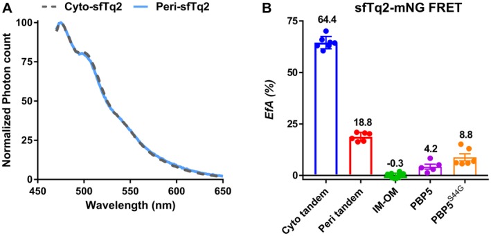
In vivo FRET using the sfTq2‐mNG FRET pair. A. Periplasmic sfTq2 does not show altered spectra compared to cytoplasmic sfTq2. LMC500 grown to steady state in minimal medium at 28°C were induced to express sfTq2 freely in the cytoplasm or the periplasm as the OM‐bound OmpA‐sfTq2 fusion. After 2 MDs, the cells were fixed, washed with PBS and set to an OD450 value of 1.000 ± 0.002 before measuring the fluorescence spectra. The normalized fluorescence spectra show the same shape suggesting that the spectral properties of sfTq2 are the same in the cytoplasmic and periplasmic compartments. B. EfA values calculated for the sfTq2‐mNG FRET pair show a large difference for the cytoplasmic and periplasmic tandem. The technical negative control (IM‐OM) did not show any energy transfer. The activity‐related conformational changes of the PBP5 dimers previously observed with the mNG‐mCh FRET pair (Meiresonne et al., 2017) were confirmed. The error bars represent the mean and standard deviation. The control EfA values were calculated from experiments in LMC500 and CS109 ∆dacA and the PBP5 dimerization experiments were exclusively performed in CS109 ∆dacA. Table S2 shows the FRET efficiencies measured with the plate reader. Representative unmixing data are shown in Fig. S14.
Next, we tested how the new donor performed in FRET experiments with mNG. Cytoplasmic mNG‐sfTq2 was capable of high rates of energy transfer with EfA values of 64.5 ± 3.0% but the periplasmic mNG‐sfTq2 tandem only reached 18.8 ± 2.1%. A technical negative control, consisting of non‐interacting proteins between the IM and OM, yielded −0.3 ± 1.4% energy transfer. The previously described (Meiresonne et al., 2017) biological homodimerization interaction of the active and inactive D,D‐carboxypeptidase PBP5 was confirmed with EfA values of 4.2 ± 2.7% and 8.8 ± 4.2%, respectively (Fig. 4B and Table 1). Although the sfTq2‐mNG FRET pair could be used successfully to measure periplasmic protein interactions, the discrepancy between the cytoplasmic and periplasmic rates of energy transfer indicated sfTq2 did not reach its full potential. For comparison, our mNG‐mCh FRET assay allows the detection of the cytoplasmic and periplasmic protein interactions with more comparable EfA values up to 19 and 16%, respectively (Meiresonne et al., 2017).
Table 1.
FRET efficiencies obtained with mTq2 variants measured by fluorometry.
| Donor FP to mNG | Fusion position | EfA (%) | SD | Repeats | |
|---|---|---|---|---|---|
| Cytoplasmic FRET | |||||
| Positive control tandems | |||||
| mNG‐mTq2 | mTq2 | Free floating | 61.3 | 6.4 | 4 |
| mNG‐sfTq2 | sfTq2 | Free floating | 64.5 | 3.0 | 7 |
| mNG‐sfTq2C70V | sfTq2ox | Free floating | 66.4 | 3.1 | 8 |
| Periplasmic FRET | |||||
| Positive control tandems | |||||
| mNG‐sfTq2 | sfTq2 | OmpA177 (OM) | 18.8 | 2.1 | 6 |
| mNG‐sfTq2C48S | C48S | OmpA177 | 32.9 | 1.9 | 2 |
| mNG‐sfTq2C70V | sfTq2ox | OmpA177 | 39.1 | 3.8 | 11 |
| mNG‐sfTq2C48S‐C70V | C48S‐C70V | OmpA177 | 40.3 | 1.2 | 2 |
| mNG‐sfTq2C70V | sfTq2ox | OmpA177* | 42.5 | 3.1 | 6 |
| mNG‐sfTq2C70V | sfTq2ox | LpoB73 (OM) | 39.8 | 2.1 | 5 |
| mNG‐sfTq2C70V | sfTq2ox | NlpAss (IM) | 41.2 | 2.4 | 7 |
| mNG‐sfTq2C70V | sfTq2ox | MalFss‐mss (IM) | 42.9 | 2.5 | 3 |
| Negative controls | |||||
| mNG‐sfTq2 (IM‐OM) | sfTq2 | PBP5, OmpA177 | –1.4 | 1.4 | 4 |
| mNG‐sfTq2C70V (IM‐OM) | sfTq2ox | PBP5, OmpA177 | –1.3 | 2.2 | 13 |
| mNG‐sfTq2C70V (IM, IM) | sfTq2ox | NlpAss, FtsB | –0.7 | 1.7 | 10 |
| Biological interactions | |||||
| PBP5 + PBP5 | sfTq2 | IM, IM | 4.2 | 2.7 | 5 |
| PBP5S44G + PBP5S44G | sfTq2 | IM, IM | 8.8 | 4.2 | 6 |
| FtsB + FtsL | sfTq2ox | IM, IM | 19.2 | 4.5 | 8 |
| FtsL + FtsB | sfTq2ox | IM, IM | 15.6 | 2.6 | 8 |
| FtsLm4 + FtsB | sfTq2ox | IM, IM | 3.1 | 2.1 | 3 |
| FtsLm4 + FtsBm4 | sfTq2ox | IM, IM | 3.0 | 3.4 | 3 |
SS, signal sequence; mss, membrane spanning sequence, LpoB73 indicates residue 1‐73 of LpoB, OmpA177 indicates residue 1‐177 of OmpA, * indicates a different linker between mNG and sfTq2ox (EF instead of EL due to differences in cloning). Table S2 shows the FRET efficiencies for the new mTq2 variants measured with the plate reader. Representative unmixing data are shown in Fig. S14; the plate reader unmixing data are shown in Fig. S15.
The periplasm is an oxidative environment and the presence of cysteines can cause the formation of non‐fluorescent oligomers (Costantini et al., 2015). The introduced superfolder mutations in sfTq2 may not necessarily fully protect its two native cysteines from promiscuous disulfide bridge formation in the periplasm. Therefore, we set out to replace the cysteines of sfTq2 by site‐directed and random mutagenesis and tested the resulting FPs in the periplasm.
Development and optimization of sfTq2ox
To improve the expression of sfTq2 in the periplasm, site‐directed mutagenesis was performed to replace sfTq2’s C48 with serine and C70 with serine, methionine or valine. Serine closely resembles cysteine with a hydroxyl instead of a sulfhydryl group. Methionine also contains a sulphur that is not nucleophilic and therefore should not participate in disulfide bond formation. Valine should resemble the more hydrophobic state of cysteine better than serine and was shown to aid fluorescence of oxBFP in the eukaryotic ER (Costantini et al., 2015).
Single and double cysteine mutants of sfTq2 performed better than sfTq2 in the periplasm with reduced toxicity and improved fluorescence (Figs 5 and S4). They were expressed as protein fusions in the periplasm of E. coli under non‐toxic conditions and the fluorescence signals of living, fixed and fixed and matured samples were quantified. sfTq2 mutants C48S, C70S, C70V and C48S‐C70V showed much brighter signals in the periplasm (Fig. 5B and C). The methionine sfTq2 variants did not perform better than sfTq2 in terms of fluorescence (Fig. S4). Western blotting showed that all cysteine replacement mutants were better expressed in the periplasm regardless of fluorescence signals (Fig. 5D). This is in line with the secretory improvement of GFP to the eukaryotic ER by cysteine replacements (Jain et al., 2001). The enhanced periplasmic production but dim fluorescence of some sfTq2 variants suggests poor chromophore maturation.
Figure 5.
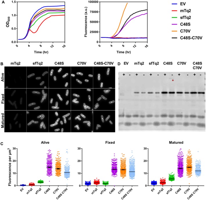
Cysteine‐replaced sfTq2 variants perform better in the periplasm. A. Periplasmic expression of sfTq2‐PBP5 variants in LMC500 grown in rich medium at 37°C at relatively non‐toxic induction conditions results in large differences in cyan fluorescence. The C70V variant performs better than C48S and the double cysteine mutant version. B. Microscopy of LMC500 grown in rich medium and induced with 15 µM IPTG shows strong fluorescence for the sfTq2‐PBP5 variants in the periplasm. For comparison, the greyscale of all photographs are the same (80–7000) and the scale bar represents 2 µm. C. Quantification of the images confirms bright fluorescence signals from the single C48S and C70V sfTq2 variants. Differences in fluorescence with the prolonged plate reader induction suggest that folding difficulties may still be a problem. The error bars at the mean represent the 95% confidence interval. The number of cells measured were between: Alive 500–1000, Fixed 1000–1500 and Matured 1000–2000 except for EV‐Alive n = 327 and C48S‐C70V‐Fixed n = 775. D. mTq2 is produced at similar levels as sfTq2 but does not fold properly; cysteine mutants of sfTq2 are produced at higher levels. Anti‐GFP immunoblotting of the corresponding samples shows that the fusion proteins are intact and that C48S has a minor propensity to form higher order complexes (asterisk). The + and signs, respectively, indicate the presence or absence of the reducing agent DTT in the sample.
Site‐directed random mutagenesis of sfTq2 cysteines revealed that residue 48 does not allow much variation besides the original cysteine or serine while a variety of amino acids are accepted as residue 70. Several brightly fluorescing sfTq2 variants were found but none of them showed improved periplasmic fluorescence as compared to C70V (Supplementary text 1, Fig. S5). Cysteine mutants in the parental mTq2 did not increase periplasmic fluorescence, although a reduction in expression toxicity was observed (Fig. S6). sfGFP mutant sfGFPC70V showed similar periplasmic fluorescence compared to its predecessor and expression was slightly less toxic (Fig. S7). This shows that cysteines are involved in translocation toxicity and subsequent low production of periplasmic fusion proteins.
All experiments showed that sfTq2C70V was the best periplasmic variant to be used in the bacterial periplasm in terms of reduced toxicity, fluorescence intensity and fast folding or maturation. sfTq2C70V was named sfTq2ox. Subsequent comparisons of sfTq2 and sfTq2ox as periplasmic fusions to several IM and OM localized proteins confirmed the superior performance of sfTq2ox (Figs S8–S10).
The superior periplasmic properties of sfTq2ox come without a trade‐off
Introducing superfolder mutations in mTq2 allowed fluorescence in the periplasm of E. coli. The additional C70V mutation in sfTq2ox resulted in a reduction in expression toxicity and the brightest periplasmic fluorescence. Interestingly, mTq2, sfTq2 and sfTq2ox were equally bright when expressed in the cytoplasm (Fig. S11). To verify the in vivo spectroscopic properties of the mTq2 variants, the fluorescence lifetime and cellular brightness were determined.
The quantum yield of cyan FPs is directly related to their fluorescence lifetime (Goedhart et al., 2010; 2012). Therefore, the lifetimes of mTq2 and sfTq2 and sfTq2ox were measured from the cytoplasm of eukaryotic (HeLa) cells using frequency domain fluorescence lifetime imaging microscopy (FLIM). This resulted in an identical distribution of lifetimes with an average of 3.9 ± 0.1 ns suggesting no differences in QY between the three variants (Fig. 6A). The fluorescence lifetime of sfTq2 and sfTq2ox expressed in the periplasm also showed a similar average lifetime of 3.9 ± 0.1 ns showing that periplasmic conditions did not alter their lifetime and suggesting an unaltered QY (Fig. 6B). Agar colonies of E. coli expressing cytoplasmic mTq2, sfTq2 or sfTq2ox were imaged and gave a similar average lifetime of 3.8 ± 0.2 ns (Fig. S11).
Figure 6.
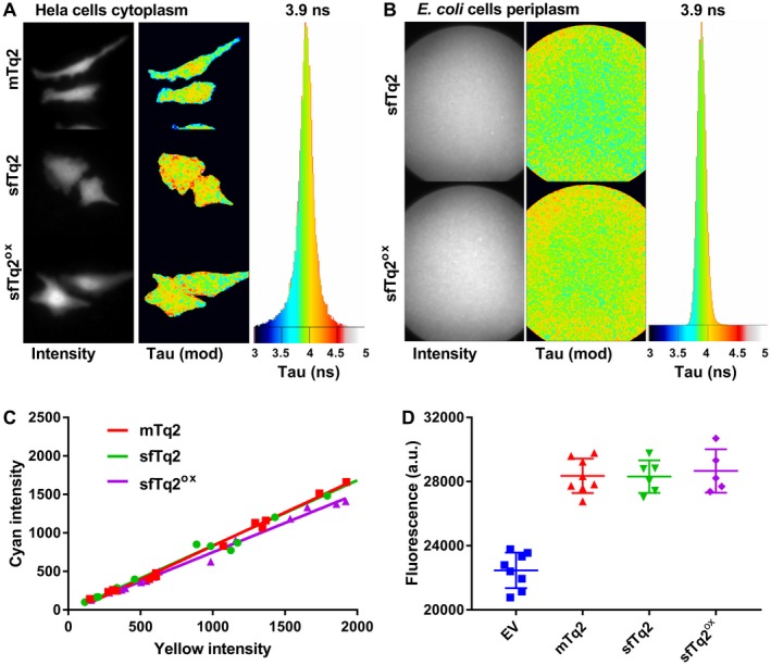
sfTq2ox comes without a trade‐off. A. Frequency domain FLIM of HeLa cells expressing mTq2, sfTq2 or sfTq2ox in the cytoplasm reveals a similar average fluorescence lifetime of 3.9 ns. B. FLIM of a suspension of E. coli cells expressing periplasmic OmpA‐sfTq2 or OmpA‐sfTq2ox also show a similar fluorescence lifetime of 3.9 ns. C. The cellular brightness of mTq2, sfTq2 and sfTq2ox is equal in Hela cells. The slope coefficients of the fits are, respectively: 0.87, 0.84 and 0.76. D. Fixed samples of LMC500 cells at OD450 = 1.00 expressing cytoplasmic mTq2, sfTq2 or sfTq2ox show equal amounts of fluorescence.
To analyse the cellular brightness of sfTq2 and sfTq2ox, self‐cleaving viral peptide (2A)‐linked tandems with super yellow FP 2 (sYFP2) were expressed in HeLa cells (Mastop et al., 2017). The equal expression of both FPs (Kim et al., 2011) showed a similar correlation for the three mTq2 variants reflecting an equal cellular brightness (Fig. 6C). Quantification of mTq2, sfTq2 and sfTq2ox expressed in the cytoplasm of E. coli further supports this (Figs 6D and S11).
Having determined that the lifetime and cellular brightness of sfTq2ox are the same as that of mTq2 and sfTq2, it is reasonable to assume that the extinction coefficient is also similar. The unaltered lifetime also implies that the spectroscopic properties (quantum yield) of mTq2 may be used for calculating R 0 and the quantification of EfA values.
mTq2 is monomeric (Cranfill et al., 2016) and will therefore not obscure FRET experiments by dimerization or oligomerization. The superfolder and C70V mutations may have impacted this property. Therefore, the tendency of sfTq2 and sfTq2ox to form undesired oligomers was assessed by the OSER assay (Costantini et al., 2012) showing equal results for both proteins, suggesting the same low propensity to aggregate as mTq2 (Fig. S12).
Further confirmation that sfTq2 and sfTq2ox have the same spectral properties as mTq2 comes from their FRET efficiencies in the cytoplasm. Tandem fusions of mNG with mTq2, sfTq2 or sfTq2ox resulted in significantly similar EfA values of 61 ± 6%, 65 ± 3% and 66 ± 3%, respectively (Table 1). The same constructs expressed in agar colonies of E. coli gave an average lifetime of 2.5 ± 0.1 ns, a 35% reduction in fluorescence lifetime compared to their single donor FPs (Fig. S11). This is similar to the 33% energy transfer observed for mNG‐mTq2 in eukaryotic cells that were measured using the same technique (Mastop et al., 2017).
Taken together, these results strongly indicate that the engineering of sfTq2ox comes without a trade‐off compared to its parent mTq2 with the added benefits of periplasmic expression, bright fluorescence and reduced toxicity.
High dynamic range periplasmic FRET with sfTq2ox
mTq2 forms a good FRET pair with mNG with an R 0 of 6.0 nm. The cytoplasmic tandems of mTq2 and sfTq2 gave similar rates of energy transfer. Yet, the periplasmic mNG‐sfTq2 tandem gave an EfA value of 18.8 ± 2.1% suggesting differences in terms of periplasmic functionality. The bright periplasmic fluorescence of cysteine‐replaced sfTq2 variants suggested that they would make better donors to mNG for periplasmic FRET. mNG‐sfTq2 tandems containing the cysteine mutants, associated with the OM through OmpA, were tested for their in vivo FRET efficiencies. This indeed greatly improved EfA values to 32.3 ± 1.9, 42.5 ± 3.1% and 40.3 ± 1.2% for sfTq2C48S, sfTq2ox or sfTq2C48S‐C70V, respectively. No degradation or cleavage products were detected by western blotting (Fig. S13).
Since sfTq2ox showed the best periplasmic behaviour compared to all other cysteine mutant variants, mNG‐sfTq2ox fusions were made to other periplasmic proteins to exclude localization effects. Tandems were associated with the OM or IM through the translocation and lipidation signals of lipoproteins LpoB and NlpA, respectively. A third tandem was localized to the IM through the first membrane spanning sequence of MalF (Fig. 7). All periplasmic tandems resulted in EfA values of 40% (Table 1) suggesting that this is the highest attainable FRET efficiency for mNG‐sfTq2ox in the periplasm regardless the location of the fusion. Negative controls, assaying energy transfer by crowding conditions in the IM or between the IM and OM, resulted in EfA values of −0.7 ± 1.7% and −1.3 ± 2.2%, respectively (Fig. 7). All assayed EfA values are shown in Table 1. The measured FRET efficiencies were confirmed using a separate 96‐wells plate reader set up showing that the throughput of FRET assays using the sfTq2ox‐mNG pair can be greatly increased (Table S2).
Figure 7.
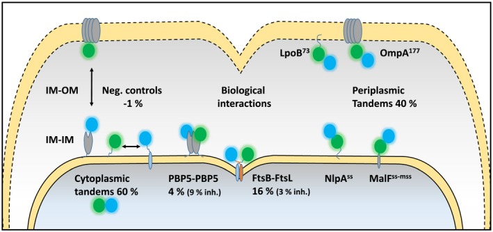
Schematic overview showing the subcellular localization of the periplasmic protein interactions and controls assayed with the sfTq2ox‐mNG FRET pair. PBP5 dimerization interactions were assayed with sfTq2‐mNG. Inh. refers to the inactive mutants PBP5S44G, FtsBm4 and FtsLm4. A full list of EfA values is shown in Table 1.
In vivo interactions of putative antibiotic targets in the periplasm
With the periplasmic sfTq2ox‐mNG FRET pair established, biological interactions could be assessed. The periplasm contains many relevant potential antibiotic targets including the cell division subcomplex of FtsB, FtsL and FtsQ (FtsBLQ) (den Blaauwen et al., 2014). All three components are essential and are thought to form the regulating link between the cytoplasmic and periplasmic machineries of cell division (Liu et al., 2015; Boes et al., 2019). Assaying the interactions in this complex could allow for the screening of novel antibiotics in vivo, determining the efficacy and specificity at the same time. FRET experiments assaying the interaction between FtsB‐sfTq2ox and FtsL‐mNG or FtsB‐mNG and FtsL‐sfTq2ox resulted in EfA values of 19.2 ± 4.5% and 15.6 ± 2.6%, respectively (Table 1 and Fig. 7). A negative control for IM‐associated proteins of FtsB‐sfTq2ox with NlpAss‐mNG resulted in an EfA value of −0.7 ± 1.7%. FtsB and FtsL were reported to interact through a leucine zipper‐like motif that can be disrupted by leucine to alanine substitutions (Robichon et al., 2011). The leucine zipper mutants of FtsB, FtsBm4 and FtsL, FtsLm4 are good controls as they are able to localize at the division site and complement their respective depletion strains (Robichon et al., 2011). The interactions of wild‐type FtsB‐mNG or mutant FtsBm4‐mNG with mutant FtsLm4‐sfTq2ox showed decreased EfA values of 3.1 ± 2.1% and 3.0 ± 3.4%, respectively. Expression without degradation of wild‐type and mutant FtsB and FtsL constructs was confirmed by western blotting (Fig. S13). The weakening of the leucine zipper motif clearly reduces the interaction between the two proteins given the decrease in EfA values. This observed reduced affinity between the two proteins may reflect a reduction in their average interaction time. In conclusion, the new FRET pair sfTq2ox‐mNG can be used to detect periplasmic protein interactions in vivo and paves the way for functional screening of essential antibiotic targets.
Discussion
In vivo visualization of periplasmic proteins by fusions to FPs is difficult since most do not fold or mature due to the oxidizing environment and the necessity of transport. Additionally, over‐expressing periplasmic FP fusions can lead to toxicity and counter selection (Meiresonne et al., 2017). The limited choice of FPs is especially problematic for applications that use multiple FPs, such as multiplex imaging and FRET studies. For FRET studies, a pair of FPs with a high R 0 is desired. The theoretical R 0 value, however, does not take into account the proper folding and maturation of FPs in vivo. Therefore, the optimal FP for specific conditions should be experimentally determined. We aimed to improve our previously developed periplasmic FRET assay using mNG‐mCh (R 0 = 5.5 nm) by searching alternatives for mCherry, which has a modest quantum yield (QY) and subsequent brightness. However, testing bright cysteine‐less red FPs showed that mCherry is still the preferred FP in the periplasm due to its favourable folding and maturation properties.
mNG has a high QY and extinction coefficient and can therefore function as a donor or acceptor FP (Shaner et al., 2013). It forms an efficient FRET pair with mTq2 (R 0 = 6.0 nm) and allows for highly efficient energy transfer (Shaner et al., 2013; Mastop et al., 2017). Since mTq2 did not fluoresce in the periplasm, sfTq2 was engineered by introducing superfolder mutations S30R, Y39N, N105T F99S, M153T, V163A and I171V based on the related sfGFP, which is fluorescent in the periplasm (Aronson et al., 2011). sfTq2 was functional in the periplasm but allowed only moderate periplasmic FRET with mNG (Fig. 4 and Table 1).
The oxidative environment of the periplasm facilitates disulfide bond formation between exposed cysteines in close proximity (Aronson et al., 2011). Aequorea victoria‐derived FPs are adapted for cytoplasmic conditions and their native cysteines are thus prone to promiscuous disulfide bonding in the periplasm resulting in non‐fluorescent oligomers. Site‐directed random mutagenesis of C48 and C70 in sfTq2 resulted in greatly increased periplasmic fluorescence and reduced expression toxicity with the single C70V variant (sfTq2ox) giving the best results (Figs 5, S4, and S5). sfTq2ox came without an observed trade‐off as the same QY and brightness values were found for mTq2 and sfTq2 (Fig. 6). The development of sfTq2ox shows that periplasmic fluorescence of FPs first depends on their efficient folding and then on the prevention of cysteine disulfide bond formation.
Employing the mNG‐sfTq2ox FRET pair in the periplasm resulted in energy transfer rates of 40% for periplasmic tandems (Fig. 7). FRET efficiencies of 60% were observed for cytoplasmic mTq2, sfTq2 or sfTq2ox fusions to mNG (Table 1). Based on the R 0 value (6.0 nm), the FRET efficiency of 60 and 40% would correspond to a distance between the fluorophores of 5.6 and 6.4 nm, respectively. Both values are plausible given the minimal distance of ± 3 nm between the two chromophores of the FRET pair. Yet, it cannot be excluded that the transport to the periplasm still hampers subsequent folding of a fraction of the FPs resulting in a lower FRET efficiency.
A factor influencing FRET efficiency (and R 0 values) is the dipole orientation of the donor and acceptor chromophore. This would contribute to the FRET efficiency difference if the environment of the cytoplasm or the periplasm would restrict the rotational freedom of the tandem differently. Presently, no evidence is available to support such a difference. The comparison of mTq2 with several acceptor FPs shows that mNG is an exceptional FRET acceptor, even compared to FPs with similar spectroscopic properties (Mastop et al., 2017). Indeed, mNG is an outlier with its chordate origin compared to the usual cnidarian‐derived FPs with different sequence homologies and possibly different behaviours (Shaner et al., 2013; Steiert et al., 2018).
Protein interactions often change significantly when the activity of one of the partners is inhibited by antibiotics (van der Ploeg et al., 2015). The Gram‐negative periplasm facilitates a myriad of essential protein interactions waiting to be investigated in vivo. Using the new periplasmic FRET pair, we demonstrated the direct interaction and disruption of essential cell division proteins FtsB and FtsL with high rates of energy transfer. Interactions of the FtsBLQ complex are considered to be the link between the cytosolic and periplasmic parts of cell division setting off constriction (Liu et al., 2015; Boes et al., 2019). Exactly how and in what stoichiometry the complex forms and functions is still a matter of debate (Karimova et al., 2005; Villanelo et al., 2011; Robichon et al., 2011; Glas et al., 2015; Condon et al., 2018). Interactions between FtsB and FtsL seem to rely on their coiled‐coil transmembrane helices (Robichon et al., 2011; Condon et al., 2018) while the interaction of FtsQ and FtsB requires their periplasmic soluble domains and forms independently of FtsL (van den Berg van Saparoea et al., 2013; Glas et al., 2015; Kureisaite‐Ciziene et al., 2018; Choi et al., 2018; Boes et al., 2019). Our findings confirm the former interaction in vivo and provide a tool for the further dissection of FtsBLQ interactions.
The ability of FPs to fold and mature under non‐native conditions is a property of increasing interest. Co‐translational expression of fusion proteins in the periplasm is a good assay to select for optimized FPs. With the development of sfTq2ox, the palette of periplasmic functional FPs is broadened to include members emitting at cyan wavelengths. This allows for the in vivo observation of (at least) three colours of periplasmic fusion proteins. Moreover, sfTq2ox forms a FRET pair with mNG that can detect periplasmic protein interactions in vivo at double the detection range of previous bacterial FRET assays. mNG‐sfTq2ox is therefore the preferred FRET pair for the detection of periplasmic as well as cytoplasmic protein interactions paving the way for the screening of novel antibiotics that may inhibit them.
Experimental procedures
Bacterial strains and culturing conditions
The Escherichia coli K12 strains used are presented in (Table 2). The cells were cultured in rich medium (TY: 10 g Tryptone (Bacto laboratories, Australia), 5 g yeast extract (Duchefa, Amsterdam, The Netherlands) and 5 g NaCl (Merck, Kenilworth, NJ) per litre) supplemented with 0.5% glucose (Merck) or in glucose minimal medium (Gb1: 6.33 g K2HPO4 (Merck), 2.95 g KH2PO4 (Riedel de Haen, Seelze, Germany), 1.05 g (NH4)2SO4 (Sigma, St. Louis, MO), 0.10 g MgSO4·7H2O (Roth, Karlsruhe, Germany), 0.28 mg FeSO4·7H2O (Sigma), 7.1 mg Ca(NO3)2·4H2O (Sigma), 4 mg thiamine (Sigma), and 4 g glucose per litre, pH 7.0) at 28°C while shaking at 205 rpm. For growth in Gb1 of LMC500‐ and CS109‐based strains, 50 mg/l lysine (Sigma) was added. Growth in rich and poor medium was at 37 and 28°C, respectively. Expression of constructs was induced with 15 µM isopropyl β‐D‐1‐thiogalactopyranoside (IPTG, Promega, Madison, WI) unless stated otherwise. Plasmids were maintained in the strains by addition of 100 µg/ml ampicillin (Sigma) or 25 µg/ml chloramphenicol (Sigma). Growth was measured by absorbance at 600 or 450 nm with a Biochrom Libra S70 spectrophotometer (Harvard Biosciences, Holliston, MA) for TY or Gb1 cultures, respectively. Fixation was done with a final concentration of 2.8% formaldehyde and 0.04% glutaraldehyde in the shaking water bath for 15 min, after which the cells were harvested. After fixation, the cells were washed three times with 1 ml PBS.
Table 2.
Strains used in this study
| Strain | Relevant characteristics | References |
|---|---|---|
| DH5α | F‐, supE44, hsdR17, recA1, endA1, gyrA96, thi1, relA1 | Bethesda Research Laboratories (1986) |
| LMC500 | MC4100, F‐, araD139, ∆(argF‐lac)U169, deoC1, flbB5301, lysA1, ptsF25, rbsR, relA1, rpsL150 | Taschner et al. (1988) |
| CS12‐7 | CS109 ΔdacA | Denome et al. (1999) |
Plasmid construction
An overview of the plasmids used in this study and their cloning strategies are given in Table S3. The sfTq2 nucleotide sequence (encoding mTurquoise2 with S30R, Y39N, F99S, N105T and I171V) was obtained by gene synthesis (MWG biotech). New constructs were created by restriction ligation cloning or site‐directed mutagenesis. Inserted fragments were amplified by PCR from plasmid or the E. coli (LMC500) chromosome template, purified and restricted. For mutagenesis, a whole plasmid template was amplified by PCR using primers that contain the desired mutation. The resulting product was treated with DpnI to digest the methylated template plasmid. All restriction enzymes used were purchased from New England Biolabs Inc. (Ipswich, MA). For all PCR amplifications for cloning, the high‐fidelity polymerase pfux7 was used (Nørholm, 2010) as per the manufacturer’s instructions. An overview of the used primers is shown in Table S4.
Imaging and image analysis
For imaging the cells were immobilized on 1% agarose in water slabs on object glasses as described (Koppelman et al., 2004) and photographed with a Hamamatsu ORCA‐Flash‐4.0LT (Hamamatsu, Naka‐ku, Japan) CMOS camera mounted on an Olympus BX‐60 fluorescence microscope (Tokyo, Japan) through a UPlanApo 100×/N.A. 1.35 oil Iris Ph3 objective. Images were acquired using the Micro Manager 1.4 plugin for ImageJ (Edelstein et al., 2010). In all experiments, the cells were first photographed in phase contrast mode and then in fluorescence mode. The fluorescence filter cubes used were: U‐MNG (red and orange, ex560/40, dic585LP, em630/75), EN‐GFP (green, ex470/40, dic495LP, em525/50) and Cyan‐GFPv2 (cyan, ex436/20, dic455LP, em480/40). Fluorescence backgrounds were subtracted using the modal values from the fluorescence images. Quantifications of cellular fluorescence were done using the ObjectJ plug‐in of ImageJ (Vischer et al., 2015). Living cells were imaged directly from the growing cultures and fixed cells directly after fixation and PBS washing. After fixation, the samples were allowed to mature overnight at RT, washed once more with 1 ml PBS and were then imaged. These samples are indicated in the text as ‘matured’.
Western blot analysis
Samples for SDS–polyacrylamide gel electrophoresis and western blotting were prepared by pelleting live cells and equalizing biomass in MilliQ and subsequent boiling (95°C) in a sample buffer (0.0625 M Tris‐Hcl pH 6.8, SDS 2%, glycerol 10%, bromphenol blue 0.001%) with and without dithiothreitol (0.1 M) reducing agent. Samples were loaded on 10% SDS‐PAGE gels (stacking buffer (0.5 M Tris‐HCl pH 6.8, 0.4% SDS), separating buffer (1.5 M Tris‐HCl pH 8.7, 0.4% SDS), separated by electrophoresis and transferred onto a nitrocellulose membrane (Biorad) by wet blotting; bound antibodies were detected with the Odyssey FC scanner (LI‐COR). The used antibodies were polyclonal rabbit α‐GFP (1:2,000, Fisher Scientific) and polyclonal goat α‐rabbit IRDye® 680LT (1:20,000, LI‐COR).
FRET experiments and spectral measurements
FRET experiments were performed as described in (Meiresonne et al., 2018) with modifications for the sfTq2‐mNG FRET pair. Acceptor and donor emission spectra were collected with a fluorometer (Photon Technology International, NJ) through 6 nm slit widths with 1 s integration time per scanned nm and three times averaging. For the acceptor (mNG) channel, samples were excited by the monochromator set at 504 nm through a 500 ± 10 nm single band pass (BP) filter (BrightLine, Semrock, Rochester, NY) and emission wavelengths from 512 to 650 nm at 1 nm increments were measured through a 510 nm long‐pass (LP) filter (Chroma technology corp., Bellow falls, VT). This spectrum was used to determine the amount of mNG in the sample. For the donor (mTq2, sfTq2 or sfTq2ox) channel, samples were excited by the monochromator set at 450 nm through a 435 ± 40 nm BP filter (Semrock) and emission wavelengths from 470 to 650 nm at 1 nm increments were collected through a 458 nm LP filter (Semrock). Knowing the amount of mNG present in the sample and the shape of the mNG and mTq2 variant reference spectra and background fluorescence spectrum in the cells, the sample spectra were unmixed into their separate components: background fluorescence, mTq2, mNG and sensitized emission (FRET). The FRET efficiencies were calculated using the published algorithms (Alexeeva et al., 2010; Meiresonne et al., 2018), using the spectral properties of mTq2 and mNG (Goedhart et al., 2012; Shaner et al., 2013). Spectral measurements using a multimode plate reader (BIOTEK Synergy MX, BioTek Instruments Inc., Winooski, VT) were performed as described (Meiresonne et al., 2017; 2018) with the mTq2 variant donor channel acquisition set at 450 nm and emission scanning from 470 to 650 nm with minimal slit widths of 9 nm.
Frequency domain fluorescence lifetime measurements
FLIM experiments were essentially performed as described before (Goedhart et al., 2012). For periplasmic samples in the solution, the exposure time was 500 ms, and the number of phase steps 18.
OSER assay
The OSER assay uses an ER localization sequence (CytERM) that was inserted in the multiple cloning site of pmTurquoise2‐N1 to obtain CytERM‐mTurquoise2 (Addgene plasmid Plasmid #98833). The coding sequence of mTurquoise2 was replaced by the superfolder variants. After transfection of the plasmids, the localization of the fusion was visualized by confocal microscopy. The OSER assay was performed as described before (Costantini et al., 2012).
Molecular brightness assay
The cellular brightness assay was performed in HeLa cells as described before (Mastop et al., 2017).
Author contributions
N.Y.M. and T.D.B. designed the study. N.Y.M., E.C., L.M.Y.M., J.G. and A.C. conducted the experiments and analysed the data. N.Y.M. and T.d.B. wrote the paper. All authors contributed to the final version of the manuscript.
Conflict of interest
The authors declare no competing financial interests.
Data availability
Mammalian plasmids sfTq2ox‐N1, sfTq2‐N1 and sfTq2‐C1 are available at Addgene: https://www.addgene.org/117930/, https://www.addgene.org/117931/ and https://www.addgene.org/117932/. Periplasmic expression plasmids sfTq2‐PBP5 and sfTq2ox‐PBP5 are available at Addgene: https://www.addgene.org/117959/ and https://www.addgene.org/117960.
Supporting information
Acknowledgements
We thank Eva Asmus and Alejandro Mónton Silva (University of Amsterdam, the Netherlands) for help with the experiments. Plasmids pCR119 and pCR124 were kind gifts from Daniel Ladant (Institut Pasteur, France). mScarlet‐C1, mScarletI‐C1 and mScarletH‐C1 were kind gifts from Theodorus W.J. Gadella Jr. (University of Amsterdam, the Netherlands). N.Y.M. was supported by the NWO, ALW open program (82202019) and HFSP Program RGP0034/2013. E.C. and L.M.Y.M. were supported by the European Union’s Horizon 2020 research and innovation programme under grant agreement No. 721484 (Train2Target).
References
- Alexeeva, S. , Gadella, T.W.J. , Verheul, J. , Verhoeven, G.S. and den Blaauwen, T. (2010) Direct interactions of early and late assembling division proteins in Escherichia coli cells resolved by FRET. Molecular Microbiology, 77, 384–398. [DOI] [PubMed] [Google Scholar]
- Aronson, D.E. , Costantini, L.M. and Snapp, E.L. (2011) Superfolder GFP is fluorescent in oxidizing environments when targeted via the Sec translocon. Traffic, 12, 543–548. [DOI] [PMC free article] [PubMed] [Google Scholar]
- Bethesda Research Laboratories . (1986) E. coli DH5 alpha competent cells. Focus (Bethesda Research Laboratories), 8, 9. [Google Scholar]
- den Blaauwen, T. , Andreu, J.M. and Monasterio, O. (2014) Bacterial cell division proteins as antibiotic targets. Bioorganic Chemistry, 55, 27–38. [DOI] [PubMed] [Google Scholar]
- Boes, A. , Breukink, E. and Terrak, M. (2019). Regulation of the peptidoglycan polymerase activity of PBP1b by antagonist. mBio, 10, e01912–18. [DOI] [PMC free article] [PubMed] [Google Scholar]
- Choi, Y. , Kim, J. , Yoon, H.‐J. , Jin, K.S. , Ryu, S. and Lee, H.H. (2018) Structural insights into the FtsQ/FtsB/FtsL complex, a key component of the divisome. Scientific Reports, 8, 18061. [DOI] [PMC free article] [PubMed] [Google Scholar]
- Condon, S.G.F. , Mahbuba, D.‐A. , Armstrong, C.R. , Díaz‐Vázquez, G. , Craven, S.J. , LaPointe, L.M. , et al (2018) The FtsLB subcomplex of the bacterial divisome is a tetramer with an uninterrupted FtsL helix linking the transmembrane and periplasmic regions. Journal of Biological Chemistry, 293, 1623–1641. [DOI] [PMC free article] [PubMed] [Google Scholar]
- Costantini, L.M. , Fossati, M. , Francolini, M. and Snapp, E.L. (2012) Assessing the tendency of fluorescent proteins to oligomerize under physiologic conditions. Traffic, 13, 643–649. [DOI] [PMC free article] [PubMed] [Google Scholar]
- Costantini, L.M. , Baloban, M. , Markwardt, M.L. , Rizzo, M. , Guo, F. , Verkhusha, V.V. , et al. (2015) A palette of fluorescent proteins optimized for diverse cellular environments. Nature Communications, 6, 7670. [DOI] [PMC free article] [PubMed] [Google Scholar]
- Cranfill, P.J. , Sell, B.R. , Baird, M.A. , Allen, J.R. , Lavagnino, Z. , de Gruiter, H.M. , et al. (2016) Quantitative assessment of fluorescent proteins. Nature Methods, 13, 557–562. [DOI] [PMC free article] [PubMed] [Google Scholar]
- Denome, S.A. , Elf, P.K. , Henderson, T.A. , Nelson, D.E. and Young, K.D. (1999) Escherichia coli mutants lacking all possible combinations of eight penicillin binding proteins: viability, characteristics, and implications for peptidoglycan synthesis. Journal of Bacteriology, 181, 3981–3993. [DOI] [PMC free article] [PubMed] [Google Scholar]
- Edelstein, A. , Amodaj, N. , Hoover, K. , Vale, R. and Stuurman, N. (2010) Computer control of microscopes using µManager. Current Protocols in Molecular Biology, Chapter 14, Unit14.20. [DOI] [PMC free article] [PubMed] [Google Scholar]
- Fukuda, H. , Arai, M. and Kuwajima, K. (2000) Folding of green fluorescent protein and the cycle3 mutant. Biochemistry, 39, 12025–12032. [DOI] [PubMed] [Google Scholar]
- Glas, M. , Van Den Berg, Bart , Van Saparoea, H. , McLaughlin, S.H. , Roseboom, W. , Liu, F. , et al (2015) The soluble periplasmic domains of Escherichia coli cell division proteins FtsQ/FtsB/FtsL form a trimeric complex with submicromolar affinity. Journal of Biological Chemistry, 290, 21498–21509. [DOI] [PMC free article] [PubMed] [Google Scholar]
- Goedhart, J. , Weeren, L.Van , Hink, M.A. , Vischer, N.O.E. , Jalink, K. and Gadella, T.W.J. (2010) Bright cyan fluorescent protein variants identified by fluorescence lifetime screening. Nature Methods, 7, 137–139. [DOI] [PubMed] [Google Scholar]
- Goedhart, J. , von Stetten, D. , Noirclerc‐Savoye, M. , Lelimousin, M. , Joosen, L. , Hink, M.A. , et al. (2012) Structure‐guided evolution of cyan fluorescent proteins towards a quantum yield of 93%. Nature Communications, 3, 751. [DOI] [PMC free article] [PubMed] [Google Scholar]
- Harvey, B.R. , Georgiou, G. , Hayhurst, A. , Jeong, K.J. , Iverson, B.L. and Rogers, G.K. (2004) Anchored periplasmic expression, a versatile technology for the isolation of high‐affinity antibodies from Escherichia coli‐expressed libraries. Proceedings of the National Academy of Sciences, 101, 9193–9198. [DOI] [PMC free article] [PubMed] [Google Scholar]
- Jain, R.K. , Joyce, P.B. , Molinete, M. , Halban, P.A. and Gorr, S.U. (2001) Oligomerization of green fluorescent protein in the secretory pathway of endocrine cells. The Biochemical Journal, 360, 645–649. [DOI] [PMC free article] [PubMed] [Google Scholar]
- Karimova, G. , Dautin, N. and Ladant, D. (2005) Interaction network among Escherichia coli membrane proteins involved in cell division as revealed by bacterial two‐hybrid analysis. Journal of Bacteriology, 187, 2233–2243. [DOI] [PMC free article] [PubMed] [Google Scholar]
- Kim, J.H. , Lee, S.‐R. , Li, L.‐H. , Park, H.‐J. , Park, J.‐H. , Lee, K.Y. , et al. (2011) High cleavage efficiency of a 2A peptide derived from porcine teschovirus‐1 in human cell lines, Zebrafish and Mice. PLoS ONE, 6, e18556. [DOI] [PMC free article] [PubMed] [Google Scholar]
- Koppelman, C.‐M. , Aarsman, M.E.G. , Postmus, J. , Pas, E. , Muijsers, A.O. , Scheffers, D.‐J. , et al. (2004) R174 of Escherichia coli FtsZ is involved in membrane interaction and protofilament bundling, and is essential for cell division. Molecular Microbiology, 51, 645–657. [DOI] [PubMed] [Google Scholar]
- Kureisaite‐Ciziene, D. , Varadajan, A. , McLaughlin, S.H. , Glas, M. , Montón Silva, A. , Luirink, R. , et al. (2018) Structural analysis of the interaction between the bacterial cell division proteins FtsQ and FtsB. mBio, 9(5), 321. [DOI] [PMC free article] [PubMed] [Google Scholar]
- Liu, B. , Persons, L. , Lee, L. and de Boer, P.A.J. (2015) Roles for both FtsA and the FtsBLQ subcomplex in FtsN‐stimulated cell constriction in Escherichia coli . Molecular Microbiology, 95, 945–970. [DOI] [PMC free article] [PubMed] [Google Scholar]
- Mastop, M. , Bindels, D.S. , Shaner, N.C. , Postma, M. , Gadella, T.W.J. and Goedhart, J. (2017) Characterization of a spectrally diverse set of fluorescent proteins as FRET acceptors for mTurquoise2. Scientific Reports, 7, 11999. [DOI] [PMC free article] [PubMed] [Google Scholar]
- Meiresonne, N.Y. , van der Ploeg, R. , Hink, M.A. and den Blaauwen, T. (2017) Activity‐related conformational changes in D, D‐carboxypeptidases revealed by in vivo periplasmic förster resonance energy transfer assay in escherichia coli. mBio, 8, e01089‐17. [DOI] [PMC free article] [PubMed] [Google Scholar]
- Meiresonne, N. , Alexeeva, S. , van der Ploeg, R. and den Blaauwen, T. (2018) Detection of protein interactions in the cytoplasm and periplasm of Escherichia coli by Förster resonance energy transfer. Bio‐Protocol, 7, 1–24. [DOI] [PMC free article] [PubMed] [Google Scholar]
- Nørholm, M.H. (2010) A mutant Pfu DNA polymerase designed for advanced uracil‐excision DNA engineering. BMC Biotechnology, 10, 21. [DOI] [PMC free article] [PubMed] [Google Scholar]
- Pédelacq, J.‐D. , Cabantous, S. , Tran, T. , Terwilliger, T.C. and Waldo, G.S. (2006) Engineering and characterization of a superfolder green fluorescent protein. Nature Biotechnology, 24, 79–88. [DOI] [PubMed] [Google Scholar]
- Piston, D.W.D.W. and Kremers, G.‐J.G.J. (2007) Fluorescent protein FRET: the good, the bad and the ugly. Trends in Biochemical Sciences, 32, 407–414. [DOI] [PubMed] [Google Scholar]
- van der Ploeg, R. , Goudelis, S.T. and den Blaauwen, T. (2015) Validation of FRET assay for the screening of growth inhibitors of Escherichia coli reveals elongasome assembly dynamics. International Journal of Molecular Sciences, 16, 17637–17654. [DOI] [PMC free article] [PubMed] [Google Scholar]
- Robichon, C. , Karimova, G. , Beckwith, J. and Ladant, D. (2011) Role of leucine zipper motifs in association of the Escherichia coli cell division proteins FtsL and FtsB. Journal of Bacteriology, 193, 4988–4992. [DOI] [PMC free article] [PubMed] [Google Scholar]
- Shaner, N.C. , Lambert, G.G. , Chammas, A. , Ni, Y. , Cranfill, P.J. , Baird, M.A. , et al. (2013) A bright monomeric green fluorescent protein derived from Branchiostoma lanceolatum . Nature Methods, 10, 407–409. [DOI] [PMC free article] [PubMed] [Google Scholar]
- Steiert, F. , Petrov, E.P. , Schultz, P. , Schwille, P. and Weidemann, T. (2018) Photophysical behavior of mNeonGreen, an evolutionarily distant green fluorescent protein. Biophysical Journal, 114, 2419–2431. [DOI] [PMC free article] [PubMed] [Google Scholar]
- Taschner, P.E. , Huls, P.G. , Pas, E. and Woldringh, C.L. (1988) Division behavior and shape changes in isogenic ftsZ, ftsQ, ftsA, pbpB, and ftsE cell division mutants of Escherichia coli during temperature shift experiments. Journal of Bacteriology, 170, 1533–1540. [DOI] [PMC free article] [PubMed] [Google Scholar]
- van den Berg van Saparoea, H. , Glas, M. , Vernooij, I.G.W.H. , Bitter, W. , Blaauwen, T.D. and Luirink, J. (2013) Fine‐mapping the contact sites of the Escherichia coli cell division proteins FtsB and FtsL on the FtsQ protein. Journal of Biological Chemistry, 288, 24340–24350. [DOI] [PMC free article] [PubMed] [Google Scholar]
- Villanelo, F. , Ordenes, A. , Brunet, J. , Lagos, R. and Monasterio, O. (2011) A model for the Escherichia coli FtsB/FtsL/FtsQ cell division complex. BMC Structural Biology, 11, 28. [DOI] [PMC free article] [PubMed] [Google Scholar]
- Vischer, N.O.E. , Verheul, J. , Postma, M. , van den Berg van Saparoea, B. , Galli, E. , Natale, P. , et al. (2015) Cell age dependent concentration of Escherichia coli divisome proteins analyzed with ImageJ and ObjectJ. Frontiers in Microbiology, 6, 586. [DOI] [PMC free article] [PubMed] [Google Scholar]
- Weiner, J.H. and Li, L. (2008) Proteome of the Escherichia coli envelope and technological challenges in membrane proteome analysis. Biochimica et Biophysica Acta, 1778, 1698–1713. [DOI] [PubMed] [Google Scholar]
Associated Data
This section collects any data citations, data availability statements, or supplementary materials included in this article.
Supplementary Materials
Data Availability Statement
Mammalian plasmids sfTq2ox‐N1, sfTq2‐N1 and sfTq2‐C1 are available at Addgene: https://www.addgene.org/117930/, https://www.addgene.org/117931/ and https://www.addgene.org/117932/. Periplasmic expression plasmids sfTq2‐PBP5 and sfTq2ox‐PBP5 are available at Addgene: https://www.addgene.org/117959/ and https://www.addgene.org/117960.


