Figure 7.

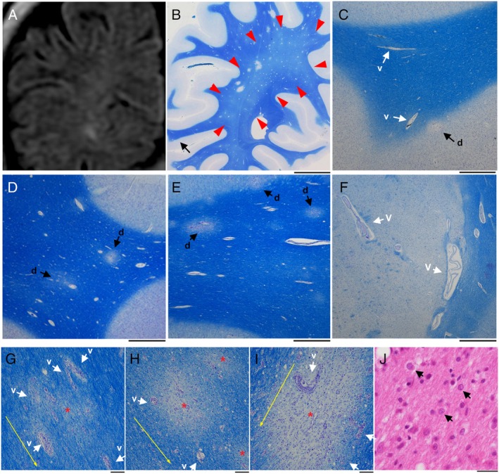
Early and small demyelinating lesions with perivascular edema. (A) An MRI FLAIR image (1.5 T). A coronal view of the right occipital lobe on day 147 suggests unclear signal intensity in the deep white matter. (B‐I) Semimacroscopic images of autopsied brain sections stained with Klüver‐Barrera. The lesions are scattered, and red arrows indicate myelin pallor (B). At a higher magnification of a part of panel B (black arrow in panel B), the lesions are present at the corticomedullary border (indicated by “d” and black arrow), and mildly enlarged blood vessels are also seen in this region (indicated by “v” and white arrow) (C). The lesions are also seen in the frontal lobe (indicated by “d” and black arrows) along with mildly enlarged blood vessels (D, E). Blood vessels in the left thalamus are enlarged and perivascular edema is seen. This resembles a lacular infarct (indicated by “v” and white arrows) (F). The lesions are present along with vascular proliferation in the surrounding area (indicated by “v” and white arrows), and red asterisks and yellow arrows indicate the centers of demyelinating lesions and the direction of nerve fiber extension, respectively (G‐I). (J) A microscopic image of a brain section stained with hematoxylin‐eosin. Oligodendroglia‐like cells with mildly enlarged nuclei are present (arrows) around early and small demyelinating lesions. Some of these cells bear dot‐shaped intrauclear structures. Scale bars: 1 cm (B), 1 mm (C‐F), 100 μm (G‐I), 25 μm (J).
