

Abstract
Introduction:
Achilles tendon injuries usually occur with abrupt movements at the level of the ankle and foot, and the consequence is the overload of the Achilles tendon.
Aim:
Examine the Achilles tendon load as a function of the landing angle, and find the critical point at which the tendon overload begins and when a further increase in the landing angle can lead to rupture.
Methods:
The study has a prospective character. The input data represent the anthropometric values of the respondents, who are professional basketball players in the senior national team of Bosnia and Herzegovina and were processed in the CATIA v5-6 software solution. Software data processing analyzed the landing angles and the transfer of force to the Achilles tendon. The end result is a regression curve, which projects the angle at which the Achilles tendon is overloaded, and indicates an increased risk of possible injury to the tendon itself.
Results:
The onset of overloading starts at an angle of 32.28° and at an angle of 35.75° the overloaded load occurs, indicating the need for the subject to change the position of the foot to prevent damage to the tendon itself.
Conclusion:
An angle of 35.75° is the critical point at which the Achilles tendons are overloaded at the very landing. Prevention of injury should go in the direction of practicing the feet for a particular position at the time of the landing, and in the direction to develop adequate footwear that would mitigate the angle at the landing.
Keywords: Achilles tendon, injury, rupture, regression analysis
1. INTRODUCTION
Prevention of injuries is one of the most important issues in modern sports medicine. Multidisciplinary approach to this issue is imperative along with understanding of overloading of some parts of lower extremities. The degree of loading of certain tendons is the subject of research, and the search for the critical point at which the rupture itself occurs must be individualized, because, despite the development of modern medicine, that critical point still cannot be found. The increasing number of recreational athletes who exercise regularly, and the increasing number of amateurs who exercise at irregular intervals, is affecting the increase in injuries throughout the whole locomotor system. The incidence of injury to the Achilles tendon is increasing, but the frequency of need for surgical treatment is decreasing due to the increase of the prevention of injury itself, with the increasing development of conservative treatment, depending on the type of rupture (1, 2).
Adequate load is a must in today’s world of planning the sports training. However, even in high elite sports injuries occur (for example, one of the best Bosnian-Herzegovinian basketball player Jusuf Nurkic (Portland Trail Blazers, National Basketball Association) was injured during landing in a match against Brooklyn Nets on March 26, 2019.
The incidence of injury to the Achilles tendon is increasing, but the frequency of need for surgical treatment is decreasing due to the increase of the prevention of injury itself, with the increasing development of conservative treatment, depending on the type of rupture (2). The etiology of the injury is still unknown. It is understood in two directions, first that the injury is the culmination of the degenerative process, and second that it is due to an overload on the Achilles tendon itself (2, 3). Acute Achilles tendon injuries affect a healthy population as well as increasingly well-trained professional athletes (although it is very common in under-trained individuals, recreational athletes) (1). The predisposing factor can also be sought in external etiologic causes, first of all using fluoroquinolones, corticosteroids or opiates (4). Gender (more common in the male sex), as well as biomechanical changes in the tendon itself that are formed with age, are believed to increase the incidence of acute injury (5, 6). The Achilles tendon is the strongest tendon in the human body, it is part of the gastrocnemius and soleus, and connects them to the calcaneus. The narrowest part of the tendon is 4 cm, and the length of the tendon is about 15 cm (the size varies greatly from person to person and the physical constitution of the person, is wrapped around the fascia and is behind the bone elements, while the space between them is filled with connective and adipose tissue) (7). At the adhesion to the heel bone, it is slightly wider, narrows towards the center, and gradually begins to expand as it approaches the muscles (7). Although it is thought that during the activity, the Achilles tendon can withstand a load of 3500 N, its rupture often occurs at the least effort, at a certain position, and in trained persons, even much stronger forces do not cause its injury (2, 3, 8–10). About 90% of its content is made up of type I (10) collagen, which forms the structure of fibrils, fibers, and fascicles bound together by small matrix molecules (proteoglycans) (11), while the adhesions themselves are made of type II, IX and X collagens (type X localized in the mineralized zone and type IX distributed throughout) (12). The task of collagen fibers is to withstand loads, but because they are much longer in diameter, they are not effective in transferring compressive loads, and they act as springs and thus store energy by pulling the fiber (11).
When the force ceases to act, the stored energy is used to return the fibers to their initial position (10, 11). The elastic properties of the tendon are thought to dominate (12). These tendons can also store and deliver energy with very high efficiency and this is especially evident with the Achilles tendon at walking, and the ability of the tendon to stretch allows the muscle to work with little or no effort by varying in length which enables it to create a greater force (13). The mechanical properties of tendons depend on the diameter and orientation of the collagen fibers and vary depending on the requirements that the tendon must meet (12, 13).
At rest, collagen fibers are lined up in parallel, and when stretched they are straightened to the elastic limit and return to their initial state after removal of load (13). It can be restored to its original state by slight unloading (13). The tendon consumes more energy to lengthen during rapid loading, and releases less energy when it is slowly unloaded (13). The rupture treatment can be surgical and conservative, and the surgical can be performed into four categories of open repair, percutaneous repair, mini-open repair, and augmentative repair (14). Surgical treatment is an option for the younger population (depending on whether it is partial or total rupture), and for those patients who require normal tolerance in daily life (14). After surgical treatment, the imperative is placed on adequate physical treatment, with an attempt to restore the Achilles tendon properties to the stage they were before the injury (14). Prevention in professional sport itself is imperative, as it is believed that after an injury, the performance of the athlete is diminished, even with a protocol-based repair (15). Increasingly, tendinopathies and prevention of the injury itself through physical exercise, adequate sports training and an individual approach to developing footwear that can alleviate the loads of the Achilles tendon and prevent accidental injury (15) are increasingly in focus.
At which angle, at which jump or landing, at which position, at what force does the injury occur, there are contradictory views, although the common site of rupture is considered to be in the region 3 to 6 cm above the os calcis (16), during strong, explosive movement from the resting state (10, 16), and in the eccentric phase of the muscular work, when the tendon is elongated and due to excessive tension and added force not to it, often during dorsal flexion of the feet that had previously been in plantar flexion (17) . Histologically, higher collagen III and a smaller amount of collagen I have been observed in those tendons that have ruptured (18).
2. AIM
The aim of this study is to examine the tendon load as a function of landing angle, and to find the critical point at which the tendon overload begins, when further increase of the landing angle can result in damage to the Achilles tendon.
3. METHODS
The study has a prospective character, and the data were processed in the CATIA v5-6 software solution (Dassault Systémes, Vélizy-Villacoublay, France). The input data represent anthropometric values of the respondents, who are professional basketball players in the senior national team of Bosnia and Herzegovina. Software analysis of the height and weight of the subjects, as well as the length of the feet, and analyzed the angles of incidence and the transfer of force to the Achilles tendon. The final result is a regression curve (obtained with our own software solution, using MATLAB (version 9.4, MathWorks, Natick, Massachusetts, United States of America) and Microsoft Excel (version 11, Microsoft Corporation, Redmond, Washington, USA), which indicates to the landing angle of the Achilles tendon itself. RULA analysis (CATIA v5-6) was used to confirm the results obtained.
4. RESULTS
Table 1 shows the anthropometric measures of the respondents. The position of the basketball player during the shot was analyzed (Figure 1) and an identical model was presented in the CATIA software (Figure 2). A model of the average respondent, who is 200 cm tall, 97 kg in weight, with a foot length of 28 cm, was obtained.
Table 1. Respondents’ anthropometric parameters .
| No. | The height of the respondents (cm) | The body mass of respondents(kg) | Foot length (cm) |
|---|---|---|---|
| 1. | 179 | 80 | 26 |
| 2. | 200 | 94 | 28.8 |
| 3. | 180 | 75 | 24.1 |
| 4. | 208 | 114 | 31.1 |
| 5. | 200 | 91 | 28.5 |
| 6. | 206 | 100 | 26.9 |
| 7. | 192 | 82 | 25.5 |
| 8. | 208 | 127 | 29 |
| 9. | 214 | 107 | 31 |
| 10. | 206 | 109 | 30.8 |
| 11. | 207 | 100 | 28 |
| 12. | 203 | 95 | 28.1 |
| 13. | 204 | 90 | 28 |
| 200.5385±14.504 | 97.2308±19.764 | 28.1385±2,.899 |
Figure 1. Position of the basketball player being analyzed – left foot.
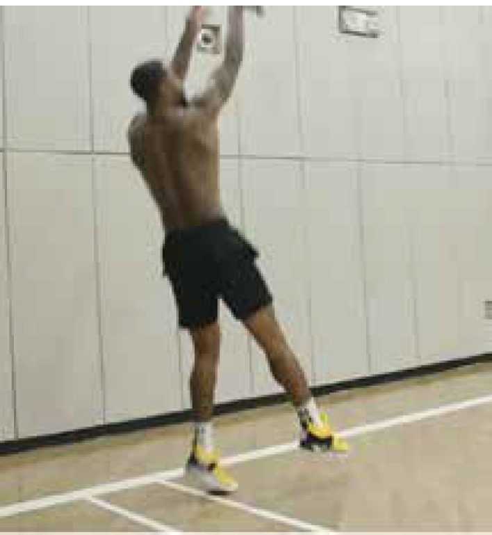
Figure 2. Identical model of the basketball player being analyzed – left foot.

Height of respondent h=200 cm
Mass of respondent Qt=97 kg Mo = Bo + B1*M + B2*M (kg) Pattern Donskij Zacijorskij of the foot:
-0.829 + 0.0077 97 + 0.0073 200 = 1.3779 kg, adopted
1.4 kg; (weight 14 N).
One foot: 0.7 kg; (weight 7 N)
To determine the stress, it is necessary to first determine the forces in the tendon, and the necessary reaction force of the substrate was obtained from CATIA software, anthropometric values were entered at the beginning of the process. Then from the equilibrium conditions where the reaction of the substrate (R) is known, the weight of the foot (G, Donskij-Zaciorskij pattern), the half body weight (Q), and the force in the tendon depending on the angle (T) can be determined, so that angle of 10° have load T10 =-9N, for angle of 20° have load T20 =-717N, for angle 35,75° the load of T35 =519N.
In order to solve the set system, or to determine the regression coefficients, the MATLAB system can be used. The same solution can be achieved with the „Polynomial Trend/Regression Type“ function of the Microsoft Office Excel module.
The fact that the chart of the function is correct is confirmed by the load on the Achilles tendon as a function of angle, via RULA analysis. For the load angle a α= 10°, the musculature (tendon) is colored orange (load within permitted limits, scale 5, with small corrections), which coincides with the function graph.
For a loading angle of a α= 20°, the musculature (tendon) is colored in orange, which corresponds to a scale of 6 (load within the allowed limits, but with large adjustments to the foot position) which coincides with the function graph.
Finally, in Figure 7 for the load angle α = 35.75°, the musculature (tendon) is colored red (overload, scale 7), so the position of the foot should be changed immediately to avoid damage to the tendon, which also coincides with the graph functions.
Figure 7. Rula analysis for an angle of 35,750.

Angle range: aα = 32,28°-35,75° represents the range in which the Achilles tendon load becomes more severe, and represents the range that can lead to possible tendon rupture.
5. DISCUSSION
This study presents the calculation of the forces and strains of the Achilles tendon at different basketball landing angles. Although the Achilles tendon is considered to be the strongest tendon it has the risk of rupture especially in athletes during training or long-term effort during matches. Modern software programs, and one of them is the Catia software program, which allows us to find the load forces of the Achilles tendon for any angle at landing in a basketball player according to the values of its average weight, average height and average foot length, is the found angle, respectively, a range of angles, between 32.28°-35.75°, which leads to an increased load, that is, an overload of the tendon, and in the case of stronger force, rupture could occur, and the effect of chronic force of moderate intensity would lead to degenerative changes that would predispose to possible rupture. Stretching is thought to lead to changes that could eventually lead to rupture. The configuration is lost if the elongation exceeds 2% but recovers if maintained below 4%, but if the 8% elongation results in macroscopic rupture (20).
Mafulli et al. stated that tendon vasculature, gastrocnemius – soleus dysfunction, age, sex, weight and height, pes cavus, and lateral side stability are intrinsic factors for rupture formation (20). They state that this rupture usually occurs 2-6 cm above the site of calcaneus attachment, and states that the incidence of rupture is increasing, primarily because of the change in the lifestyle of modern humans, and that as much as 75% of all ruptures occur during recreational activities (20). Extrinsic factors refer to the form of training itself, poor exercise technique, previous injuries, footwear, and the very background on which the activity is performed (21, 22).
The formation of tendinopathies is also conditioned by pathohistological changes, in the form of collagen degeneration, thinning in tendinopathy is characterized by collagen degeneration and an increase in the amount of glycosaminoglycans (22). Klatte-Schulz et al. have shown a higher concentration of adipose tissue in tendinopathies than in acute ruptures, and in acute ruptures there was less blood flow than intact tendons, between collagen fibrils, and very low blood flow in acute ruptures around the edges (18).
A number of inflammatory cells have been observed in intact tendons, but which were under chronic inflammatory process (18). All this indicates that acute rupture can also occur conditionally in a healthy tendon, regardless of the absence of degenerative changes, and that the degenerative changes themselves lead to chronic tendon alteration and predispose to a possible rupture (2, 8, 10, 12, 18).
The angle we get obtained represent the angle at which the load occurs and lead to these degenerative changes, and that angle could also be one of the predisposing factors leading to the rupture. On the other hand, the inputs represent measures of professional athletes, and certainly continued research could be based on differences in load angle between healthy populations and professional athletes. Although the force acting on the load angle range is much smaller, compared to the possible load from the literature, this supports a large number of unexplained ruptures (either partial or total) even at a not so large load (23).
Thanks to the biomechanical analysis performed in the study, the feet can be practiced in relation to the jump and landing angles, as well as input data can be provided for the stated stresses in the design of sports equipment, e.g. sneakers, in order to attenuate these stresses, decreased the likelihood of Achilles tendon deformity. Reducing the angle between the sole and the feet of the individual could be a solution and therapeutic effect.
6. CONCLUSION
The onset of overload begins with an angle of 32.28°, and at an angle of 35.75° the overload occurs, indicating the need for the subject to change the position of the foot to prevent the damage to the tendon itself. Prevention should go in the direction of practicing the feet for a particular position at the very point of the landing, and in the direction to develop adequate footwear that would mitigate the angle at the landing itself, as well as eccentric strengthening of the tendon.
Figure 3. Skeleton and foot lever with shown forces.

Figure 4. Substrate reaction for angle 35.750.

Figure 5. Substrate response for angle 200 (left) and angle of 100 (right).

Figure 6. Polynomial regression analysis, chart of Achilles tendon function (X-axis-angle, Y-axis values of tendon strain force).

Table 2. Tension force of the tendon at certain angles.
| ANGLE (°) | The force; in the tendon(N) |
|---|---|
| 10 | -9 |
| 20 | -717 |
| 35.75 | 519 |
Author’s contribution:
R.K. contributed equally to this work with F.V.. F.V., R.K., S.B., S.B., E.B., Dz.J., and I.M. gave substantial contribution to the conception or design of the work and in the acquisition, analysis and interpretation of data for the work. R.K, E.B., A.V., S.B. and F.V. had main role in drafting the work and revising it critically for important intellectual content. Each author gave final approval of the version to be published and they agree to be accountable for all aspects of the work in ensuring that questions related to the accuracy or integrity of any part of the work are appropriately investigated and resolved.
Conflicts of interest:
There are no conflicts of interest.
Financial support and sponsorship:
Nil.
REFERENCES
- 1.Yang X, Meng H, Quan Q, Peng J, Lu S, Wang A. Management of acute Achilles tendon ruptures: A review. Bone Joint Res. 2018;7(10):561–69. doi: 10.1302/2046-3758.710.BJR-2018-0004.R2. [DOI] [PMC free article] [PubMed] [Google Scholar]
- 2.Egger AC, Berkowitz MJ. Achilles tendon injuries. Curr Rev Musculoskelet Med. 2017;10(1):72–80. doi: 10.1007/s12178-017-9386-7. [DOI] [PMC free article] [PubMed] [Google Scholar]
- 3.Arner O, Lindholm A, Orell SR. Histologic changes in subcutaneous rupture of the Achilles tendon; a study of 74 cases. Acta Chir Scand. 1959;116(5-6):484–490. [PubMed] [Google Scholar]
- 4.Sode J, Obel N, Hallas J, Lassen A. Use of fluroquinolone and risk of Achilles tendon rupture: a population-based cohort study. Eur J Clin Pharmacol. 2007;63(5):499–503. doi: 10.1007/s00228-007-0265-9. [DOI] [PubMed] [Google Scholar]
- 5.Vosseller JT, Ellis SJ, Levine DS, et al. Achilles tendon rupture in women. Foot Ankle Int. 2013;34:49–53. doi: 10.1177/1071100712460223. [DOI] [PubMed] [Google Scholar]
- 6.Ganestam A, Kallemose T, Troelsen A, Barfod KW. Increasing incidence of acute Achilles tendon rupture and a noticeable decline in surgical treatment from 1994 to 2013. A nationwide registry study of 33,160 patients. Knee Surg Sports Traumatol Arthrosc. 2016;24:3730–3737. doi: 10.1007/s00167-015-3544-5. [DOI] [PubMed] [Google Scholar]
- 7.Lin Y, Yang L, Yin L, Duan X. Surgical Strategy for the Chronic Achilles Tendon Rupture. Biomed Res Int. 2016;2016:1416971. doi: 10.1155/2016/1416971. [DOI] [PMC free article] [PubMed] [Google Scholar]
- 8.Fukashiro S, Komi PV, Jarvinen M, Miyashita M. In vivo Achilles tendon loading during jumping in humans. Eur J Appl Physiol Occup Physiol. 1995;71:453–458. doi: 10.1007/BF00635880. [DOI] [PubMed] [Google Scholar]
- 9.Maffulli N. Rupture of the Achilles tendon. J Bone Joint Surg Am. 1999;81:1019–1036. doi: 10.2106/00004623-199907000-00017. [DOI] [PubMed] [Google Scholar]
- 10.Freedman BR, Gordon JA, Soslowsky LJ. The Achilles tendon: fundamental properties and mechanisms governing healing. Muscles Ligaments Tendons J. 2014;4(2):245–255. [PMC free article] [PubMed] [Google Scholar]
- 11.Longo UG, Ronga M, Maffulli N. Achilles tendinopathy. Sports Med Arthrosc. 2009;17:112–126. doi: 10.1097/JSA.0b013e3181a3d625. [DOI] [PubMed] [Google Scholar]
- 12.Fukuta S, Oyama M, Kavalkovich K, Fu FH, Niyibizi C. Identification of types II, IX and X collagens at the insertion site of the bovine achilles tendon. Matrix Biol. 1998;17:65–73. doi: 10.1016/s0945-053x(98)90125-1. [DOI] [PubMed] [Google Scholar]
- 13.Peltonen J, Cronin NJ, Stenroth L, Finni T, Avela J. Viscoelastic properties of the Achilles tendon in vivo. Springerplus. 2013;2:212. doi: 10.1186/2193-1801-2-212. [DOI] [PMC free article] [PubMed] [Google Scholar]
- 14.Egger AC, Berkowitz MJ. Achilles tendon injuries. Curr Rev Musculoskelet Med. 2017;10(1):72–80. doi: 10.1007/s12178-017-9386-7. [DOI] [PMC free article] [PubMed] [Google Scholar]
- 15.Amin NH, McCullough KC, Mills GL, et al. The Impact and Functional Outcomes of Achilles Tendon Pathology in National Basketball Association Players. Clin Res Foot Ankle. 2016;4(3):205. doi: 10.4172/2329-910X.1000205. [DOI] [PMC free article] [PubMed] [Google Scholar]
- 16.Beddy P, Dunne R, de Blacam C. Achilles wiiitis. AJR Am J Roentgenol. 2009;192:W79. doi: 10.2214/AJR.08.1654. [DOI] [PubMed] [Google Scholar]
- 17.Maquirriain J. Achilles tendon rupture: avoiding tendon lengthening during surgical repair and rehabilitation. Yale J Biol Med. 2011;84(3):289–300. [PMC free article] [PubMed] [Google Scholar]
- 18.Klatte-Schulz F, Minkwitz S, Schmock A, et al. Different Achilles Tendon Pathologies Show Distinct Histological and Molecular Characteristics. Int J Mol Sci. 2018;19(2):404. doi: 10.3390/ijms19020404. [DOI] [PMC free article] [PubMed] [Google Scholar]
- 19.Donskij DD, Zacijorskij VM. Biomehanika Izdateljstvo Fizkultura i sport. Moskva, Rusija: 1979. [Google Scholar]
- 20.Maffulli N, Sharma P, Luscombe KL. Achilles tendinopathy: aetiology and management. J R Soc Med. 2004;97(10):472–476. doi: 10.1258/jrsm.97.10.472. [DOI] [PMC free article] [PubMed] [Google Scholar]
- 21.Selvanetti ACM, Puddu G. Overuse tendon injuries: basic science and classification. Operative Techniques Sports Med. 1997;5:110–117. [Google Scholar]
- 22.Leadbetter WB. Cell-matrix response in tendon injury. Clin Sports Med. 1992;11:533–578. [PubMed] [Google Scholar]
- 23.Longo UG, Ronga M, Maffulli N. Acute ruptures of the Achilles tendon. Sports Med Arthrosc. 2009;17(2):127–138. doi: 10.1097/JSA.0b013e3181a3d767. [DOI] [PubMed] [Google Scholar]