Abstract
Oncocytic sialolipoma is a very rare tumor of which only three cases have been reported. This entity is considered to be a variant of sialolipoma which harbours oncocytic nodules within a well-circumscribed lipomatous mass. We report a parotid mass in 73-year-old female that was difficult to diagnose in imaging and on biopsy. Ultrasonography and MRI demonstrated a mass with features thought to be consistent with lipoma. Twice needle core biopsies were performed. Both were indefinite for diagnosis. The first report favoured a lipoma and the second report suggested the lesion represented oncocytic hyperplasia or an oncocytoma. The microscopic examination of the excised surgical specimen demonstrated typical features of oncocytic sialolipoma, characterized by a predominately lipomatous component, sparse normal-appearing salivary gland tissue and multiple oncocytic nodules. This is the second case of oncocytic sialolipoma reported to occur in the parotid gland.
Keywords: Oncocytic, Sialolipoma, Parotid
Introduction
Salivary gland neoplasms account for less than 3% of all neoplasms of head and neck region [1]. The majority of both benign and malignant tumors occur in the parotid gland. Although the majority of the tumors are of epithelial cell origin, approximately 2–5%, develop from mesenchymal cells [2]. Lipoma can be present in the salivary gland, but it is very rare, and such lesions account for less than 0.5% of all tumours that occur in the parotid gland [3]. Apart from conventional lipoma, other variants of lipoma have been reported in salivary gland, including fibrolipoma [4], angiolipoma [5], spindle cell lipoma [6] and the recent entity, sialolipoma [7]. The name ‘sialolipoma’ was coined in 2001, characterized by a well-circumscribed and encapsulated tumor with a predominately adipocytic component and entrapped normal salivary gland tissue [7]. In 2009, Pusiol et al. described a sialolipoma containing oncocytic nodules and this tumor was named ‘Oncocytic sialolipoma’ [8]. From 2009 until present, two cases have been added to this unique entity [9, 10]. This case report will represent the fourth case of oncocytic sialolipoma.
Case Report
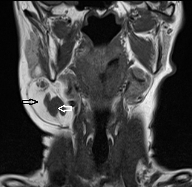
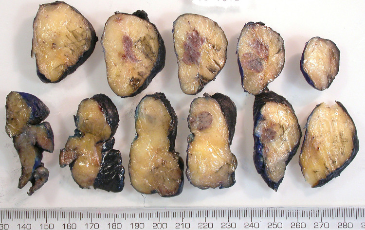
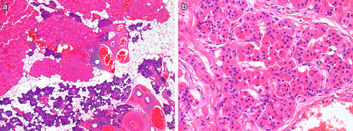
The patient was 73-year-old female with history of epilepsy for 60 years and she was a non-smoker. On examination, she had an irregularly nodular mass, measuring 4.7 × 4 × 4 cm in right parotid area extending inferiorly to lateral neck. Ultrasonography showed what was thought to be a lipoma in right parotid gland which appeared to extend inferiorly into the lateral neck and submandibular space. At the inferior level, the echogenicity of the lesion was similar to surrounding fat and, therefore, difficult to differentiate from adjacent tissue. MRI was subsequently performed. T1w coronal (Fig. 1) and T1w axial images of the head and neck demonstrated an exophytic mass extending inferiorly from the right tail of parotid gland (Fig. 1). The lesion lay posterior to the submandibular gland and anterior to the sternocleidomastoid muscle with lateral bulging of the superficial cervical fascia. The mass demonstrated predominantly T1w high signal (black open arrow in Fig. 1, which was isointense to fat and nulled on other fat suppressed sequences), whilst there was an additional lobulated T1w isointense component (white open arrow in Fig. 1).The patient underwent needle core biopsy twice. The first specimen showed parotid type salivary gland tissue with benign adipose tissue. Focal oncocytic change associated with mild lymphocytic infiltrate was noted. Therefore lipoma could not be confirmed on this biopsy. The histologic findings of the second biopsy demonstrated closely packed oncocytic cells which were surrounded by benign fibroadipose tissue. The features suggested as an oncocytoma or dominant nodule in oncocytic hyperplasia. After MDM discussion, Parotidectomy to remove the mass in deep lobe of right parotid gland was performed. Gross examination of the surgical specimen revealed a well-circumscribed and lobulated soft yellow fatty mass, measuring 7.5 cm in maximum dimension. The cut surface revealed two separate oval shaped light tan nodules in the fatty tissue (Fig. 2). The microscopic examination confirmed a partially encapsulated mass with a fibrous capsule of variable thickness. The majority of the mass consisted of mature adipocytes with sparse normal parotid gland tissue, containing lobules of serous acini, salivary ducts and myoepithelial cells. The normal salivary gland tissue was located at the periphery of the mass and adjacent to the two oncocytic nodules. The oncocytic cells were arranged in lobules with intervening by fibrous septa (Fig. 3a). The oncocytes possessed monotonous round nuclei with granular chromatin, conspicuous nucleoli and abundant granular eosinophilic cytoplasm (Fig. 3b). A lymphocytic infiltrate was present within oncocytic nodules and in the normal salivary gland tissue. Focal atrophic change and fibrosis were also present. No sebaceous differentiation was identified. The fat component was estimated approximately 85% of the total tumor volume. Immunohistochemistry was not considered necessary because the histologic features were sufficient for the diagnosis of ‘Oncocytic sialolipoma’.
Fig. 1.

T1-weighted magnetic resonance imaging of head and neck. T1w coronal image showed an exophytic mass extending inferiorly from the tail of right parotid. The mass demonstrated T1w high signal (black open arrow), which was isointense to fat and there was an additional lobulated T1w isointense component (white open arrow)
Fig. 2.
Gross pathology. The tumor had yellow fatty cut surface with two separate well-circumscribed light tan nodules
Fig. 3.
Microscopy demonstrated a nodule, comprising oncocytes arranged in lobules. Benign appearing salivary gland tissue was located near the oncocytic nodule (a hematoxylin and eosin, magnification × 100). The oncocytes possessed monotonous round nuclei and abundant granular eosinophilic cytoplasm (b hematoxylin and eosin, magnification × 200)
Discussion
Lipomatous lesions of salivary gland are divided into two categories, true lipomatous neoplasms, or typical lipoma, and lipomatous tumors with epithelial component, called ‘lipoepithelial’ neoplasms. The latter group has been further classified into sialolipoma and oncocytic lipoadenoma [10]. Sialolipoma was firstly described in 2001 by Nagao et al. This neoplasm is characterized by a well-defined and encapsulated lipomatous tumor with entrapped normal salivary gland tissue morphologically similar to normal salivary gland parenchyma [7]. Sialolipoma can arise in either major or minor salivary glands, and the parotid gland is the most common location to occur [11]. At present, 30 cases of parotid sialolipoma have been reported (summarized in Table 1) [7, 10–28]. The age of patients ranged from 6 weeks to 74 years old (mean age: 39.4 years) with men and women almost equally affected. The tumor more commonly occurred on the left side than on the right side (n = 18; 64.3%). Tumor sizes ranged from 10 to 90 mm (mean size: 46.2 mm). All tumors were encapsulated by a thin fibrous capsule. In all cases, the tumors were composed of mature adipose tissue with entrapped salivary gland tissue, including salivary ducts and serous acini with underlying myoepithelial cells. The proportion of fatty tissue within the mass was variable, ranging from 50% to more than 90% (mean: 82.3%). The other morphologic findings reported in this entity were lymphocytic infiltrate (n = 14, 56%), atrophic change of salivary duct and acini (n = 12, 48%), fibrosis (n = 10, 40%), dilated salivary ducts (n = 5, 20%), sebaceous differentiation (n = 6, 24%), oncocytic metaplasia (n = 5, 20%), squamous metaplasia (n = 2, 8%) and oncocytic nodule (n = 2, 8%). A sialolipoma containing an oncocytic nodule was first reported in 2009 by Pusiol et al. and this tumor was called ‘oncocytic sialolipoma’ [8]. Our case demonstrated the typical morphologic features previously described for oncocytic sialolipoma [8, 9]. There was a large proportion of mature adipocytic tissue, sparse normal-appearing salivary gland parenchyma and oncocytic nodules.
Table 1.
Reported cases of parotid sialolipoma
| Case # | Reference | Age | Sex | Site | Size (mm) | % fat | Additional findings |
|---|---|---|---|---|---|---|---|
| 1 | Nagao et al. [7] | 20 | M | Rt | 35 | 90 | Atrophy, squamous metaplasia, sebaceous differentiation |
| 2 | 45 | F | Lt | 60 | 90 | Atrophy | |
| 3 | 67 | M | Rt | 17 | 90 | Atrophy, lymphocytic infiltration | |
| 4 | 66 | F | Lt | 60 | > 90 | Atrophy, oncocytic metaplasia | |
| 5 | 42 | M | Lt | 60 | > 90 | Atrophy | |
| 6 | Hornigold et al. [12] | 7 wk | F | Lt | 35 | ND | Atrophy, fibrosis, dilated duct, lymphocytic infiltration |
| 7 | Kadivar et al. [13] | 3 | F | Lt | 30 | ND | Atrophy, fibrosis, dilated duct, squamous metaplasia, sebaceous differentiation, lymphocytic infiltration |
| 8 | Mazlumoglu et al. [14] | 10 wk | F | Lt | 40 | ND | Atrophy, fibrosis, dilated duct, lymphocytic infiltration |
| 9 | Arakeri et al. [15] | 1 | M | ND | 80 | ND | Fibrosis, lymphocytic infiltration |
| 10 | Michaelidis et al. [16] | 44 | M | Rt | 35 | ND | No |
| 11 | Doğan et al. [17] | 33 | M | Lt | 26 | ND | Atrophy, fibrosis, dilated duct |
| 12 | Eldamati et al. [18] | 38 | F | Lt | 48 | 90 | No |
| 13 | Baker et al. [19] | 44 | M | Rt | 10 | ND | ND |
| 14 | Walts et al. [20] | 48 | M | Lt | 35 | ND | ND |
| 15 | 65 | M | Lt | 26 | ND | ND | |
| 16 | Lee et al. [21] | 65 | F | Rt | 30 | ND | ND |
| 17 | Ranjan et al. [22] | 52 | M | Lt | 90 | 50 | Oncocytic metaplasia, lymphocytic infiltration |
| 18 | Agaimy et al. [29] | 74 | M | Lt | 15 | 70 | Fibrosis, lymphocytic infiltration |
| 19 | 18 | F | ND | 40 | 80 | Sebaceous differentiation | |
| 20 | 49 | F | Lt | 43 | > 90 | Fibrosis, sebaceous differentiation, lymphocytic infiltration | |
| 21 | 47 | F | Lt | 25 | > 90 | Fibrosis, sebaceous differentiation, lymphocytic infiltration | |
| 22 | 55 | M | ND | 27 | 70 | Oncocytic nodule, sebaceous differentiation, fibrosis, lymphocytic infiltration | |
| 23 | Khazaeni et al. [23] | 45 | F | Rt | 75 | ND | Atrophy |
| 24 | 18 | F | Lt | 50 | ND | Atrophy, lymphocytic infiltration | |
| 25 | Bansal et al. [24] | 11 | M | Lt | 70 | ND | Dilated duct, lymphocytic infiltration |
| 26 | Kidambi et al. [25] | 6 wk | M | Lt | 65 | ND | ND |
| 27 | Qayyum et al. [11] | 69 | M | Rt | 27 | 90 | Oncocytic metaplasia, lymphocytic infiltration |
| 28 | Pandey et al. [26] | 45 | F | Rt | 70 | 70 | Oncocytic metaplasia |
| 29 | Ghafar et al. [27] | 40 | F | Lt | 38 | ND | No |
| 30 | Fritzsche et al. [28] | 43 | M | Rt | 65 | ND | ND |
| 31 | Current case | 73 | F | Rt | 75 | 85 | Atrophy, fibrosis, oncocytic metaplasia, oncocytic nodule, lymphocytic infiltration |
ND no data available
The main differential diagnosis for oncocytic sialolipoma is oncocytic lipoadenoma. The other possible differential diagnosis is nodular oncocytic hyperplasia but this entity has no surrounding capsule. It has been suggested that oncocytic sialolipoma and oncocytic lipoadenoma represent part of the spectrum of the same disease. However, the lesions are distinguishable histologically and epidemiology of sialolipoma, including oncocytic sialolipoma, is different from oncocytic lipoadenoma (summarized in Table 2). The relationship with smoking has not been clearly established for these lesions. The histological features do overlap and descriptions in the literature make it difficult to reliably discriminate oncocytic lipoadenoma from oncocytic sialolipoma or even sialolipoma containing oncocytic metaplasia, which sometimes cause incorrect categorization as seen in a case of oncocytic lipoadenoma reported by Hirokawa et al. that actually was sialolipoma containing oncocytic metaplasia [30]. Both lesions are encapsulated. In oncocytic lipoadenoma the oncocytic component predominates and adipocytes are admixed within oncocytic nests. Normal salivary gland parenchyma associated with oncocytic metaplasia is not usually observed. Usually in oncocytic lipoadenoma, there is a dominant oncocytic nodule. In rare cases, multifocal nodules are described. Therefore, we suggest encapsulated lesions with dominant oncocytic nodule and sparse or absent salivary gland tissue is categorized as oncocytic lipoadenoma. Whereas, those encapsulated lesions with dominant lipomatous component together with one or multiple oncocytic nodules and normal salivary gland component are classified as oncocytic sialolipoma.
Table 2.
Difference in epidemiology between oncocytic lipoadenoma and sialolipoma
| Oncocytic lipoadenoma | Sialolipoma | |
|---|---|---|
| Site | Major gland | Major and minor glands |
| Recurrence | No | Yes |
| Congenital case | No | Yes |
| Sex | M:F = 5:2 | M:F = 8:13 |
The duct and acini present in sialolipoma were shown to have a normal cellular phenotype by both immunohistochemistry and ultrastructural examination by electron microscopy [7]. These findings support the idea that the glandular component is entrapped in the mass during adipocytic proliferation [7]. The histogenesis of sialolipoma was proposed by Akrish et al. who suggested that the development of sialolipoma was associated with dysfunction of salivary gland. The features that support this theory include the long standing duration of disease, acinar and duct component in the tumor exhibiting atrophic change, fibrosis, dilated salivary ducts, oncocytic metaplasia and squamous metaplasia [31]. In oncocytic sialolipoma, the two previously reported cases of oncocytic sialolipoma described the oncocytic nodules as being located adjacent to an entrapped salivary gland component [8, 9] This finding was also present in our case and this demonstrated the relationship between normal entrapped salivary ducts and oncocytes in oncocytic nodules. Thus, multifocal oncocytic nodules are considered to be the consequence of both hyperplastic change and oncocytic metaplasia of entrapped salivary ducts [8, 9]. The immunohistochemical profile of oncocytes in oncocytic sialolipoma is different from the profile of oncocytes in oncocytic lipoadenoma; in terms of, oncocytes in the sialolipoma express CK19 intensely [8] but it is negative in oncocytic lipoadenoma [32, 33]. Interestingly, these CK19-positive oncocytes are also found in oncocytic metaplasia in inflammatory driven condition [34]. This finding might support metaplastic process rather than neoplastic origin. The other proposed pathogenesis of sialolipoma was hamartomatous process [35]. The findings that supported this theory were presence of tortuous thick-walled arterial and venous vessels, resembling in arterio-venous malformation, and numerous nerve bundles [35]. This hypothesis might suggest that congenital sialolipoma is “hamartomatous lesion” instead of a true lipomatous neoplasm.
In our case, the needle core biopsies were not diagnostic but possibly useful in retrospect for the diagnosis oncocytic sialolipoma, in term of retrieving both lipomatous tissue and a part of an oncocytic nodule. Thus, if a salivary needle core biopsy contains both fat and oncocytic parenchymal tissue and the imaging suggests a mass with features suggestive of lipoma, oncocytic sialolipoma should be included in the differential diagnosis.
Acknowledgements
We would like to express our gratitude to Dr. Ann Sandison for providing the pathologic figures and her valuable advices in all the time of gathering information and critical analysis as well as comments on the final manuscript.
Author Contributions
KR gathered information, reviewed literatures and wrote the manuscript. SC interpreted the imaging study and provided imaging pictures with their figure legends. RO performed the surgery and provided clinical information.
Funding
No funding was received.
Compliance with Ethical Standards
Conflict of interest
The authors have no conflicts of interest to declare.
Contributor Information
Komkrit Ruangritchankul, Phone: 662-256-4235, Email: komkritruang@gmail.com.
Steve Connor, Email: Steve.Connor@gstt.nhs.uk.
Richard Oakley, Email: Richard.Oakley@gstt.nhs.uk.
References
- 1.Eveson JW, Cawson RA. Salivary gland tumours. A review of 2410 cases with particular reference to histological types, site, age and sex distribution. J Pathol. 1985;146(1):51–58. doi: 10.1002/path.1711460106. [DOI] [PubMed] [Google Scholar]
- 2.Ilie M, Hofman V, Pedeutour F, Attias R, Santini J, Hofman P. Oncocytic lipoadenoma of the parotid gland: immunohistochemical and cytogenetic analysis. Pathol Res Pract. 2010;206(1):66–72. doi: 10.1016/j.prp.2009.02.008. [DOI] [PubMed] [Google Scholar]
- 3.Ellis GL, Auclair PL. Tumor of the salivary glands. Atlas of tumor pathology. Washington, DC: Armed Forces Institute of Pathology; 1996. [Google Scholar]
- 4.Hatziotis JC. Lipoma of the oral cavity. Oral Surg Oral Med Oral Pathol. 1971;31:511–524. doi: 10.1016/0030-4220(71)90348-3. [DOI] [PubMed] [Google Scholar]
- 5.Reilly JS, Kelly DR, Royal SA. Angiolipoma of the parotid: case report and review. Laryngoscope. 1988;98:818–821. doi: 10.1288/00005537-198808000-00005. [DOI] [PubMed] [Google Scholar]
- 6.Christopoulos P, Nicolatou O, Patrikiou A. Oral spindle cell lipoma: report of a case. Int J Oral Maxillofac Surg. 1989;18:208–209. doi: 10.1016/S0901-5027(89)80054-2. [DOI] [PubMed] [Google Scholar]
- 7.Nagao T, Sugano I, Ishida Y, Asoh A, Munakata S, Yamazaki K, et al. Sialolipoma: a report of seven cases of a new variant of salivary gland lipoma. Histopathology. 2001;38:30–36. doi: 10.1046/j.1365-2559.2001.01054.x. [DOI] [PubMed] [Google Scholar]
- 8.Pusiol T, Franceschetti I, Scialpi M, Piscioli I. Oncocytic sialolipoma of the submandibular gland with sebaceous differentiation: a new pathological entity. Indian J Pathol Microbiol. 2009;52(3):379–382. doi: 10.4103/0377-4929.55000. [DOI] [PubMed] [Google Scholar]
- 9.Ahn D, Park TI, Park J, Heo SJ. Oncocytic sialolipoma of the submandibular gland. Clin Exp Otorhinolaryngol. 2014;7(2):149–152. doi: 10.3342/ceo.2014.7.2.149. [DOI] [PMC free article] [PubMed] [Google Scholar]
- 10.Agaimy A, Ihrier S, Märkl B, Lell M, Zenk J, Hartmann A, et al. Lipomatous salivary gland tumors: a series of 31 cases spanning their morphologic spectrum with emphasis on sialolipoma and oncocytic lipoadenoma. Am J Surg Pathol. 2013;37(1):128–137. doi: 10.1097/PAS.0b013e31826731e0. [DOI] [PubMed] [Google Scholar]
- 11.Qayyum S, Meacham R, Sebelik M, Zafar N. Sialolipoma of the parotid gland: case report with literature review comparing major and minor salivary gland sialolipomas. J Oral Maxillofac Pathol. 2013;17(1):95–97. doi: 10.4103/0973-029X.110687. [DOI] [PMC free article] [PubMed] [Google Scholar]
- 12.Hornigold R, Morgan PR, Pearce A, Gleeson MJ. Congenital sialolipoma of the parotid gland first reported case and review of the literature. Int J Pediatr Otorhinolaryngol. 2005;69:429–434. doi: 10.1016/j.ijporl.2004.10.014. [DOI] [PubMed] [Google Scholar]
- 13.Kadivar M, Shahzadi SZ, Javadi M. Sialolipoma of the parotid gland with diffuse sebaceous differentiation in a female child. Pediatr Dev Pathol. 2007;10(2):138–141. doi: 10.2350/06-05-0091.1. [DOI] [PubMed] [Google Scholar]
- 14.Mazlumoglu MR, Altas E, Oner F, Ucuncu H, Calik M. Congenital sialolipoma in an infant. J Craniofac Surg. 2015;26(8):e696–e697. doi: 10.1097/SCS.0000000000002160. [DOI] [PubMed] [Google Scholar]
- 15.Arakeri SU, Banga S. Sialolipoma of parotid gland in a 1-year-old male child: a case report and review of literature. J Oral Maxillofac Pathol. 2018;22(1):57–59. doi: 10.4103/jomfp.JOMFP_38_15. [DOI] [PMC free article] [PubMed] [Google Scholar]
- 16.Michaelidis IG, Stefanopoulos PK, Sambaziotis D, Zahos MA, Papadimitriou GA. Sialolipoma of the parotid gland. J Craniomaxillofac Sur. 2006;34(1):43–46. doi: 10.1016/j.jcms.2005.08.008. [DOI] [PubMed] [Google Scholar]
- 17.Doğan S, Can IH, Unlü I, Süngü N, Gönültaş MA, Samim EE. Sialolipoma of the parotid gland. J Craniofac Surg. 2009;20(3):847–848. doi: 10.1097/SCS.0b013e3181a2ef7d. [DOI] [PubMed] [Google Scholar]
- 18.Eldamati A, Niaz R, Tanveer S, Zakaria H, AlJazzan N, El-Sharkawy T, et al. Sialolipoma of the superficial lobe of the parotid gland: a case report and literature review. Saudi J Med Med Sci. 2016;4:38–41. doi: 10.4103/1658-631X.170893. [DOI] [PMC free article] [PubMed] [Google Scholar]
- 19.Baker SE, Jensen JL, Correll RW. Lipomas of the parotid gland. Oral Surg Oral Med Oral Pathol. 1981;52:167–171. doi: 10.1016/0030-4220(81)90315-7. [DOI] [PubMed] [Google Scholar]
- 20.Walts AE, Perzik SL. Lipomatous lesions of the parotid area. Arch Otolaryngol. 1976;102:230–232. doi: 10.1001/archotol.1976.00780090072010. [DOI] [PubMed] [Google Scholar]
- 21.Lee PH, Chen JJ, Tsou YA. A recurrent sialolipoma of the parotid gland: a case report. Oncol Lett. 2014;7(6):1981–1983. doi: 10.3892/ol.2014.2026. [DOI] [PMC free article] [PubMed] [Google Scholar]
- 22.Ranjan R, Jha RK, Malua S, Bodra P, Murari K. Sialolipoma of parotid gland, a rare benign tumor. Int J Med Dent Sci. 2015;4(2):886–890. [Google Scholar]
- 23.Khazaeni K, Jafarian AH, Khajehahmadi S, Rahpeyma A, Asadi L. Sialolipoma of salivary glands: two case reports and review of the literature. Dent Res J. 2013;10(1):93–97. doi: 10.4103/1735-3327.111807. [DOI] [PMC free article] [PubMed] [Google Scholar]
- 24.Bansal B, Ramavat AS, Gupta S, Singh S, Sharma A, Gupta K, et al. Congenital sialolipoma of parotid gland: a report of rare and recently described entity with review of literature. Pediatr Dev Pathol. 2007;10(3):244–246. doi: 10.2350/06-09-0170.1. [DOI] [PubMed] [Google Scholar]
- 25.Kidambi T, Been MJ, Maddalozzo J. Congenital sialolipoma of the parotid gland: presentation, diagnosis, and management. Am J Otolaryngol. 2012;33(2):279–281. doi: 10.1016/j.amjoto.2011.07.008. [DOI] [PubMed] [Google Scholar]
- 26.Pandey D, Vats M, Akhtar A, Pathania OP. Sialolipoma of the parotid gland: a rare entity. BMJ Case Rep. 2015. [DOI] [PMC free article] [PubMed]
- 27.Ghafar MA, Tuang G, Mohammad NY, Nadarajan Ch, Abdullah B. Sialolipoma of the parotid gland: an uncommon lipoma variant of salivary gland. Acta Med Bulg. 2018;45(1):39–41. doi: 10.2478/amb-2018-0007. [DOI] [Google Scholar]
- 28.Fritzsche FR, Bode PK, Stinn B, Huber GF, Noske A. Sialolipoma of the parotid gland. Pathologe. 2009;30(6):478–480. doi: 10.1007/s00292-009-1233-1. [DOI] [PubMed] [Google Scholar]
- 29.Agaimy A. Fat-containing salivary gland tumors: a review. Head Neck Pathol. 2013;7:90–96. doi: 10.1007/s12105-013-0459-7. [DOI] [PMC free article] [PubMed] [Google Scholar]
- 30.Hirokawa M, Shimizu M, Manabe T, Ito J, Ogawa S. Oncocytic lipoadenoma of the submandibular gland. Hum Pathol. 1998;29(4):410–412. doi: 10.1016/S0046-8177(98)90125-3. [DOI] [PubMed] [Google Scholar]
- 31.Akrish S, Leiser Y, Shamira D, Peled M. Sialolipoma of the salivary gland: two new cases, literature review, and histogenetic hypothesis. J Oral Maxillofac Surg. 2011;69(5):1380–1384. doi: 10.1016/j.joms.2010.05.010. [DOI] [PubMed] [Google Scholar]
- 32.Lau SK, Thompson LD. Oncocytic lipoadenoma of the salivary gland: a clinicopathologic analysis of 7 cases and review of the literature. Head Neck Pathol. 2015;9(1):39–46. doi: 10.1007/s12105-014-0543-7. [DOI] [PMC free article] [PubMed] [Google Scholar]
- 33.Klieb HBE, Perez-Ordoñez B. Oncocytic lipoadenoma of the parotid gland with sebaceous differentiation: study of its keratin profile. Virchows Arch. 2006;449:722–725. doi: 10.1007/s00428-006-0317-z. [DOI] [PubMed] [Google Scholar]
- 34.Rangel ALCA, León JE, Jorge J, Lopes MA, Vargas PA. Oncocytic metaplasia in inflammatory fibrous hyperplasia: Histopathological and immunohistochemical analysis. Med Oral Patol Oral Cir Bucal. 2008;13(3):E151–E155. [PubMed] [Google Scholar]
- 35.Parente P, Longobardi G, Bigotti G. Hamartomatous sialolipoma of the submandibular gland: case report. Br J Oral Maxillofac Surg. 2008;46:599–600. doi: 10.1016/j.bjoms.2008.02.006. [DOI] [PubMed] [Google Scholar]


