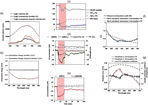
Fig. 2.
An example of the optical data from a piglet (LWP483). (a) Incident () and transmitted light spectra ( and ) through the brain during baseline and HI. The significantly reduced photon count in and is due to the optical properties of the piglet brain. The increased peak count in relative to is due to less absorption during HI. (b) Attenuation spectra measured at baseline and insult. There is no major change in the attenuation spectrum relative to the first measurement at baseline (). However, during HI, there is a significant change in the magnitude and the shape of the attenuation spectrum due to the physiological changes in the brain, i.e., reduction in blood flow and oxygenation (). (c) Changes in the systemic physiological data at baseline, HI, and recovery. (d) and (e) Changes in the bNIRS measurements baseline, HI, and the recovery period as measured by miniCYRIL based on second derivative spectroscopy for pathlength change measurement. The shaded area indicates the HI period. There is no change in the optical pathlength during HI and recovery. (f) Measured attenuation spectrum at HI and back-calculated attenuations from two- and three-chromophore fit. (g) Residual errors from two- and three-chromophore fit. The two-chromophore fit residual is similar to the extinction coefficient of oxCCO, whereas the three-chromophore fit residual error is approximately zero.

