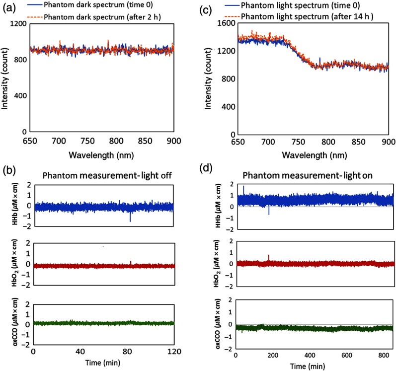
Fig. 6.
Phantom measurements in dark and light showing the stability of the miniCYRIL system. (a) and (b) The spectral data and concentration changes in a tissue-like phantom ( and , ) for 2 h when the shutter of the light source is closed. (c) and (d) The spectral data and concentration measurements for 14 h, when the same phantom is illuminated (shutter open). Integration time for both measurements is 1 s and SDS is 4 cm.

