Abstract
Enterococcus faecalis is a lactic acid bacterium characterized by its tolerance of very diverse environmental conditions, a property that allows it to colonize many different habitats. This species can be found in food products, especially in fermented foods where it plays an important role as a biopreservative and influences the development of organoleptic characteristics. However, E. faecalis also produces the biogenic amines tyramine and putrescine. The consumption of food with high concentrations of these compounds can cause health problems. The present work reports the construction, via homologous recombination, of a double mutant of E. faecalis in which the clusters involved in tyramine and putrescine synthesis (which are located in different regions of the chromosome) are no longer present. Analyses showed the double mutant to grow and adhere to intestinal cells normally, and that the elimination of genes involved in the production of tyramine and putrescine has no effect on the expression of other genes.
Subject terms: Applied microbiology, Molecular biology, Applied microbiology, Molecular biology
Introduction
Enterococcus faecalis is a Gram-positive bacterium of the phylum Firmicutes. It is tolerant of very diverse environmental conditions, which allows it to colonize many different habitats, including water, soil, the gastrointestinal tract (GIT) of different animals (from insects to mammals, including humans)1, and food products, especially fermented foods. E. faecalis is a member of the lactic acid bacteria (LAB), which are responsible for numerous food fermentations. These bacteria therefore play a role as biopreservatives. Unlike for most LAB, however, allowing the presence of E. faecalis in food is controversial. In artisanal cheeses, E. faecalis is believed involved in the ripening process via its proteolytic activity, and in the development of desirable aromas and flavours. It is also an important producer of bacteriocins with activity against pathogenic bacteria and food spoilage microorganisms, making it a potential biopreservative2. Some strains of E. faecalis are even considered to benefit human health3. For example, E. faecalis Symbioflor 1 has been marketed as a probiotic for more than 50 years4. However, some strains have virulence factors and are resistant to antibiotics, and have caused serious infections, especially in the hospital environment5. Indeed, vancomycin-resistant enterococci are regarded as a serious threat to human health by the World Health Organization. In fermented foods such as cheese, E. faecalis is also the bacterium largely responsible for the accumulation of tyramine and putrescine6–8, biogenic amines (BA) that can reach concentrations so high that they cause headaches, migraines and even hypertensive crises9. Putrescine and especially tyramine10,11 are also cytotoxic at concentrations easily reached in cheese. Furthermore, tyramine is genotoxic for intestinal cells in vitro; it might therefore play a role in the promotion of intestinal cancer12.
In E. faecalis, putrescine is made via the agmatine deiminase pathway, in which agmatine is deiminated to putrescine with the concomitant formation of CO2, ATP and ammonium ions13. This route provides energy to the cell in the form of ATP as well resistance to acid stress14. The genes involved in this pathway make up the agdi cluster; aguD codes for the agmatine/putrescine antiporter, aguA for agmatine deiminase, aguB for putrescine transcarbamylase, and aguC for a specific carbamate kinase. These genes are cotranscribed in a polycistronic mRNA, the formation of which is induced by the presence of agmatine (the substrate for the reaction), and via AguR, a transcription regulator encoded by aguR located upstream of aguD but with the opposite orientation14,15.
Tyramine is produced by the decarboxylation of the amino acid tyrosine, via the action of tyrosine decarboxylase (TdcA). Tyramine is secreted from the cytoplasm in exchange for tyrosine by the antiporter TyrP. This mechanism helps E. faecalis to adapt to acidic environments such as the stomach or fermented foods by maintaining its intracellular pH16. The proteins involved in this pathway are encoded in the tdc cluster, which contains four genes in the following order: tyrS, an aminoacyl transfer RNA (tRNA) synthetase-like gene; tdcA, which codes for the decarboxylase TdcA; tyrP, which encodes the antiporter TyrP; and nhaC-2, which codes for a protein thought to be an Na+/H+ antiporter, although its role in the synthesis of tyramine remains uncertain. This gene has been found in all the LAB tdc clusters so far examined17. tyrS is transcribed as monocistronic mRNA, while tdcA, tyrP and nhaC-2 are co-transcribed as a polycistronic mRNA16.
It is generally believed that the synthesis of BA by LAB is a strain characteristic, and screening for non-BA producing strains with good biotechnological or probiotic traits is routinely performed18,19. E. faecalis, however, shows the species-specific trait of producing both tyramine and putrescine13; screening for non-BA producing strains for technological or biomedical uses is, therefore, not an option. The possibility of generating and screening large collections of spontaneous mutants has recently been suggested for finding those without the capacity to produce tyramine; strains with this characteristic might be of use in food fermentations20. Unfortunately, this is a long and laborious business and the result could be the finding of a non-BA-producing strain that has also lost desirable phenotypic characteristics naturally associated with BA production, such as good growth performance21. Thus the overall involvement of the BA formation pathways in the physiology of the target strain or species should be known before setting out to follow such a long and tedious strategy.
The aim of the present work was to construct, by homologous recombination, a double mutant of E. faecalis via the deletion of the agdi and tdc clusters, and to check how this affects the fitness of the bacterium and its capacity to colonize the GIT. In addition, the effect of the deletion of these clusters on the expression of other genes was examined using transcriptional microarrays.
Results
The proteins involved in the putrescine biosynthesis pathway of E. faecalis are encoded in the agdi cluster
The involvement of the tdc cluster genes in the synthesis of tyramine in E. faecalis was already known; our group reported how a mutant in which the tdc cluster had been deleted was unable to produce tyramine16. In the present work, the production of the E. faecalis V583 Δagdi deletion strain (an intermediate in the production of the non-BA producer double mutant), confirmed the function of the putative agdi cluster (as identified by its similarity to the nucleotide sequence of this cluster in other LAB). Its inability to produce putrescine, as determined by UHPLC analysis of the cultures supplemented with agmatine (data not shown), revealed the genes of the agdi cluster of E. faecalis to be responsible for putrescine production.
Once this was confirmed, the tdc cluster was deleted in the E. faecalis V583 Δagdi strain. The generated double deletion mutant was named E. faecalis V583 ΔagdiΔtdc, and it was confirmed that this double mutant produced neither tyramine nor putrescine (data not shown).
Putrescine and tyramine production slightly improves the growth of E. faecalis
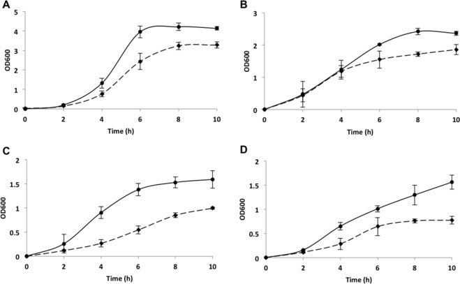
The influence of the simultaneous production of putrescine and tyramine on the growth of E. faecalis was studied by monitoring the OD600 of wt and ΔagdiΔtdc cultures in GM17 supplemented with agmatine and tyrosine. The mutant strain achieved a maximum OD600 of 3.3, while the wt strain reached a value of 4.1 (Fig. 1A). The effect of BA production was also determined under carbon source depletion by growing the strains in M17 with agmatine and tyrosine and a reduced concentration of glucose. Figure 1B shows the maximum OD600 achieved by both the wt and ΔagdiΔtdc strains to be reduced compared to the standard carbon source condition (Fig. 1A). The mutant strain returned a reduced OD600 value (1.8 vs. 2.3), indicating a role for the BA pathways in growth under this stress condition.
Figure 1.
Growth curves for E. faecalis V583 (continuous line) and E. faecalis V583 ΔagdiΔtdc (discontinuous line) in media supplemented with 20 mM agmatine and 10 mM tyrosine. (A) influence of putrescine and tyrosine synthesis on cultures grown in GM17. (B) effect of limiting the carbon source on cells cultured in M17 supplemented with glucose at 1 g L−1. (C) influence of acidic pH on the growth of strains propagated in GM17 adjusted to an initial pH of 5.0. (D) growth under a combination of carbon source depletion and acidic pH in M17 with glucose 1 g L−1 and pH adjusted to 5.0. The OD600 was monitored over 10 h.
The strains were then grown in GM17 with the same substrates but with the initial pH adjusted to 5 to examine the influence of the BA pathways in an acidic environment. While the wt strain reached an OD600 of 1.6, the mutant strain only managed an OD600 of 1 and showed a less steep exponential phase slope (Fig. 1C). Finally, the growth of the wt and ΔagdiΔtdc strains was monitored in M17 with reduced glucose and an acidic initial pH in the presence of agmatine and tyrosine. The OD600 values obtained (Fig. 1D) were similar to those recorded for the acidic conditions (Fig. 1C).
These outcomes show that, despite the lack of BA biosynthesis, the ΔagdiΔtdc mutant was still able to grow reasonably well.
The mutant strain E. faecalis ΔagdiΔtdc tolerates transit through an in vitro gastrointestinal tract model
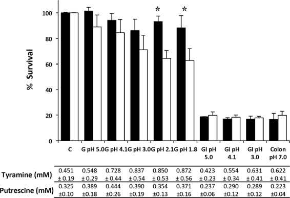
The wt and ΔagdiΔtdc strains were challenged with GI conditions in the presence of agmatine and tyrosine (Fig. 2). Greater concentrations of tyramine were recorded in the more acidic gastric conditions (pH 3.0, 2.1 and 1.8), while greater putrescine accumulation was seen at pH 4.1.
Figure 2.

Gastrointestinal transit simulation for E. faecalis V583 (black bars) and E. faecalis V583 ΔagdiΔtdc (white bars). Survival (%) of strains under gastric (G), gastrointestinal (GI) and colonic stresses in the presence of 20 mM agmatine and 10 mM tyrosine. C, untreated cells (control). Survival was measured using the LIVE/DEAD® BacLight fluorescent stain kit. Values are expressed as a percentage of the control value. Cells from cultures grown with 20 mM agmatine and 10 mM tyrosine for 6 h were used in all cases. An asterisk indicates a significant difference (p < 0.05; Student t test). The putrescine and tyramine concentrations accumulated by the wt strain under each condition are indicated below the graph.
The viability of the mutant strain was some 10–35% lower than for the wt strain under gastric stress conditions (Fig. 2). This reduction became significant at pH 2.1 and 1.8, coinciding with the conditions under which the wt strain accumulated tyramine more strongly. Under the conditions simulating the end phase of digestion in the colon, approximately 65% of the ΔagdiΔtdc cells survived.
The survival of both the wt and ΔagdiΔtdc populations was reduced to approximately 17% under the gastric and colonic stresses. Although the wt strain was able to synthesise both BA under both of these conditions, no significant differences between the strains were observed in terms of survival. These findings indicate that, although the mutant survived the acidic environment less well than did the wt strain, a significant proportion of the population can tolerate GI stress and reach the colon.
The adhesion of E. faecalis to intestinal epithelial cells is not affected by the synthesis of biogenic amines
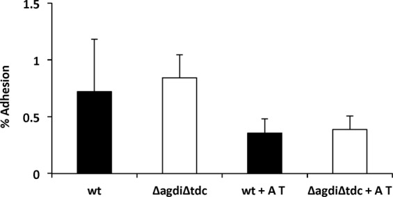
Figure 3 shows that 0.80% of both the wt and mutant bacterial cells adhered to the Caco-2 intestinal cells when agmatine and tyrosine were absent. In their presence, this figure was reduced to about 0.50% for both strains. During its incubation with the Caco-2 cells the wt strain produced 0.19 mM of putrescine and 0.14 mM of tyramine (as determined by UHPLC).
Figure 3.

Adhesion of E. faecalis V583 (wt, black bars) and E. faecalis V583 ΔagdiΔtdc (ΔagdiΔtdc, white bars) to Caco-2 cells in the absence or presence of 20 mM agmatine (A) and 10 mM tyrosine (T) after 5 h of co-culture. The adhesion level is expressed as a percentage of total bacteria after 5 h co-culture with Caco-2 cells in DMEM.
No BA was produced by the Caco-2 cells. These findings suggest that the intestinal adhesion capacity of the ΔagdiΔtdc strain is similar to that of the wt strain.
E. faecalis ΔagdiΔtdc is able to form biofilms
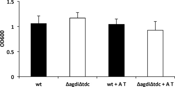
To further confirm that BA production had no role in the above-mentioned adhesion, the ability of the wt and mutant strains to form biofilms on polystyrene surfaces was examined. No significant differences were observed between the BA-producer and non-producer strains in the presence, or in the absence, of agmatine and tyrosine, revealing BA formation not to be involved in the biofilm formation capacity of E. faecalis (Fig. 4).
Figure 4.

Biofilm formation by E. faecalis V583 (wt, black bars) and E. faecalis V583 ΔagdiΔtdc (ΔagdiΔtdc, white bars) on polystyrene microtitre plates in GM17 in the absence or presence of 20 mM agmatine (A) and 10 mM tyrosine (T) after 16 h. Biofilms were assessed by crystal violet staining and the data corrected for the OD600.
The transcriptome of E. faecalis is unaffected by the deletion of the tdc and agdi clusters
To determine whether the deletion of the tdc and agdi clusters affected the expression of other genes, the transcriptomes of E. faecalis ΔagdiΔtdc and wt grown in GM17 were compared using the DNA microarray.
The results showed no gene outside of the deleted clusters to be differentially expressed in the mutant strain (Table 1). Since there is some tyrosine but no agmatine in GM17, the wt might have been able to express the genes of the tyrosine decarboxylase pathway. As expected, the expression of the deleted genes tyrS, tdcA and tdcP was null in the mutant strain. However, the nhaC-2 gene appeared to be slightly overexpressed. The same has been reported for E. faecalis Δtdc and is a consequence of the construction of the mutant and the microarray’s design (Perez et al., 201636): (i) the mutant strain was constructed keeping the promoter of the first gene of the cluster (tyrS) and the 3′ end of the last gene in the cluster (nhaC-2), and (ii) one of the two nhaC-2 gene probes designed for the array hybridizes with the 3′ end of the remaining region of nhaC-2. Thus, in the presence of tyrosine, a polar effect causes the apparent expression of nhaC-2 in the Δtdc mutant.
Table 1.
Genes expressed significantly different in the transcriptome of ΔagdiΔtdc compared to wt after 6 h of culture in GM17.
| Gene | Locus | Function | Fold change | p-value |
|---|---|---|---|---|
| tyrS | EF0633 | Tyrosyl-tRNA synthetase | −93.44 | 0.014 |
| tdcA | EF0634 | Tyrosine decarboxylase | −9.78 | 0.016 |
| tdcP | EF0635 | Tyrosine/tyramine exchanger | −4.32 | 0.020 |
| nhac-2 | EF0636 | Na+/H+ antiporter | 27.41 | 0.026 |
Discussion
Allowing the presence of E. faecalis in food is controversial: some strains are used in the fermentation of foods2 and even as probiotics3, yet pathogenic strains responsible for serious nosocomial infections also exist5. An undesirable characteristic of this species is that it produces the biogenic amines tyramine and putrescine13, the accumulation of which in fermented foods can pose a risk to health9. In this work, a double mutant was constructed in which the clusters of genes responsible for the biosynthesis of these BA (tdc and agdi respectively) were absent. Its performance suggests the method followed might be appropriate for designing strains with desired technological properties but which are safer for consumers.
The similarity of the agdi cluster in the wt strain to those of others previously reported, and the inability of the Δagdi mutant to produce putrescine in the presence of agmatine, confirm this cluster to be responsible for the biosynthesis of this BA. The involvement of the tdc cluster in tyramine production and its physiological role in protection against acid stress, were already known16.
Since the decarboxylation of amino acids consumes protons, this reaction affords a mechanism of resistance to acid stress in prokaryotes22. Moreover, the BAs thus synthesized, which possess a more positive charge than their precursor amino acids, are secreted from the cell, causing the net displacement of positive charges towards the exterior. This generates a proton-motive force that could be used by the cell to generate ATP via F1F0 ATPase23,24. Both functions have been demonstrated in Enterococcus faecium25. The decarboxylation of amino acids thus affords an advantage to microorganisms that face acidic environments such as those that occur in the stomach or in fermented foods.
The AGDI route has also been linked to acid stress resistance via the production of NH3. This also generates ATP, which could be used for the expulsion of protons from the cytoplasm via F1F0 ATPase22. In the LAB Lactococcus lactis, the AGDI route has been shown to enlist agmatine as a source of energy and to provide resistance to acid stress, counteracting the acidification of the cytosol21,26.
The present results obtained with the E. faecalis ΔagdiΔtdc mutant show that it grows and resists acidic pHs less well than the wt strain (Fig. 1), but that it does survive acidic pH both in broth and under simulated gastric conditions (Figs 1 and 2). This is not very surprising, since the BA-producing pathways are only a piece of the acid stress resistance mechanisms. Indeed, it survived the conditions it would meet during GIT transit, with live bacteria reaching the colon in numbers similar to those seen for the wt strain (Fig. 2), and without their adhesion capacity (Fig. 3) or ability to form biofilms on polystyrene affected (Fig. 4). The influence of BA on cellular adhesion has been previously analysed in other BA-producing LAB strains having obtained different results. While the presence of tyrosine increases the adhesion of E. durans IPLA655 to Caco-2 cells27, it was not detected any influence of the tyramine and putrescine BA biosynthetic pathways on L. brevis adhesion capability28.
Importantly, the deletion of the clusters did not affect the expression of any non-related gene (Table 1), including any genes encoding pathogenicity factors. It should be remembered that the model strain used, E. faecalis V583, has several pathogenicity genes29, the expression of which could potentially have been affected. Thus, the gene cluster inactivation strategy followed could be used to construct E. faecalis strains with good technological characteristics but that do not produce BA, and perhaps even strains with good probiotic potential.
In the latest EFSA risk analysis of BAs, one of the strategies proposed to reduce BA concentrations in fermented food was the use of starter cultures with no BA-producing capacity (EFSA, 2011). Although this strategy could be used for those starter species in which the production of BA is a strain-dependent trait, in the case of E. faecalis, tyramine and putrescine production are species-dependent traits (Ladero et al., 201213); this type of screening is therefore unviable. One alternative is the screening of spontaneous or induced mutants that have lost both characters - a tedious business requiring a large collection of mutants and with no guarantee of success. The present mutant construction strategy, however, does not suffer so strongly from these drawbacks.
In conclusion, the deletion of the agdi and tdc clusters gave rise to a strain of E. faecalis that does not produce the BAs putrescine and tyramine. Although the resulting mutant strain, E. faecalis ΔagdiΔtdc, grew more poorly in the presence of agmatine and tyrosine than the wt strain, it survived in vitro GIT-like conditions. Importantly, the expression of no gene outside the removed clusters was affected. Taking into account (i) that the production of tyramine and putrescine is a species-level characteristic in E. faecalis13, (ii) that these BA have toxic and genotoxic in vitro effects10,12, and (iii) the results of this work, the double mutant approach would allow to obtain safer probiotic and starter E. faecalis strains.
Materials and Methods
Bacterial strains
The wild-type strain E. faecalis V583 (hereafter referred to as ‘wt’) was used as a model bacterium since its entire genome sequence is known. The strain was obtained from the American Type Culture Collection (accession number ATCC 700802). For all fermentation assays, overnight cultures of E. faecalis strains were used (0.1% v/v inoculum).
Escherichia coli Gene-Hogs (Invitrogen, UK) was used as an intermediate host for cloning30 in the construction of the knock-out mutant.
DNA extraction
The GenElute Bacterial Genomic DNA Kit (Sigma-Aldrich, Spain) was used to extract total DNA from 2 mL of overnight cultures, following the manufacturer’s instructions. Plasmid extraction was performed following standard procedures31.
PCR reaction and DNA sequencing
PCR reactions were performed in 25 µL reaction volumes with 1 µL of DNA as a template (typically 200 ng), 400 nM of each primer, 200 µM of dNTP (GE Healthcare, UK), the reaction buffer, and 1 U of Taq polymerase (Phusion High-Fidelity DNA Polymerase, Thermo Scientific, Spain). Reactions were performed in a MyCycler device (Bio-Rad, CA) with the program: 94 °C for 5 min, 35 cycles of 94 °C for 30 s, 55 °C for 45 s, 72 °C for 1 min, and a final extension step at 72 °C for 5 min. Table 2 shows the primers used (all synthesized by Macrogen [Seoul, Korea]). Primers were designed based on the E. faecalis V583 genome sequence (GenBank number: AE016830). PCR fragments were purified using the GenElute PCR Clean-Up Kit (Sigma-Aldrich) when needed. Sequencing of the PCR amplicons was performed at Macrogen.
Table 2.
Primers used in this work.
| Primer | Function | Sequence (5′-3′) | Reference |
|---|---|---|---|
| P1F | Deletion agdi | CTGCACCGACCATTATCTTATACTATGAAGGAA | This work |
| P2R | Deletion agdi | ATCGCCGTCTTCTCGCTGGCATGGTTTATTGGTGGGCTAAGCATTGGTTTCGGTGT | This work |
| P3R | Deletion agdi | AACCATGCCACGAGAAGACGGCGAT | This work |
| P4F | Deletion agdi | CATCAACTGTTTGGCTGTTTCTTCGTCATAATAAC | This work |
| AguR1F | agdi deletion check | ACTCCCAAAAATGATCGTAAAAACATG | This work |
| KagV5R | agdi deletion check | CAAAACGACCGATGTCCTACTCTCTAACG | This work |
| CardF | tdc deletion check | GATGATAGTGTCTTGGCTGCTTTAAAGG | 13 |
| EF0637R | tdc deletion check | GACTCGCTTGTGAAGTTGTCGCTGCAG | 13 |
Construction of the Enterococcus faecalis ΔagdiΔtdc mutant
E. coli was routinely cultured at 37 °C with aeration in Luria-Bertani medium (Green and Sambrook, 2012[31]) supplemented with 1 mg L−1 ampicillin (USB Corporation,USA) when necessary. A mutant strain of E. faecalis V583 with the tdc and agdi clusters deleted (rendering it a non-tyramine and non-putrescine producer) was obtained by two subsequent steps of double-crossover homologous recombination using the cloning vector pAS222 as previously described30. The deletion of the agdi cluster was completed first. Sequence overlap extension PCR (SOE-PCR)32 was used to amplify the flanking fragments of the cluster. Two PCR reactions were performed with the primers P1 F and P2 R, and P3 F and P4 R (Table 2). The fragments were purified and then mixed to be used as the template for PCR amplification with the outer primers P1 F and P4 F. P3 R, the inner primer, carried regions of homology necessary for the fusion step. The amplicon was cloned into the SnaBI (Fermentas, Lithuania) site of pAS222 to generate pAS222 AGDI, which was propagated in E. coli Gene-Hogs cells. Electrocompetent E. faecalis V583 cells16 were transformed with pAS222 AGDI and the cells harbouring the plasmid grown in GM17 under previously described conditions to allow double-crossover recombination33. The formation of the intermediate mutant E. faecalis V583 with the deleted agdi cluster (hereafter referred to as ‘Δagdi’) was confirmed by PCR with the primers AguR1F and KagV5R and amplicon sequencing, and by checking the lack of putrescine accumulation in overnight cultures in GM17 supplemented with 20 mM agmatine.
To effect tdc cluster deletion, electrocompetent cells of Δagdi were produced and transformed with the plasmid pAS222 TDC, previously obtained by the same technique16. After double-crossover recombination, the deletion of the tdc cluster was confirmed via PCR with the primers CardF and EF0637R and further sequencing. The deletion of the tdc cluster encompassed the interval from tyrS (793 nt downstream of its start codon) to nhaC-2 (691 nt downstream of its start codon), and agdi cluster deletion covered the interval from aguR (469 nt downstream of its stop codon) to aguA (785 nt downstream of its start codon). The confirmed deletion mutant E. faecalis V583 ΔagdiΔtdc (hereafter referred to as ‘ΔagdiΔtdc’) was used in further analyses.
Growth of the wt and mutant strains under different conditions
E. faecalis V583 and the derived mutant ΔagdiΔtdc were grown at 32 °C without aeration in M17 (Oxoid, UK) supplemented 5 g L−1 glucose (Merck, Germany) (GM17). To determine the effect of the carbon source concentration, the same medium was supplemented with 1 g L−1 glucose. The influence of acidic pH was analyzed in GM17 adjusted to an initial pH of 5.0. Whenever necessary, media were supplemented with 20 mM agmatine and 10 mM tyrosine (Sigma-Aldrich). The optical density at 600 nm (OD600) was monitored over 10 h. For all the experiments, overnight cultures of E. faecalis strains were used (0.1% v/v inoculum).
Resistance to gastrointestinal conditions
The assay described by Fernández de Palencia, et al.34, with the modifications of Perez, et al.16, was followed for the simulation of bacterial transit through the GIT. Briefly, approximately 1010 cfu mL−1 of the wt and ΔagdiΔtdc strains from late exponential phase cultures (in GM17 supplemented with 20 mM agmatine and 10 mM tyrosine) were collected and mixed with the electrolyte solution (supplemented with the same concentrations of substrates). Cells were exposed first to lysozyme and then pepsin plus a successive reduction in pH to simulate gastric stress conditions. Gastrointestinal stress was mimicked by exposure of samples incubated at pH 5, 4.1, 3.0, 2.1 and 1.8 (gastric conditions), followed by their incubation in the presence of bile salts and pancreatin at pH 8 (small intestine conditions, GI). Colonic stress was simulated with the sample originally at pH 3 adjusted to pH 7 and incubated overnight. For each condition, cell viability was measured using the LIVE/DEAD® BacLight fluorescent stain (Molecular Probes, Netherlands) as previously described34. The values provided are the mean of three independent replicates, expressed as a percentage of the untreated control. BA accumulation at the end of the assay was quantified as described below.
Adhesion to the intestinal epithelium
The adhesion of the strains to the intestinal epithelium was studied in an in vitro model with Caco-2 cells obtained from the human cell bank at the Centro de Investigaciones Biológicas (Madrid, Spain), following the protocol previously described 34 with minor modifications. These cells were grown in Men-Alpha Medium (Invitrogen) supplemented with 10% (v/v) heat-inactivated foetal bovine serum at 37 °C in a 5% CO2 atmosphere, and then seeded in 24-well tissue culture plates (Falcon Microtest™, Becton Dickinson, USA) at 4 × 10⁴ cells per well, and grown for 15 days to obtain a monolayer of differentiated, polarized cells. The bacterial strains were grown in GM17 supplemented with 20 mM agmatine and 10 mM tyrosine until the end of the exponential phase. They were then washed with phosphate buffered saline pH 7.1 (PBS) and resuspended in Dulbecco’s Modified Eagle medium (DMEM) (Invitrogen). The Caco-2 cells and bacterial strains were then co-cultured at a ratio of 1:100 in DMEM in the presence or absence of 20 mM agmatine and 10 mM tyrosine. After 5 h of incubation at 37 °C in a 5% CO2 atmosphere, the cells were washed three times with 500 µL PBS and resuspended in 0.1 mL of PBS. Non-washed wells were used as a control. The Caco-2 cells were detached by the addition of 500 µL trypsin-EDTA (0.05%) (Gibco, USA) for 10 min at 37 °C; the reaction was inactivated by adding 500 µL PBS at 4 °C. The total number of bacteria was determined by serial dilutions and plate counts, and the number adhered to the intestinal cells calculated as a percentage of the total bacteria in the unwashed controls. Each adhesion assay was performed in triplicate. BAs produced at the end of the assay were detected as indicated below.
Measurement of biofilm-forming capacity
The capacity of the strains to form a biofilm was tested on polystyrene microtitre plates (TC Microwell 96U, Thermo Scientific)35. Overnight cultures in GM17 with or without 20 mM agmatine and 10 mM tyrosine were diluted 1:40 in 200 µL of the same medium. After 16 h of incubation in the microtitre plates, the cells were washed, stained with crystal violet, and their OD600 determined.
DNA microarrays and data analysis
Agilent eArray v5.0 software (Agilent Technologies, USA) was used to design a DNA microarray for E. faecalis V583. The array harboured duplicate spots of two different 60-mer probes specific for each of the 3182 coding DNA sequences (CDSs) in the chromosome of the above strain (GenBank accession no. AE016830)29. The microarray design was added to the Gene Expression Omnibus (GEO) database (Platform GPL21449). Total RNA was extracted from late exponential phase cultures of the wt and ΔagdiΔtdc mutant grown in 30 ml of GM17 supplemented with 20 mM of agmatine and 10 mM of tyrosine as previously described36,37. 20 µg of RNA were used to synthesize cDNA employing the SuperScript III Reverse Transcriptase Kit (Life Technologies, The Netherlands). cDNA (20 mg) was labeled with DyLight 550 or DyLight 650 using the DyLight Amine-Reactive Dyes Kit (Thermo Scientific). The hybridization step was carried out with 900 ng each of DyLight 550- and DyLight 650-labeled cDNA for 17 h at 60 °C on the E. faecalis V583 DNA microarray using the In situ Hybridization Kit Plus, a hybridization gasket slide, and a G2534 A microarray hybridization chamber36, all from Agilent Technologies. Slides were scanned using a GenePix 4200 A Microarray Scanner (Molecular Devices, USA) and images acquired and analyzed using GenePix Pro v.6.0 software. The standard routines provided by GENOME2D software (http://genome2d.molgenrug.nl/index.php/analysispipeline) were used for background subtraction and locally weighted scatterplot normalization. Microarray data were obtained from two independent cultures and one technical replicate that included a dye swap. Expression ratios were calculated from the comparison of four spots per gene per microarray.
Genes returning a significant difference (p ≤ 0.05) in their wt and mutant expressions, plus an expression fold-change of at least double, were considered differentially expressed. The microarray data is available in the GEO database under the Accession no. GSE136953. Functional analysis was executed using Gene Set Enrichment Analysis (GSEA) routines as provided by GENOME2D (http://server.molgenrug.nl/index.php/gsea-pro).
Quantification of biogenic amine synthesis
Samples obtained during the above experiments were centrifuged and the supernatants recovered and filtered through 0.45 µm polytetrafluoroethylene (PTFE) filters (VWR, Spain), The detection and quantification of the BAs and their substrates was performed by UHPLC. The compounds were derivatized with diethyl ethoxymethylenemalonate (Sigma-Aldrich) and separated in a UPLC system (Waters, USA) under the conditions described 38. Chromatograms were obtained and analysed using Empower 2 software (Waters). The results are the means at least three replicates.
Statistical analysis
Data are presented as the means ± standard deviations calculated from at least three independent replicates. Means were compared by the Student t test using SPSS software v.15.0 (SPSS, Inc., USA). Significance was set at p ≤ 0.05.
Acknowledgements
This work was performed with the financial support of AEI under grant number AGL2016-78708-R, (AEI/FEDER, UE) and the Plan for Science, Technology and Innovation 2013–2017 of the Principality of Asturias, co-funded by FEDER (GRUPIN IDI/2018/000114). The authors thank Adrian Burton for language and editing assistance.
Author contributions
M.P. performed some of the experiments and participated in data interpretation. M.C.E. constructed some of the mutants, B.R. performed BA analysis. B.d.R., M.C.M. and M.F. participated in study design and data interpretation. A.d.J. and O.P.K. participated in study design and data interpretation of the microarray experiments, V.L. participated in data interpretation and manuscript writing. M.A.A. provided the general concept, participated in study design, manuscript writing and data interpretation, and supervised the work and the manuscript. All authors contributed to the discussion of the research and approved the final manuscript.
Competing interests
The authors declare no competing interests.
Footnotes
Publisher’s note Springer Nature remains neutral with regard to jurisdictional claims in published maps and institutional affiliations.
References
- 1.Lebreton F, et al. Tracing the Enterococci from paleozoic origins to the hospital. Cell. 2017;169:849–861 e813. doi: 10.1016/j.cell.2017.04.027. [DOI] [PMC free article] [PubMed] [Google Scholar]
- 2.Giraffa G. Functionality of enterococci in dairy products. Int. J. Food Microbiol. 2003;88:215–222. doi: 10.1016/S0168-1605(03)00183-1. [DOI] [PubMed] [Google Scholar]
- 3.Franz CM, Huch M, Abriouel H, Holzapfel W, Galvez A. Enterococci as probiotics and their implications in food safety. Int J Food Microbiol. 2011;151:125–140. doi: 10.1016/j.ijfoodmicro.2011.08.014. [DOI] [PubMed] [Google Scholar]
- 4.Fritzenwanker, M. et al. Complete genome sequence of the probiotic Enterococcus faecalis Symbioflor 1 Clone DSM 16431. Genome announcements1, 10.1128/genomeA.00165-12 (2013). [DOI] [PMC free article] [PubMed]
- 5.Van Tyne D, Gilmore MS. Friend turned foe: evolution of enterococcal virulence and antibiotic resistance. Annu. Rev.Microbiol. 2014;68:337–356. doi: 10.1146/annurev-micro-091213-113003. [DOI] [PMC free article] [PubMed] [Google Scholar]
- 6.Linares, D. M. et al. Factors influencing biogenic amines accumulation in dairy products. Front.Microbiol. 3 (2012). [DOI] [PMC free article] [PubMed]
- 7.O’Sullivan DJ, et al. High-throughput DNA sequencing to survey bacterial histidine and tyrosine decarboxylases in raw milk cheeses. BMC Microbiol. 2015;15:266. doi: 10.1186/s12866-015-0596-0. [DOI] [PMC free article] [PubMed] [Google Scholar]
- 8.Ladero Victor, Cañedo Elena, Pérez Marta, Martín María Cruz, Fernández María, Alvarez Miguel A. Multiplex qPCR for the detection and quantification of putrescine-producing lactic acid bacteria in dairy products. Food Control. 2012;27(2):307–313. doi: 10.1016/j.foodcont.2012.03.024. [DOI] [Google Scholar]
- 9.Ladero V, Calles-Enríquez M, Fernández M, Alvarez MA. Toxicological effects of dietary biogenic amines. Curr. Nut. Food Sci. 2010;6:145–156. doi: 10.2174/157340110791233256. [DOI] [Google Scholar]
- 10.del Rio B, et al. The biogenic amines putrescine and cadaverine show in vitro cytotoxicity at concentrations that can be found in foods. Sci. Rep.-UK. 2019;9:120. doi: 10.1038/s41598-018-36239-w. [DOI] [PMC free article] [PubMed] [Google Scholar]
- 11.Linares DM, et al. Comparative analysis of the in vitro cytotoxicity of the dietary biogenic amines tyramine and histamine. Food Chem. 2016;197:658–663. doi: 10.1016/j.foodchem.2015.11.013. [DOI] [PubMed] [Google Scholar]
- 12.del Rio B, et al. An altered gene expression profile in tyramine-exposed intestinal cell cultures supports the genotoxicity of this biogenic amine at dietary concentrations. Sci. Rep.UK. 2018;8:17038. doi: 10.1038/s41598-018-35125-9. [DOI] [PMC free article] [PubMed] [Google Scholar]
- 13.Ladero V, et al. Is the production of the biogenic amines tyramine and putrescine a species-level trait in enterococci? Food Microbiol. 2012;30:132–138. doi: 10.1016/J.Fm.2011.12.016. [DOI] [PubMed] [Google Scholar]
- 14.Suarez C, Espariz M, Blancato VS, Magni C. Expression of the agmatine deiminase pathway in Enterococcus faecalis is activated by the AguR regulator and repressed by CcpA and PTS(Man) systems. PloS One. 2013;8:e76170. doi: 10.1371/journal.pone.0076170. [DOI] [PMC free article] [PubMed] [Google Scholar]
- 15.Linares DM, et al. An agmatine-inducible system for the expression of recombinant proteins in Enterococcus faecalis. Microb. Cell Fact. 2014;13:169. doi: 10.1186/S12934-014-0169-1. [DOI] [PMC free article] [PubMed] [Google Scholar]
- 16.Perez M, et al. Tyramine biosynthesis is transcriptionally induced at low pH and improves the fitness of Enterococcus faecalis in acidic environments. Appl. Microbiol. Biot. 2015;99:3547–3558. doi: 10.1007/s00253-014-6301-7. [DOI] [PubMed] [Google Scholar]
- 17.Ladero, V. et al. In Microbial Toxins in Dairy Products. 94–131 (Wiley Blackwell, 2016).
- 18.Ladero Victor, Martín María Cruz, Redruello Begoña, Mayo Baltasar, Flórez Ana Belén, Fernández María, Alvarez Miguel A. Genetic and functional analysis of biogenic amine production capacity among starter and non-starter lactic acid bacteria isolated from artisanal cheeses. European Food Research and Technology. 2015;241(3):377–383. doi: 10.1007/s00217-015-2469-z. [DOI] [Google Scholar]
- 19.Linares DM, Cruz Martin M, Ladero V, Alvarez MA, Fernandez M. Biogenic amines in dairy products. Crit. Rev. Food Sci. Nutr. 2011;51:691–703. doi: 10.1080/10408398.2011.582813. [DOI] [PubMed] [Google Scholar]
- 20.Stroman P, Sorensen KI, Derkx PMF, Neves AR. Development of tyrosine decarboxylase-negative strains of Lactobacillus curvatus by classical strain improvement. J Food Prot. 2018;81:628–635. doi: 10.4315/0362-028X.JFP-17-301. [DOI] [PubMed] [Google Scholar]
- 21.del Rio B, et al. Putrescine production via the agmatine deiminase pathway increases the growth of Lactococcus lactis and causes the alkalinization of the culture medium. Appl. Microbiol. Biot. 2015;99:897–905. doi: 10.1007/s00253-014-6130-8. [DOI] [PubMed] [Google Scholar]
- 22.Lund P, Tramonti A, De Biase D. Coping with low pH: molecular strategies in neutralophilic bacteria. FEMS Microbiol Rev. 2014;38:1091–1125. doi: 10.1111/1574-6976.12076. [DOI] [PubMed] [Google Scholar]
- 23.Molenaar D, Bosscher JS, Tenbrink B, Driessen AJM, Konings WN. Generation of a proton motive force by histidine decarboxylation and electrogenic histidine histamine antiport in Lactobacillus-buchneri. J. Bacteriol. 1993;175:2864–2870. doi: 10.1128/jb.175.10.2864-2870.1993. [DOI] [PMC free article] [PubMed] [Google Scholar]
- 24.Wolken WAM, Lucas PM, Lonvaud-Funel A, Lolkema JS. The mechanism of the tyrosine transporter TyrP supports a proton motive tyrosine decarboxylation pathway in Lactobacillus brevis. J. Bacteriol. 2006;188:2198–2206. doi: 10.1128/Jb.188.6.2198-2206.2006. [DOI] [PMC free article] [PubMed] [Google Scholar]
- 25.Pereira CI, Matos D, Romao MVS, Crespo MTB. Dual role for the tyrosine decarboxylation pathway in Enterococcus faecium E17: response to an acid challenge and generation of a proton motive force. Appl. Environ. Microbiol. 2009;75:345–352. doi: 10.1128/Aem.01958-08. [DOI] [PMC free article] [PubMed] [Google Scholar]
- 26.del Rio B, et al. Putrescine biosynthesis in Lactococcus lactis is transcriptionally activated at acidic pH and counteracts acidification of the cytosol. Int J Food Microbiol. 2016;236:83–89. doi: 10.1016/j.ijfoodmicro.2016.07.021. [DOI] [PubMed] [Google Scholar]
- 27.Fernandez de Palencia P, et al. Role of tyramine synthesis by food-borne Enterococcus durans in adaptation to the gastrointestinal tract environment. Appl. Environ. Microbiol. 2011;77:699–702. doi: 10.1128/aem.01411-10. [DOI] [PMC free article] [PubMed] [Google Scholar]
- 28.Russo P, et al. Biogenic amine production by the wine Lactobacillus brevis IOEB 9809 in systems that partially mimic the gastrointestinal tract stress. BMC Microbiol. 2012;12:247. doi: 10.1186/1471-2180-12-247. [DOI] [PMC free article] [PubMed] [Google Scholar]
- 29.Paulsen IT, et al. Role of mobile DNA in the evolution of vancomycin-resistant Enterococcus faecalis. Science. 2003;299:2071–2074. doi: 10.1126/science.1080613299/5615/2071. [DOI] [PubMed] [Google Scholar]
- 30.Jonsson, M., Saleihan, Z., Nes, I. F. & Holo, H. Construction and characterization of three lactate dehydrogenase-negative Enterococcus faecalis V583 mutants. Appl Environ Microbiol75, 4901–4903, doi:AEM.00344-09 (2009). [DOI] [PMC free article] [PubMed]
- 31.Green, M. R. & Sambrook, J. Molecular cloning: a laboratory manual. 4 edn, (Cold Spring Harbor Laboratory Press, 2012).
- 32.Horton RM, Hunt HD, Ho SN, Pullen JK, Pease LR. Engineering hybrid genes without the use of restriction enzymes: gene splicing by overlap extension. Gene. 1989;77:61–68. doi: 10.1016/0378-1119(89)90359-4. [DOI] [PubMed] [Google Scholar]
- 33.Biswas I, Gruss A, Ehrlich SD, Maguin E. High-efficiency gene inactivation and replacement system for gram-positive bacteria. J Bacteriol. 1993;175:3628–3635. doi: 10.1128/jb.175.11.3628-3635.1993. [DOI] [PMC free article] [PubMed] [Google Scholar]
- 34.Fernández de Palencia P, López P, Corbí A, Peláez C, Requena T. Probiotic strains: survival under simulated gastrointestinal conditions, in vitro adhesion to Caco-2 cells and effect on cytokine secretion. Eur. Food Res. Technol. 2008;227:1475–1484. doi: 10.1007/s00217-008-0870-6. [DOI] [Google Scholar]
- 35.Perez M, et al. IS256 abolishes gelatinase activity and biofilm formation in a mutant of the nosocomial pathogen Enterococcus faecalis V583. Can. J. Microbiol. 2015;61:517–519. doi: 10.1139/cjm-2015-0090. [DOI] [PubMed] [Google Scholar]
- 36.Perez M, et al. Transcriptome profiling of TDC cluster deletion mutant of Enterococcus faecalis V583. Genom Data. 2016;9:67–69. doi: 10.1016/j.gdata.2016.06.012. [DOI] [PMC free article] [PubMed] [Google Scholar]
- 37.Perez M, et al. The Relationship among tyrosine decarboxylase and agmatine deiminase pathways in Enterococcus faecalis. Front Microbiol. 2017;8:2107. doi: 10.3389/fmicb.2017.02107. [DOI] [PMC free article] [PubMed] [Google Scholar]
- 38.Redruello B, et al. A fast, reliable, ultra high performance liquid chromatography method for the simultaneous determination of amino acids, biogenic amines and ammonium ions in cheese, using diethyl ethoxymethylenemalonate as a derivatising agent. Food Chem. 2013;139:1029–1035. doi: 10.1016/j.foodchem.2013.01.071. [DOI] [PubMed] [Google Scholar]


