



Summary
Visceral leishmaniasis (VL) is epidemic in Brazil with an increasing incidence of human cases and canine reservoirs, with host hypergammaglobulinemia. Conventional enzyme‐linked immunosorbent assay (cELISA) based on several parasitic antigens is the main method for diagnosis and indication of treatment. Dissociative ELISA (dELISA) uses acidic treatment to free immunoglobulin G (IgG) from immune complexes, and its use revealed a significant positive fraction of suspected cases with negative serology. Looking for small molecules or haptens that block IgG antibodies, we purified by molecular exclusion chromatography, 1000–3000 MW molecules from promastigote soluble extract, mostly oligosaccharides comprising 6–13 sugar residues using MALDI‐TOF analysis. Glycan‐BSA complex (GBC) was constructed by conjugating promastigote glycans to BSA molecules, allowing their use in the solid support in cELISA or dELISA. Sera from experimentally infected hamsters showed higher levels of blocked monomeric IgG during infection, mostly against GBC, which was also present in lower concentrations in the promastigote soluble extract dELISA. Those data show that most of the specific monomeric IgG in serum are blocked by haptens composed by glycans produced by the parasite, better detected in the high dilution of sera in the dELISA assays. dELISA is a useful technique for detecting blocked monomeric antibodies that could have difficult clearance from blood, which could result in hypergammaglobulinemia.
Keywords: glycan, hapten, hypergammaglobulinemia, immune complexes, Leishmania (Leishmania) infantum chagasi
Abbreviations
- BSA
bovine serum albumin
- cELISA
conventional ELISA
- CNPq
Conselho Nacional de Pesquisas
- dELISA
dissociative ELISA
- ELISA
enzyme‐linked immunosorbent assay
- FAPESP
Fundação de Amparo à Pesquisa do Estado de São Paulo
- GBC
glycan–BSA complex
- IC
immune complexes
- IgG
immunoglobulin G
- LIM
Laboratório de Investigação Médica
- LPLC
low‐pressure liquid chromatography
- NS
not significant
- PSE
promastigote soluble extract
- VL
visceral leishmaniasis
Introduction
Endemic in 88 countries on the African, American, Asian and European continents, visceral leishmaniasis (VL) is potentially fatal if left untreated and mainly affects populations in socially vulnerable conditions. Between 50 000 and 90 000 new cases occur each year, mainly in countries such as Brazil, Ethiopia, Kenya, Somalia, India, Sudan and South Sudan.1 Leishmania (Leishmania) infantum chagasi causes VL in Latin American countries, transmitted to a mammalian host by phlebotomine sandflies. This disease has great medical and veterinary importance, and is considered an anthropozoonosis; among its hosts, the domestic dog plays a prominent role in the transmission cycle, as the main reservoir responsible for the spread of the disease among the human population.2
Clinical manifestations of VL may present in different forms, ranging from asymptomatic to lethal.3 Susceptibility to VL has been related to a number of factors that influence both the severity of the disease and its prognosis, where the host immune response is crucial in determining the clinical outcome of the infection.4 The maintenance of parasitic Leishmania spp. in their hosts depends on survival strategies that involve glycoconjugates, which are part of the glycocalyx and form a protective ‘barrier’.5 The molecular composition of the surface of parasites Leishmania spp. has an important role in the evasion and/or modulation of the immune response of susceptible hosts.6
Hosts susceptible to VL have high levels of specific immunoglobulin G (IgG) antibodies, which are not effective in the immune response during infection and do not prevent the reactivation of the disease.7, 8 During active VL in men and dogs, there are high levels of IgG, IgE, IgA and IgM antibodies.9, 10 However, experimental studies consider that B cells and antibodies are of minimal importance for protective immunity during VL.11, 12 B‐cell polyclonal activation and hypergammaglobulinemia are predictive of disease exacerbation during VL, but the mechanisms involved in these processes are still unknown.13
The diagnosis of VL is considered complex because the symptomatology is not specific and requires careful evaluation, which should consider clinical, epidemiological and laboratory characteristics.14 Serological tests are widely used for laboratory diagnosis of VL, especially for the detection of specific antibodies, and have excellent levels of sensitivity and specificity.15 However, a high precision serological test for diagnosis of VL is still a problem for the medical community, as the detection of specific antibodies can be misinterpreted in symptomatic, asymptomatic and post‐treatment cases.16, 17
Recently, we devised a dissociative enzyme‐linked immunosorbent assay (dELISA), involving acid treatment and antibody recovery that results in seroconversion in a small fraction of suspected negative patients and also increases the detection of IgG in confirmed cases.18, 19 In this study, we report the presence of immune complexes (IC) composed of hapten glycan in serum during experimental hamster VL, as detected by dELISA with promastigote soluble extract (PSE). Glycan promastigote haptens were isolated, characterized and conjugated to a carrier protein, allowing their application in dELISA in these experimental model samples.
Methods
Experimental infection, culture, antigen production and samples
Methods such as experimental hamster infection, sample collection, in vitro maintenance of L. (L.) infantum chagasi, preparation and quantification of PSE are described in a previously published study.18 For serology assays, 68 serum samples from hamsters with experimental infection (15, 30, 45, 60 and 90 days) and 10 negative hamster sera were used, from the biorepository of the Laboratory of Protozoology of the Institute of Tropical Medicine of São Paulo, which also provided PSE‐hyperimmunized rabbit serum. All procedures were previously approved by the Research and Ethics Committee and the Ethics Committee in Animal Experimentation of the Instituto de Medicina Tropical de São Paulo (no. CE‐IMT/002/29042008).
Molecular exclusion chromatography
Molecular exclusion chromatography was carried out using ÄKTA prime system LPLC (GE Healthcare, Chalfont St Giles, UK) with 278 nm spectrometry and conductivity detection. For the mobile phase of Sephadex® G‐25, 0·1 m sodium carbonate pH 9·2 was used for analytical or preparative chromatography. Fractions were analysed individually for analytical purposes or pooled as excluded and/or retained fractions in preparative runs. Bio‐Gel® P‐2 chromatography with ammonium bicarbonate 0·1 m pH 7·0 for the mobile phase was used for desalting samples.
Glycan quantification
Glycan quantification was based on the periodic acid Schiff method, as reported in the literature.20 Briefly, it was performed on a 96‐well microplate adding 125 μl of sample to 120 μl of sodium metaperiodate 0·2% in 7% acetic acid/well, with incubation at 37° for 30 min. After this, 100 μl/well of Schiff reagent was added and incubated for 30 min at 37°. The 540 nm optical density was determined in a microplate reader and standard galactose curve was running.
Leishmania (L.) infantum chagasi promastigote glycan characterization by MALDI‐TOF
The fraction containing low mass molecules of the PSE was analysed by the DEMPSTER laboratory, Institute of Chemistry of the University of São Paulo. The α‐cyano‐4‐hydroxycinnamic acid matrix was prepared as a saturated solution in 50% acetonitrile and 2·5% trifluoroacetic acid. Subsequently, 1 μl of sample was spotted onto a steel target plate and allowed to dry at room temperature. Finally, 1 μl of matrix solution was added and left to air dry. Mass Spectrometry analyses were performed using an UltrafleXtreme matrix‐assisted laser desorption/ionization time‐of‐flight mass spectrometry (MALDI‐TOF) mass spectrometer (Bruker Daltonics, Bremen, Germany) operating in reflectron positive ion mode. Mass spectra were acquired in a mass range from 600 to 5000 MW with ions formed by irradiation of the smart beam using a frequency of 2000 Hz, PIE 100 ns, 7 kV lens. The voltages for the first and second ion sources were 25 kV and 23 kV, respectively.
Glycan–bovine serum albumin complex
The conjugation between bovine serum albumin (BSA) and desalted purified 1000–3000 MW glycans of L. (L.) infantum chagasi promastigote was performed as described in the literature.21 Briefly, glycans (~100 μg) were suspended in sodium metaperiodate 0·01 m in 0·1 m sodium acetate pH 6·0, vortexed and 1 mg BSA was added with constant shaking for 1 hr at room temperature. After this step, pH was changed by addition of 1 volume of 0·5 m sodium carbonate pH 9·0, with incubation and shaking for 1 hr. To stop the binding, the sample received 1 mg of sodium borohydride and was incubated for 2 hr at 4° with occasional stirring. The mixture was submitted to molecular exclusion chromatography (Sephadex® G‐25), with mobile phase with 0·1 m carbonate pH 9·0 and excluded fractions were pooled and used as Glycan–BSA complex (GBC).
ELISA
Conventional ELISA (cELISA) and dELISA12 plates were adsorbed with 100 μl of PSE (0·6 μg/ml) or GBC (0·03 μg/ml) in 0·1 m sodium carbonate pH 9·5 overnight at 4°. The concentration of PSE and GBC was standardized by dilution curve against hyperimmune rabbit serum (1 : 200) revealed by anti‐rabbit IgG peroxidase conjugate (1 : 20 000). Residual binding on wells was blocked with 200 μl/well of 0·3% skimmed milk, 0·01 m phosphate‐buffered saline (PBS), 0·05% Tween‐20 (PBSTL) for 1 hr at 37°. For dELISA assays, we added half diluted serum samples 50 μl/well, followed by 25 μl/well of 0·1 m glycine pH 2·0, stirring for 5 min and 25 μl/well of 0·5 m Tris–HCl pH 8·0 was added with 1 hr incubation at 37°. For cELISA, PBSTL was added to control wells. The anti‐hamster IgG peroxidase conjugate (1 : 20 000) 100 μl was added for 1 hr at 37°. The reaction was revealed with 100 μl/well of 3,3',5,5'‐tetramethylbenzidine substrate for 15 min and stopped with 50 μl 4 m HCl, with absorbance at 450 nm determined on a microplate reader. Data were presented as ELISA optical density or Δ values determined by the reactivity difference between wells using GBC antigen or control BSA. An ELISA test was run with duplicates and tested on three different days. There was excellent qualitative reproducibility both intra‐test and inter‐test, with a minimum variation of 10% in the quantitative analysis.
Statistical analysis
Statistical analyses were performed using graphpad prism 6.0 software (GraphPad Prism, San Diego, CA). Data of individual groups were analysed using analysis of variance with Bonferroni post‐tests. The correlation between data was performed using Pearson correlation and expressed as r 2. All comparisons were considered significant between data when the probability of equality was lower than 5% (P < 0·05). In graphs, the extent of significance is expressed as asterisks (*P < 0·05, **P < 0·01, ***P < 0·001 and ****P < 0·0001) or not significant (NS) in the absence of difference.
Results
IgG anti‐PSE cELISA and dELISA in hamster experimental infection
The PSE of L. (L.) infantum chagasi was used in cELISA and dELISA for the detection of IgG antibodies and the indirect detection of IC, respectively. The comparison between cELISA and dELISA results for the 1 : 1000 dilution (Fig. 1a) showed high significant difference at most time‐points, with greater reactivity for dELISA at all time‐points. For the higher dilution (Fig. 1b), the profile was maintained and there was a greater distinction between the methods. This reactivity was intense, including in the earlier stages, when IgG was at low detection levels even in lower dilutions. Comparison between the reactivity of the results among the dilutions tested showed a high correlation in cELISA (r 2 = 0·94) and moderate correlation in dELISA (r 2 = 0·75). Most samples were more reactive in dELISA, showing that a frequent feature in samples from this experimental model is the blocking of IgG by antigens. The reactivity of the samples and the discriminatory ability of the dELISA increased at the highest dilution tested.
Figure 1.
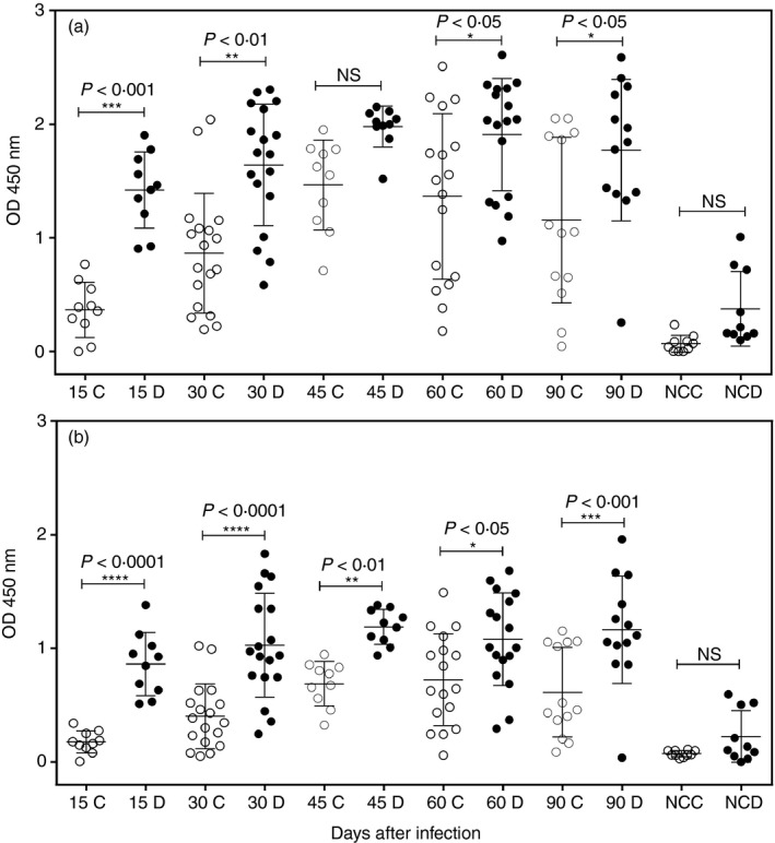
Anti‐promastigote soluble extract (PSE) IgG detection by conventional ELISA (cELISA) and dissociative ELISA (dELISA) in hamster serum samples at 1 : 1000 (a) and 1 : 2000 (b) dilutions. Open dots cELISA, solid dots dELISA. Labels show days after infection and negative controls (C = cELISA and D = dELISA). The bars represent the standard deviation and the asterisks indicate the level of statistical significance between the results using Bonferroni analysis of variance post‐tests.
Search of low‐molecular‐weight antigens in PSE
Assuming that dELISA unlocks monomeric IgG ICs, we planned to investigate low‐mass molecules in the PSE. The sample was subjected to analytical molecular exclusion chromatography in resin with an exclusion limit of 5000 MW with quantification of proteins and carbohydrates in fractions (Fig. 2a). The first finding was the higher carbohydrate concentration (~ 900 μg/ml) in samples expected to be between 1000 and 5000 MW, almost three times the amount of protein found in excluded > 5000 MW samples (~ 300 μg/ml). This protein also presented a carbohydrate reaction but less intense, suggesting a relationship between those fractions. Preparative molecular exclusion chromatography was designed to separate low‐molecular‐weight components or haptens from the PSE (Fig. 2b). These low‐molecular‐weight components or haptens were pooled and subjected to more restrictive molecular exclusion chromatography (Fig. 2c). The pooled fractions were dried for salt removal and submitted to MALDI‐TOF analysis, restricted to 800–3000 MW, and their composition showed six major peaks, ranging from 950 to 1800 MW (Fig. 2d).
Figure 2.

Chromatography purification and MALDI‐TOF analysis of 1000–3000 MW antigens from promastigote soluble extract (PSE). (a) PSE analytical G‐25 chromatogram showing A280nm (protein) and Schiff reaction (carbohydrate) of individual fractions. (b) PSE preparative G‐25 chromatogram. A = high molecular weight protein and glycoproteins; Pool P2 = pooled fractions with salts and < 3000 MW antigens. (c) Pool P2 (B chromatogram) preparative Bio‐gel P2 chromatogram. B = pooled fraction desalted and containing 1000–3000 MW antigens. (d) MALDI‐TOF analysis of pooled fractions (C chromatogram) with molecular weight and estimation of sugar residue numbers between parentheses.
Construction and testing of GBC
Conjugation of purified glycans to BSA was required for the adsorption process on ELISA microplates and IgG antibody detection (Fig. 3). Decreasing amounts of PSE, GBC and BSA were used for ELISA standardization (Fig. 4a). The reactivity detected for the PSE was consistent and its 50% reaction occurred at 0·0663 μg/ml (r 2 = 0·99), while the reactivity to GBC showed similar consistency with a reaction of 50% to 0·0047 μg/ml (r 2 = 0·98), a sustained reactivity 14 times higher than in PSE. The BSA background was evaluated and the concentration of 0·03 μg/ml showed better discrimination of the GBC‐positive samples. IgG antibody reactivity was compared between GBC and PSE antigens in experimentally infected hamster (> 60 days of infection) and hyperimmune rabbit (PSE immunization) samples as shown in Fig. 4(b). Sample reactivity curves were similar between PSE and GBC antigens, with high correlation between results (r 2 > 0·98 for all curves). The immunized rabbit sera presented the highest similarity between PSE and GBC reactivity in ELISA, whereas our experimental infection standards showed greater reactivity to PSE than to GBC ELISA, which presented the lowest titres.
Figure 3.

Glycan–bovine serum albumin complex (GBC) ELISA standardization. (a) Detection of anti‐leishmania IgG in rabbit sample using various concentrations of GBC (open points), promastigote soluble extract (PSE) (closed points) and bovine serum albumin (BSA) (open points) in solid phase. Δ values (asterisks) represent the difference between the reactivity of rabbit serum to GBC and BSA. (b) Reactivity in GBC and PSE ELISA of hamster (square) or rabbit (circle) serum dilutions for anti‐leishmania IgG detection, analysed by sigmoidal dose models. GBC (dashed line) and PSE (continuous line).
Figure 4.

IgG anti‐glycan–bovine serum albumin complex (GBC) detection by conventional ELISA (cELISA) and dissociative ELISA (dELISA) in hamster serum samples at 1 : 1000 (a) and 1 : 2000 (b) dilutions. Open dots cELISA, solid dots dELISA. Labels show days after infection or negative controls (C = cELISA and D = dELISA) The bars represent the standard deviation and the asterisks the level of statistical significance between the results using Bonferroni analysis of variance post‐tests.
Specific IgG anti‐GBC cELISA and dELISA in hamster experimental infection
The specific reactivities of anti‐leishmania IgG antibodies from hamster samples at different time‐points after experimental infection were evaluated for GBC in cELISA and dELISA. As for the PSE, the detection of IgG antibodies and IC varied for post‐infection time‐points. Comparison of results between cELISA and dELISA showed high significance of the results (P < 0·005 and < 0·0005) for both dilutions tested (Fig. 4A/B). Results between dilutions showed low correlation for ELISA (r 2 = 0·11) and moderate correlation for dELISA (r 2 = 0·79). The correlation of the results shows that higher serum dilutions affect the ability to detect antibodies in cELISA; however, the results of dELISA are not affected.
Discussion
Our data show that higher serum dilutions in the PSE dELISA reaction on solid support showed higher discrimination and high reactivity, suggesting the blockade of IgG in its monomeric form. This higher reactivity at higher dilutions suggests the presence of antigenic blockers at lower dilutions and may even completely inhibit the reaction, as reported in the literature.18 Antigenic blockers could be free or complexed to IgG molecules, which competitively inhibited their reaction with solid support PSE, at high concentrations of these complexes.
The use of PSE in ELISA increases the sensitivity of the test as a result of exposure to multiple antigenic epitopes. However, the molecular composition of this extract and also the binding interactions on the solid supports restrict the reactivity of antibodies to protein antigens. This type of assay limits the interaction of antibodies to proteins and glycoproteins, minimizing the presence of other specific antibodies, especially anti‐glycans. Protein or glycoprotein ICs (> 10 000 MW) are generally detected at low levels in VL18 or other leishmaniasis, such as post‐Kala‐azar dermal leishmaniasis.22, 23
Structural composition of antigens complexed to IgG anti‐leishmania antibodies is an important tool for the understanding of trigger mechanisms, permanence and elimination of ICs. Our search of low‐molecular‐weight antigens in PSE by sequential molecular exclusion chromatography revealed glycans (950–1800 MW) probably with 6–13 sugar residues by MALDI‐TOF. The presence of antigenic glycans is described in other studies involving Leishmania sp.24, 25The antigenicity of glycan haptens and their application in diagnostic methods in VL is little explored; references to glycans are generally in studies on the development of vaccines and related to the structural composition of molecules present in amastigote and promastigote forms and their impact on the immune response.26
Conjugation of glycans to carrier molecules is a usual tool for the immobilization of these antigens on solid supports and is used in tests for antibody detection.27 We conjugated this glycan fraction to BSA (GBC) for use in the solid support (cELISA and dELISA) and the results surprisingly showed a higher reactivity in dELISA at higher dilutions and lower reactivity in cELISA, showing that high concentrations of IgG are blocked by glycans in VL. The dELISA demonstrated that glycan antibody blockade occurs after the early stage of the infection and is maintained at all subsequent stages of the experimental hamster infection.
We believe that the interaction between glycan haptens and antibodies triggers the formation of most of the ICs present in VL, resulting in the uselessness of these antibodies, affecting clearance, and resulting in hypergammaglobulinemia. As demonstrated in our results, the antigenicity of glycan haptens was previously reported in VL in a work that evaluated the immunogenicity of glycoconjugate haptens and the diagnostic potential of these molecules was evaluated.28 The presence of high levels of blocked antibodies in hamster samples with experimental infection explains the lower reactivity to dELISA using PSE in lower serum dilutions. This is a result of the lower availability of the glycans in PSE, implying less competition with the high concentration of soluble glycans dissociated from the blocked antibodies. Using high serum dilutions in dELISA, including levels higher than those used in this article, may result in potent and discriminating diagnostic methods of disease activity in VL, by revealing glycan produced by the living agent.
Dissociative ELISA allows detection or suspicion of an individual IgG blockade by a hapten of molecular weight between 1000 and 3000 MW. The blockade of the variable fraction of IgG by the hapten prevents its recognition by eventual anti‐idiotype, one of the possible forms of serum IgG clearance, according to the idiotype network model.29 Hypergammaglobulinemia has always been attributed to polyclonal activation or multiple antigens, but may be due only to the presence of monomeric IgG blocking haptens in a new type of immune involvement. In our experiments it was possible to isolate and identify antigenic glycans present in promastigotes of L. (L.) infantum chagasi. Its association with carrier proteins allows the study of the role of these molecules in the antigen–antibody interactions in this disease.
Disclosure
The authors declare no conflict of interest.
Acknowledgements
We thank Maria Anita Mendes, PhD for the MALDI‐TOF analyses (FAPESP, Process: 2013/50218‐2). This study was supported by CNPq, FAPESP (Process: 2017/26558‐9) and LIM49 da Faculdade de Medicina da Universidade de São Paulo.
Contributor Information
Camila Aparecida de Carvalho, Email: camilacarvalho@usp.br.
Heitor Franco de Andrade, Júnior, Email: hfandrad@usp.br.
References
- 1. World Health Organization . Leishmaniasis. [cited 30 April 2019]. URL http://www.who.int/mediacentre/factsheets/fs375/en/ [accessed on 25 July 2019]
- 2. Carvalho AG, Luz JGG, Rodrigues LD, Dias JVL, Fontes CJF. High seroprevalence and peripheral spatial distribution of visceral leishmaniasis among domestic dogs in an emerging urban focus in Central Brazil: a cross‐sectional study. Pathog Glob Health 2018; 112:29–36. [DOI] [PMC free article] [PubMed] [Google Scholar]
- 3. Kevric I, Cappel MA, Keeling JH. New world and old world Leishmania infections: a practical review. Dermatol Clin 2015; 33:579–93. [DOI] [PubMed] [Google Scholar]
- 4. Saporito L, Giammanco GM, De Grazia S, Colomba C. Visceral leishmaniasis: host–parasite interactions and clinical presentation in the immunocompetent and in the immunocompromised host. Int J Infect Dis 2013; 17:e572–6. [DOI] [PubMed] [Google Scholar]
- 5. Mukhopadhyay S, Mandal C. Glycobiology of Leishmania donovani . Indian J Med Res 2006; 123:203–20. [PubMed] [Google Scholar]
- 6. Ferguson MA, Murray P, Rutherford H, McConville MJ. A simple purification of procyclic acidic repetitive protein and demonstration of a sialylated glycosyl‐phosphatidylinositol membrane anchor. Biochem J 1993; 291(Pt 1):51–5. [DOI] [PMC free article] [PubMed] [Google Scholar]
- 7. Murray HW, Berman JD, Davies CR, Saravia NG. Advances in leishmaniasis. Lancet 2005; 366:1561–77. [DOI] [PubMed] [Google Scholar]
- 8. Mougneau E, Bihl F, Glaichenhaus N. Cell biology and immunology of Leishmania . Immunol Rev 2011; 240:286–96. [DOI] [PubMed] [Google Scholar]
- 9. Anam K, Afrin F, Banerjee D, Pramanik N, Guha SK, Goswami RP et al Differential decline in Leishmania membrane antigen‐specific immunoglobulin G (IgG), IgM, IgE, and IgG subclass antibodies in Indian kala‐azar patients after chemotherapy. Infect Immun 1999; 67:6663–9. [DOI] [PMC free article] [PubMed] [Google Scholar]
- 10. de Freitas JC, Lopes‐Neto BE, de Abreu CR, Coura‐Vital W, Braga SL, Reis AB et al Profile of anti‐Leishmania antibodies related to clinical picture in canine visceral leishmaniasis. Res Vet Sci 2012; 93:705–9. [DOI] [PubMed] [Google Scholar]
- 11. Kumar R, Nylén S. Immunobiology of visceral leishmaniasis. Front Immunol 2012; 3:251. [DOI] [PMC free article] [PubMed] [Google Scholar]
- 12. Rodrigues V, Cordeiro‐da‐Silva A, Laforge M, Silvestre R, Estaquier J. Regulation of immunity during visceral Leishmania infection. Parasit Vectors 2016; 9:118. [DOI] [PMC free article] [PubMed] [Google Scholar]
- 13. Silva‐Barrios S, Stäger S. Hypergammaglobulinemia sustains the development of regulatory responses during chronic Leishmania donovani infection in mice. Eur J Immunol 2019; 49:1082–91. [DOI] [PubMed] [Google Scholar]
- 14. Solano‐Gallego L, Koutinas A, Miró G, Cardoso L, Pennisi MG, Ferrer L et al Directions for the diagnosis, clinical staging, treatment and prevention of canine leishmaniasis. Vet Parasitol 2009; 165:1–18. [DOI] [PubMed] [Google Scholar]
- 15. Kumar R, Pai K, Pathak K, Sundar S. Enzyme‐linked immunosorbent assay for recombinant K39 antigen in diagnosis and prognosis of Indian visceral leishmaniasis. Clin Diagn Lab Immunol 2001; 8:1220–4. [DOI] [PMC free article] [PubMed] [Google Scholar]
- 16. Srivastava P, Dayama A, Mehrotra S, Sundar S. Diagnosis of visceral leishmaniasis. Trans R Soc Trop Med Hyg 2011; 105:1–6. [DOI] [PMC free article] [PubMed] [Google Scholar]
- 17. Srividya G, Kulshrestha A, Singh R, Salotra P. Diagnosis of visceral leishmaniasis: developments over the last decade. Parasitol Res 2012; 110:1065–78. [DOI] [PubMed] [Google Scholar]
- 18. de Carvalho CA, Partata AK, Hiramoto RM, Borborema SE, Meireles LR, Nascimento Nd et al A simple immune complex dissociation ELISA for leishmaniasis: standardization of the assay in experimental models and preliminary results in canine and human samples. Acta Trop 2013; 125:128–36. [DOI] [PubMed] [Google Scholar]
- 19. de Carvalho CA, Ferrão TF, Hiramoto RM, Partata AK, de Andrade HF Jr. Indirect evidence of circulating parasite hapten immune complexes in visceral leishmaniasis. J Appl Lab Med 2019; 4 10.1373/jalm.2018.028282 [DOI] [PubMed] [Google Scholar]
- 20. Kilcoyne M, Gerlach JQ, Farrell MP, Bhavanandan VP, Joshi L. Periodic acid‐Schiff's reagent assay for carbohydrates in a microtiter plate format. Anal Biochem 2011; 416:18–26. [DOI] [PubMed] [Google Scholar]
- 21. Nakane PK, Kawaoi A. Peroxidase‐labeled antibody. A new method of conjugation. J Histochem Cytochem 1974; 22:1084–91. [DOI] [PubMed] [Google Scholar]
- 22. Datta S, Modak D, Sarkar S, Saha B, Mukhopadhyay S. Identification and glycobiological characterization of circulating immune complexes in patients with visceral leishmaniasis and post kala azar dermal leishmaniasis. Indian J Exp Biol 2015; 53:321–8. [PubMed] [Google Scholar]
- 23. Jaiswal P, Datta S, Sardar B, Chaudhuri SJ, Maji D, Ghosh M et al Glycoproteins in circulating immune complexes are biomarkers of patients with Indian PKDL: a study from endemic districts of West Bengal, India. PLoS ONE 2018; 13:e0192302. [DOI] [PMC free article] [PubMed] [Google Scholar]
- 24. Silveira TG, Kemmelmeier C. Leishmania braziliensis: isolation of carbohydrate‐containing antigen and possibility of its use in the immunodiagnosis of American cutaneous leishmaniasis. Rev Inst Med Trop Sao Paulo 1995; 37:245–52. [DOI] [PubMed] [Google Scholar]
- 25. Tabatabaee PA, Abolhassani M, Mahdavi M, Nahrevanian H, Azadmanesh K. Leishmania major: secreted antigens of Leishmania major promastigotes shift the immune response of the C57BL/6 mice toward Th2 in vitro . Exp Parasitol 2011; 127:46–51. [DOI] [PubMed] [Google Scholar]
- 26. Osanya A, Song EH, Metz K, Shimak RM, Boggiatto PM, Huffman E et al Pathogen‐derived oligosaccharides improve innate immune response to intracellular parasite infection. Am J Pathol 2011; 179:1329–37. [DOI] [PMC free article] [PubMed] [Google Scholar]
- 27. Xie ZM, Yu L, Fang LS. Preparation and characteristic of dextran‐BSA antibody and establishment of its ELISA immunoassay. J Immunoassay Immunochem 2015; 36:597–612. [DOI] [PubMed] [Google Scholar]
- 28. Anish C, Martin CE, Wahlbrink A, Bogdan C, Ntais P, Antoniou M et al Immunogenicity and diagnostic potential of synthetic antigenic cell surface glycans of Leishmania . ACS Chem Biol 2013; 8:2412–22. [DOI] [PubMed] [Google Scholar]
- 29. Civello A. On the genesis of the idiotypic network theory. J Hist Biol 2013; 46:125–58. [DOI] [PubMed] [Google Scholar]