Abstract
Granuloma annulare (GA) is usually a self-limited, benign granulomatous disease of the dermis and subcutaneous tissue. It's generalized or disseminated form is associated with underlying diabetes mellitus and at times it precedes the sign and symptoms of diabetes mellitus. We are reporting a case of a 56-year-old female, who is a known case of type 2 diabetes mellitus she presented to us with symmetric lesions on her trunk, arms, and legs. On further evaluation by the dermatologists, it was established to be lesions of GA. This case has been reported to highlight the incidence and the importance of recognition of this common but rarely diagnosed condition.
Keywords: Dermatological manifestation of diabetes, diabetes mellitus, granuloma annulare
Introduction
Granuloma annulare (GA) is usually a self-limited, benign granulomatous disease of the dermis and subcutaneous tissue.[1] The term “GA” was coined in 1902 by Radcliff-Crocker. Clinically, the condition is characterized by asymptomatic, flesh-colored, or erythematous-brown papules, frequently arranged in a ring or annular pattern on the distal extremities. GA is often localized and not associated with systemic diseases although it can be trigged by trauma, infection, drugs, and metabolic derangements.
This condition manifests as numerous (a minimum of 10 and often hundreds to thousands) small, asymptomatic, erythematous, violaceous, brown, or skin-colored papules. Lesions are distributed symmetrically on the trunk, extremities, and neck. It has a bimodal peak age and it presents in the first decade of life and subsequently between the fourth and sixth decades of life.[1] It is associated with underlying diabetes mellitus and at times it precedes the sign and symptoms of diabetes mellitus.[2] Henceforth, it becomes all the more important to the primary care physician because it may be the sole presentation with which the patient may present and timely intervention may prevent complications of both GA as well as diabetes mellitus. Many precipitating factors, such as subcutaneous injection for desensitization, Octopus bite, bacillus Calmette–Guérin vaccination, mesotherapy, and ultraviolet light exposure have been reported but never confirmed by controlled studies.[3]
Case Report
A 56-year-old female patient, who is a known case of type 2 DM for last 5 years presented to our outdoor department with complain of generalized ring like, reddish, papular lesion on both upper and lower limb and trunks. Her blood sugar fasting was 160 mg/dl and postprandial was 310 mg/dl and HbA1c 8.5%. She was taking tablet metformin 1 gm and glimepiride 2 mg daily. Her lipid profile and thyroid function test, liver function test (LFT), kidney function test (KFT) were in normal range. Her HIV, HBsAg and hepatitis C antibody test (anti-HCV) were nonreactive. She did not have any drug history or any history of chronic disease. On local examination, the lesions were present over her back, calves and dorsolateral aspects of both legs, these lesions have been depicted in Figures 1–5. The lesions were typically fleshy and expanding outward in a ring-like fashion. Further on skin biopsy from the lesion showed focal collagen degeneration with palisading histiocytes. These findings were suggestive of GA. We admitted her and gave her insulin for faster and better blood sugar control, after 1 week she was discharged on insulin with blood sugar fasting 100 and post prandial 160 mg/dl. After 3 months these lesions had markedly regressed without scarring.
Figure 1.
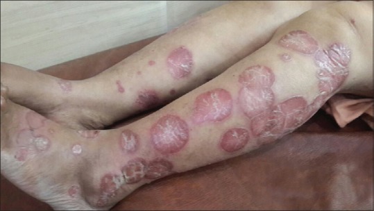
Granuloma annulare- lesions over the leg
Figure 5.
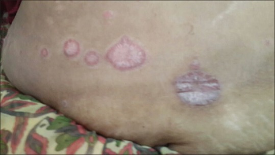
Granuloma annulare- lesions over the back
Figure 2.
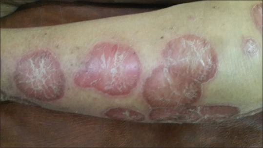
Granuloma annulare- lesions over the calf
Figure 3.
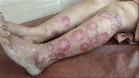
Granuloma annulare- lesions over the legs
Figure 4.
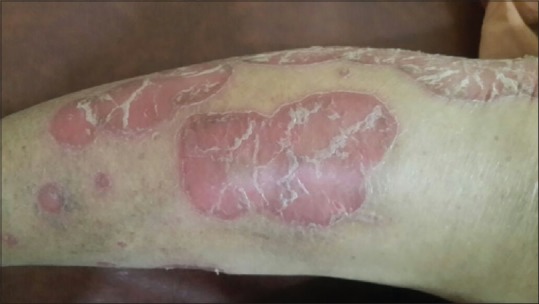
Granuloma annulare-closer look at the lesions
Discussion
GA is a benign necrobiotic condition similar to necrobiosis lipodica diabeticorum, the only difference being absence of epidermal atrophy. In its localized form it is seen in nondiabetics, but in generalized form it is usually associated with DM occurring in approximately 0.5% to 10% of such patients.[4] The skin condition is usually seen in children and young adults. Skin signs are characterized by red spots in the initial stages which expand outward in a ring-like fashion. The histologic examination of a truly classic lesion shows an infiltrate in the deep dermis and/or hypodermis of granulomas predominantly formed by palisaded histiocytes around a central region of degenerating collagen fibers (necrobiosis) and abundant mucin, best seen under alcian blue staining.[5] The presence of mucin is in fact a key histological feature that helps us to distinguish GA from other noninfectious granulomatous diseases. The hands especially the fingers, on the dorsal and lateral aspects of hand and elbow are affected. The lesions may precede the signs and symptoms of DM. Dyslipidemia is more common in patients with GA. Rarely, GA may also be complicated by nerve involvement as a result of granulomatous inflammation surrounding cutaneous nerves and perineural infiltrates of histiocytes in the dermis.[1]
GAs are usually asymptomatic and are self-limited and most often no treatment other than reassurance is required. For those patients who insist on treatment for cosmetic reasons, the options include topical or intralesional corticosteroids, imiquimod cream, topical calcineurin inhibitors (tacrolimus, pimecrolimus), cryotherapy, and pulsed dye laser. For the nodular lesion seen in subcutaneous GA surgical removal is an option. Generalized GA is often resistant to treatment and for it systemic therapy may be required. Interventions that have been used with varying degrees of success include oral corticosteroids, fumaric acid esters, dapsone, isotretinoin, hydroxychloroquine, methotrexate, cyclosporine, niacinamide, calcitriol, vitamin E, tumor necrosis factor inhibitors (adalimumab and infliximab), and several variants of phototherapy.[1]
Declaration of patient consent
The authors certify that they have obtained all appropriate patient consent forms. In the form the patient(s) has/have given his/her/their consent for his/her/their images and other clinical information to be reported in the journal. The patients understand that their names and initials will not be published and due efforts will be made to conceal their identity, but anonymity cannot be guaranteed.
Financial support and sponsorship
Nil.
Conflicts of interest
There are no conflicts of interest.
References
- 1.Leung AK, Benjamin Barankin, Hon KL. Granuloma Annulare. International Journal of Pediatrics and Child Health. 2013;1:15–18. [Google Scholar]
- 2.Sreedevi C, Car N, Renar IP. Dermatological Lesions in Diabetes Mellitus. Diabetologia Croatica. 2002;1:31–3. [Google Scholar]
- 3.Alirezaei P, Farshchian M. Granuloma annulare: Relationship to diabetes mellitus, thyroid disorders and tuberculin skin test. Clin Cosmet Investig Dermatol. 2017;10:141–5. doi: 10.2147/CCID.S129187. [DOI] [PMC free article] [PubMed] [Google Scholar]
- 4.Hanna W, Friesen D, Bombardier C, Gladman D, Hanna A. Pathologic features of diabetic thick skin. J Am Acad Dermatol. 1987;16:546–53. doi: 10.1016/s0190-9622(87)70072-3. [DOI] [PubMed] [Google Scholar]
- 5.Doshi BR, Sajjan VV, Manjunathswamy BS. Disseminated subcutaneous granuloma annulare in adult. Indian J Dermatopathol Diagn Dermatol. 2019;6:48–50. [Google Scholar]


