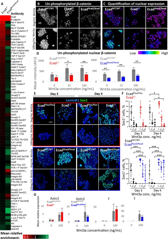
Fig. 4.
Effects of cadherin switching on β-catenin and Wnt signalling. (A) Heat map showing enrichment of protein and phospho-protein species in Ecad−/− and EcadNcad/Ncad cells compared with control cell lines; all cells were cultured in neural differentiation conditions for 24 h at time of analysis. Data generated by RPPA, n=3. (B) Example confocal images of cells at 24 h of neural differentiation cultured in varying Wnt3a concentration and stained for unphosphorylated β-catenin. (C) Quantitative visualisations of nuclear staining of the images shown in B. (D) Quantification of mean nuclear voxel intensity for unphosphorylated β-catenin in cells cultured for 24 h in neural differentiation conditions. n=4 biological replicates, each containing hundreds of cells. (E,F) Example images (E) of Sox1 expression in cells cultured with or without Wnt3a. ‘High Wnt3a’ refers to 100 ng/ml. Dot plots (F) show quantification of the percentage of Sox1-positive cells; each dot represents one field of view; n=9, three images each sampled from three biological replicates. (G) qPCR analysis of two Wnt pathway readouts in Ecad−/− and EcadNcad/Ncad cells during neural differentiation in increasing concentrations of Wnt3a. n=3 biological replicates. For all graphs, error bars represent s.d., *P≤0.05, **P≤0.01, ***P≤0.001; unpaired Student's t-test. Scale bars: 50 µm.

