Abstract
Purpose
To investigate whether reduced Sox9 function exerts neuroprotection in light-induced retinal damage in rats and to explore the potential mechanism behind it.
Methods
Retinal light damage was used as a model for retinal degeneration. Two weeks before light damage in adult Sprague Dawley (SD) rats, the Sox9-shRNA lentiviral vector was intravitreally injected. On days 3, 7, and 14, retinal function was assessed using electroretinography (ERG), and the thickness of the outer nuclear layer (ONL) was measured in hematoxylin and eosin (HE) stained sections. The protein levels of glial fibrillary acidic protein (GFAP), vimentin, nestin, and chondroitin sulfate proteoglycans (Cspgs), which are related to gliosis and extracellular matrix (ECM) remodeling, were observed using western blot analysis. The expression of GFAP was further evaluated by immunohistochemistry.
Results
On days 3, 7, and 14 after light damage, the thickness of the ONL and the amplitudes of the ERG waves were significantly better preserved in the Sox9-shRNA group when compared with the control group. The protein levels of GFAP, vimentin, nestin, and Cspgs were significantly downregulated in the Sox9-shRNA group. Furthermore, the staining intensity and the spatial distribution of GFAP in the retinas were also obviously attenuated at every studied time point.
Conclusions
Intravitreal injection of the Sox9-shRNA lentiviral vector preserved rat retinal morphology and function after light damage and downregulated GFAP, vimentin, nestin, and Cspgs, which are related to Müller cell gliosis and ECM remodeling. The results indicate that Sox9 might be a potential therapeutic target for retinal degenerative diseases.
Introduction
As the predominant glial element in the sensory retina, Müller cells are responsible for the homeostatic and metabolic support of retinal neurons and are active players in virtually all forms of retinal injury and disease [1,2]. In response to damage, the reactive changes in Müller cells, which are part of a process called gliosis, can be neuroprotective in the very early stages after damage. But when the activation is excessive, overactive gliosis becomes detrimental, forming glial scars and contributing to retinal remodeling [3]. The most sensitive nonspecific response of gliosis is the upregulation of the intermediate filaments’ glial fibrillary acidic protein (GFAP), vimentin, and nestin, which, especially in the case of GFAP, can be used as an indicator of Müller cell activation [3]. Previous research has shown that inhibiting the expression of GFAP has a neuroprotective effect: the retinas of adult mice deficient in GFAP and vimentin provide a permissive environment for grafted neurons to migrate and extend neurites [4], and mice that are deficient in GFAP and vimentin show attenuated glial reactions and photoreceptor degeneration induced by retinal detachment [5].
Recent evidence indicates that the transcription factor sex-determining region Y (SRY) box 9—also known as Sox9 and part of the SOX family [6]— regulates the glial activity of astrocytes and extracellular matrix (ECM) deposits in the central nervous system (CNS) [7-9]. Conditional Sox9 ablation in mice reduces GFAP expression, decreases the levels of chondroitin sulfate proteoglycans (Cspgs) that are the critical components of ECM, and improves motor function following spinal cord injury [7,8]. Sox9 knockout mice have shown improved recovery following a stroke [9]. In the sensory retina, it has been demonstrated that Sox9 is expressed mainly in Müller cells in adult mice [10,11], and our previous data have shown the upregulation of Sox9 in Müller cells in retinal light damage in rats [12], a model of retinal degenerative diseases. However, it is still unknown whether the downregulation of Sox9 can exert neuroprotection after retinal light damage.
In the current study, we aim to test the hypothesis that reduced Sox9 function exerts neuroprotection in light-induced retinal damage in rats. We further observe the expression levels of GFAP, vimentin, nestin, and Cspgs, exploring the possible mechanism of neuroprotection.
Methods
Animals
Adult female Sprague-Dawley (SD) rats weighing 200–220 g were treated in accordance with the ARVO Statement for the Use of Animals in Ophthalmic and Vision Research, and all procedures were approved by the Animal Care Committee of the Eye and ENT Hospital of Fudan University. The rats were randomly divided into two groups: the Sox9-shRNA group that received an intravitreal injection of the Sox9-shRNA lentiviral vector and the control group that received a scrambled shRNA containing lentiviral vector. The animals were sacrificed at different time points during the experiment for functional and histological evaluations (n = 4/time point), immunohistochemistry (n = 3/time point), and western blot analysis (n = 3/time point). The animals were anesthetized with an intramuscular injection of ketamine (80 mg/kg) and xylazine (16 mg/kg) during the examination and then euthanized with an overdose of pentobarbitone.
Construction of lentiviral vectors
Synthetic oligonucleotides containing the rat Sox9 splice variant (NCBI accession number NM080403) RNA interference target GAAGGAGAGCGAGGAAGAT was synthesized, annealed, and ligated into the pLKD-CMV-Puro-U6-shRNA lentiviral vector (Obio Biotech company, Shanghai, China) between the AgeI and EcoRI restriction enzyme sites following the U6 promoter. The oligo sequences are 5′- CCGGGAAGGAGAGCGAGGAAGATTTCAAGAGAATCTTCCTCGCTCTCCTTCTTTTTTG-3′ (sense) and 5′-AATTCAAAAAA GAAGGAGAGCGAGGAAGATTCTCTTGAAATCTTCCTCGCTCTCCTTC-3′ (antisense). The mock vector, which produces a nontargeting sequence TTCTCCGAACGTGTCACGT, was used as the negative control that is a scrambled shRNA containing lentivirus vector. The viral supernatant was harvested, filtered, and concentrated. The lentivirus was procured from Obio Biotech (Shanghai, China). The titer of the recombinant lentivirus was 1×10⁸ infectious units per ml.
Intravitreal injection and light exposure
Two weeks before light exposure, 4 μl of the lentivirus vector were intravitreally injected into one eye of each rat with a microsyringe (Hamilton, Hamilton Bonaduz AG, Switzerland) under a microscope. The Sox9-shRNA group received an intravitreal injection of the Sox9-shRNA lentiviral vector and the control group received the scrambled shRNA containing lentiviral vector. After 2 weeks of intravitreal injections, the rats were placed in separate cages. After adapting to the dark for 24 h, their pupils were fully dilated with 1% atropine and then exposed to evenly distributed blue light at an intensity of 2500 lx for 48 h, after which they were returned to a normal light–dark cycle [12,13].
Electroretinography
Electroretinography (ERG) tests were recorded after 2 weeks of intravitreal injections and on days 3, 7, and 14 after light exposure. The rats were dark-adapted for 2 h before ERG and were anesthetized—per the protocols mentioned above—under dim red light. The pupils were fully dilated with 0.5% phenylephrine hydrochloride and 0.5% tropicamide. The cornea was anesthetized with 0.4% oxybuprocaine HCL. The animals were held steady with a bite bar and nose clamp in a stereotaxic frame. A heating pad maintained the body temperature at approximately 37 °C. Retinal signals were recorded from the cornea using a silver chloride ring-recording electrode. The reference electrode was connected to the nose, and a grounding silver needle electrode was connected to the tail. The ERGs were recorded using the Espion visual electrophysiology system (Espion E3, Diagnosys UK Ltd., Cambridge, UK). The procedure was designed to record the retinal responses of both rods and cones. Under scotopic conditions, a single flash (4 ms, 0.1Hz) was applied at the intensity of 0.01 cd.s/m2 to record rod-ERG waveforms and a single flash at the intensity of 20 cd.s/m2 to record mixed rod-cone-ERG waveforms. Ten ERG waves were averaged for each measurement. After 5 min of photopic adaptation (50 cd.s/m2), the cone-ERG was recorded using a single flash (4 ms, 1Hz) at the intensity of 20 cd.s/m2 with 20 times superposition, and the flicker-ERGs were recorded using a continuous flash (4 ms, 10Hz) at the intensity of 20 cd.s/m2 with 30 times superposition. The a-wave amplitude was measured from the baseline to the peak of the negative wave, and the b-wave amplitude was measured from the trough of the negative a-wave deflection to the peak of the positive wave.
Histologic analysis
After 2 weeks of intravitreal injections and at 3, 7, and 14 days after light exposure, the rats were euthanized after the ERGs were recorded, following which their eyes were enucleated and immersion fixed in 4% paraformaldehyde (Beyotime, Shanghai, China) for 1 h and then transferred to 10% neutral-buffered formalin overnight. The paraffin-embedded tissue was cut into 5-μm-thick sections and stained with hematoxylin and eosin (HE). All eyes were cut vertically, and only the sections through the optic nerves were collected. Images of the tissue sections were captured using a photomicroscope (Leica Microsystems, Bensheim, Germany), and the thickness of the outer nuclear layer (ONL) was measured using an image-analysis system (Image-Pro Plus ver. 6.0, Media Cybernetics, MD). In total, 18 locations for each retinal section were measured, starting from either side of the optic nerve, with each segment 0.5 mm apart; the 18 measurements were then averaged for the mean thickness of the ONL.
Immunohistochemistry
After 2 weeks of intravitreal injections and at 3, 7, and 14 days after light exposure, the rats were euthanized and transcardially perfused with saline followed by 4% paraformaldehyde; then, their eyes were enucleated. The cornea and lens were removed, and the eyecups were immersed in the same fixative for 2 h. The eyecups were cryoprotected in graded sucrose solutions (20%–30% in a phosphate buffer [PB]), embedded in an optimal cutting temperature (OCT) compound (Tissue-Tek; Ted Pella, Inc., Redding, CA), snap frozen in liquid nitrogen, and then sectioned (10 μm). Air-dried slides were incubated with a blocking buffer (phosphate buffer saline [PBS] containing 5% goat serum and 0.25% Triton X-100) at room temperature for 1 h. After washing three times in 0.01 M PBS, the sections were incubated overnight at 4 °C with the following primary antibodies: rabbit polyclonal antibody against Sox9 (1:800; Millipore, MA); mouse monoclonal antibody against glutamine synthetase (GS); (a specific Müller cell marker; 1:200; Abcam, MA, USA); and mouse monoclonal antibody against GFAP (1:1000; MAB360, Millipore, MA, USA). The sections were then rinsed three times in 0.01 M PBS and incubated with Alexa Fluor 488-conjugated goat anti-mouse IgG secondary antibody (1:800; Invitrogen, Carlsbad, CA) at room temperature for 1 h. After three rinses with 0.01 M PBS, they were counterstained with DAPI (1:800, Sigma-Aldrich, St. Louis, MO) and examined using a photomicroscope (Leica Microsystems, Bensheim, Germany).
Western blot analysis
After 2 weeks of intravitreal injections and at 3, 7, and 14 days after light exposure, the eyes were enucleated from the rats while the animals were under deep anesthesia. Their anterior segments were then removed, and the retinas were isolated and shock-frozen at -80 °C within approximately 3 min of enucleation. The retinas were later homogenized in a RIPA buffer (Beyotime), treated ultrasonically, incubated in ice for 15 min, and centrifuged at 12,000 ×g for 15 min at 4 °C. The supernatants were collected, and the protein concentration was determined by the BCA protein assay (Beyotime). Equal amounts of the protein were loaded into each lane, and the proteins were separated using 10% sodium dodecyl sulfate PAGE, which was followed by transfer to Immobilon-P membranes (Millipore). The membranes were preblocked in 5% nonfat milk at room temperature for 1 h and then incubated with a primary antibody directed against Sox9 (1:1000; AB5535, Millipore), GFAP (1:1000; MAB360, Millipore), vimentin (1:1000; ab8979, Abcam, UK), nestin (1:500; MAB530, Millipore), Cspg (1:200; CS-56 [aggrecan], C8035, Sigma-Aldrich), and β-actin (1:5000; ab20272, Abcam). A peroxidase-conjugated donkey anti-rabbit or anti-mouse secondary antibody (1:5000, A0208, A0216, Beyotime) was used. The immunoblots were visualized by chemiluminescence (Pierce Biotechnology, Rockford, IL). The chemiluminescent images were captured using a camera (Kodak Image Station 4000MM PRO, Carestream, Rochester, NY). Densitometric analyses were performed with image-analysis software (Image-Pro Plus ver. 6.0, Media Cybernetics, Bethesda, MD). First, the relative expression levels of target proteins were compared with the expression levels of β-actin in the same retinas. Then the values were compared with that of the control, which was normalized to 1.0.
Data analysis
Data were analyzed using statistical software (Stata, ver. 10.0; Stata Corporation, College Station, TX). Comparisons between the two groups were made using a Student t test. p<0.05 was considered statistically significant.
Results
Validation of the Sox9 knockdown at the protein level
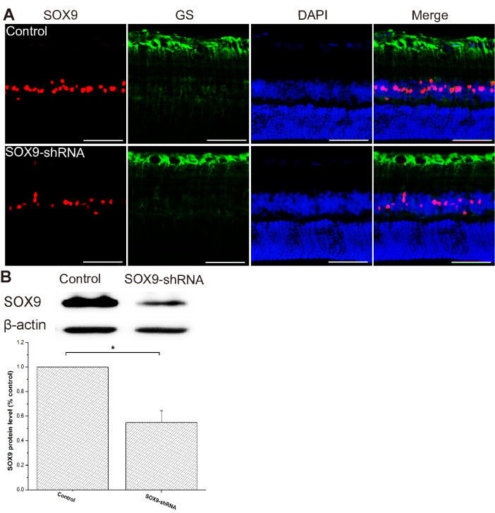
Two weeks after intravitreal injection, co-labeling immunostaining of Sox9 and GS (a specific Müller cell marker) was conducted. It was shown that Sox9 was mainly located in the Müller cell nucleus in both the control and Sox9-shRNA groups and there was relatively weaker staining of Sox9 in the Sox9-shRNA group compared with the control group (Figure 1A). Furthermore, the western blot analysis for the expression of Sox9 was performed to validate the knockdown status of the Sox9-shRNA lentiviral vector. The data showed approximately 0.55-fold lower Sox9 protein level in the Sox9-shRNA group compared with the control (n = 3, t test, p<0.05; Figure 1B, Appendix 1), in which the Sox9 had been knocked down.
Figure 1.
Validation of the Sox9 knockdown status of the Sox9-shRNA lentiviral vector after two weeks of intravitreal injection using co-labeling immunostaining and western blotting. A: Sox9 was mainly located in the Müller cell nucleus in both the control group and the Sox9-shRNA group and there was relatively weaker staining of Sox9 in the Sox9-shRNA group compared with the control group. Scale bar, 50 μm. B: Western blot analysis of the Sox9 protein in the retinas from the control and Sox9-shRNA groups. Β-actin is the loading control. Relative expression of the Sox9 protein in the Sox9-shRNA group is significantly reduced compared with the control at every studied time point. Data are expressed as a percentage of the control values presented as mean ± SD (n = 3), t test, *p<0.05.
Histologic analysis
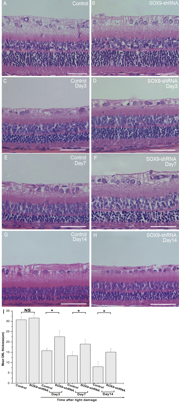
Morphological changes after light expose were measured using HE staining. The mean thickness of the ONL decreased obviously with time in both groups. However, in the Sox9-shRNA group, the mean thickness of the ONL was significantly better preserved when compared with the control at each time point (n = 4, t test, p<0.05; Figure 2). These data demonstrate that the Sox9 knockdown can preserve the loss of retinal photoreceptors induced by light damage.
Figure 2.
The Sox9 knockdown preserved retinal morphology as measured by HE staining. Representative photomicrographs show the histological appearance of retinas from the control and Sox9-shRNA groups on days 3, 7, and 14 after light expose (A-H). All the pictures were taken of the superior regions (approximately 1.0 mm from the optic disc) of the retinas. Note that the mean thickness of the ONL was better preserved in the Sox9-shRNA group (D, F, H) than the control (C, E, G) at each time point. I: The morphometric analysis of the thickness of the ONL shows a significant preservation in the Sox9-shRNA group compared with the control group. Mean ± SD (n = 4), t test, *p<0.05. Scale bar, 50 μm.
Electroretinography
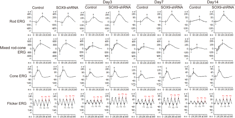
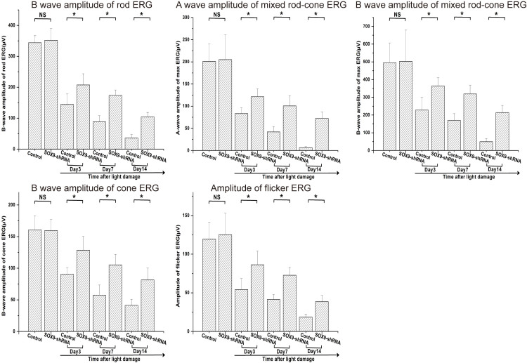
An ERG was used to evaluate the retinal function after light exposure. We recorded rod ERG, mixed rod-cone ERG, cone ERG and flicker ERG, respectively (Figure 3). The amplitudes of each ERG’s waves decreased significantly with time in both groups. However, the amplitudes of the ERG waves in the Sox9-shRNA group were significantly better preserved compared with the control at each time point (n = 4, t test, p<0.05; Figure 4). The results indicate that the downregulation of Sox9 could recuse the loss of both retinal rod and cone function induced by light damage.
Figure 3.
The Sox9 knockdown preserved retinal function as measured by ERG. The representative ERGs (rod ERG, mixed rod-cone ERG, cone ERG and flicker ERG, respectively) from the control and Sox9-shRNA groups were recorded on days 3, 7, and 14 after light expose.
Figure 4.
The Sox9 knockdown preserved retinal function as measured by ERG. The Sox9 knockdown significantly preserved amplitudes of ERG waves in the Sox9-shRNA group compared with the control on days 3, 7, and 14 after light expose. Mean ± SD (n = 4), t test, *p<0.05.
GFAP, vimentin, nestin, and Cspg changes at the protein level
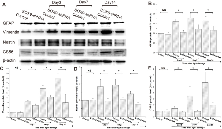
Western blot analysis was used to observe the changes of GFAP, vimentin, nestin, and Cspg proteins—which are closely related to Müller gliosis and ECM deposits—after light exposure. The expression levels of GFAP, vimentin, nestin, and Cspg proteins increased in the control group but were significantly downregulated in the Sox9-shRNA group at each studied time point after light damage (n = 3, t test, p<0.05; Figure 5, Appendix 2).
Figure 5.
The Sox9 knockdown attenuated the upregulation of GFAP, vimentin, nestin, and Cspg protein levels, as measured by western blot analysis. A: Western blot analysis was used to measure the GFAP, vimentin, nestin, and Cspg protein levels in the retinas from the control and Sox9-shRNA groups on days 3, 7, and 14 after light expose. Β-actin served as the loading control. The relative expressions of GFAP (B), vimentin (C), nestin (D), and Cspg (E) protein levels in Sox9-shRNA group are significantly downregulated compared with the control group. Data are expressed as a percentage of the control values presented as mean ± SD (n = 3), t test, *p<0.05.
Immunohistochemistry of GFAP
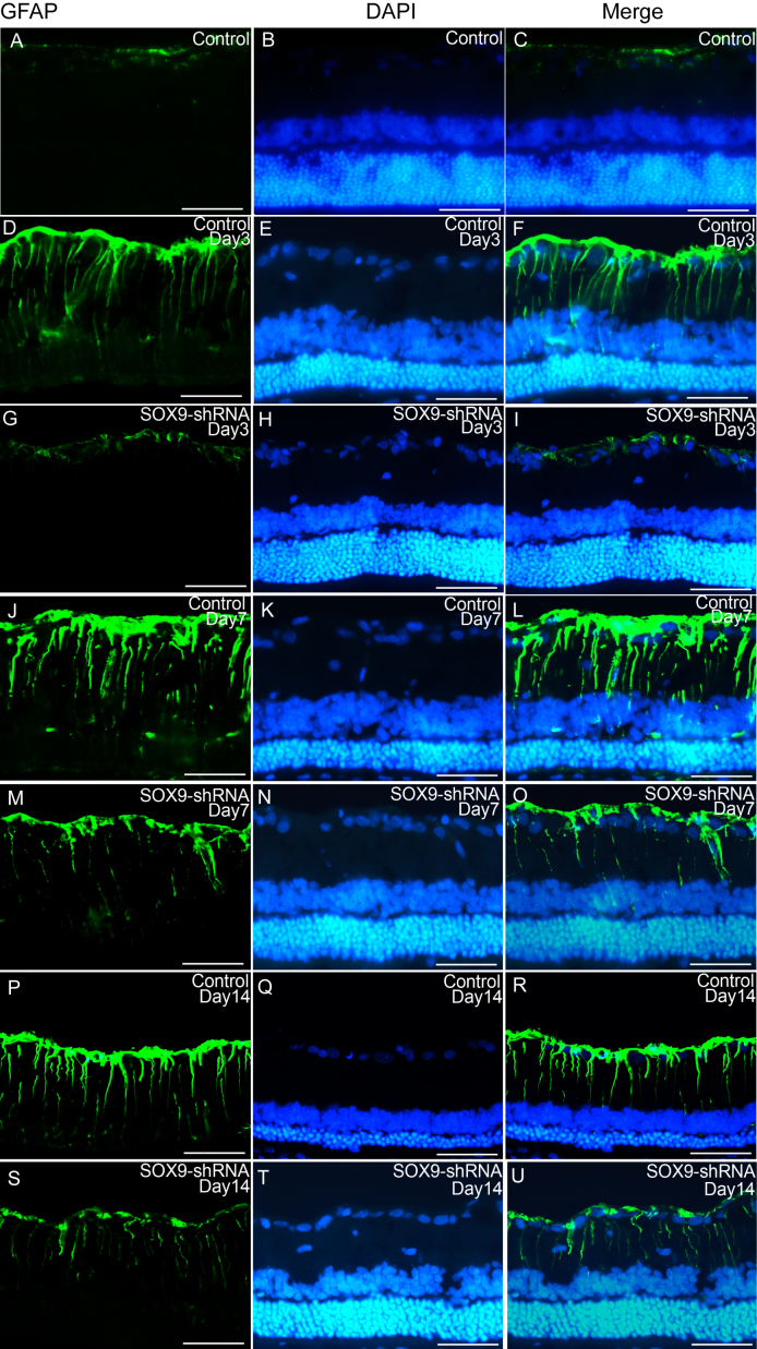
To confirm the expression changes of GFAP, the hallmark of Müller gliosis, the immunohistochemistry of GFAP was performed after light exposure. In both the control and Sox9-shRNA groups, the labeling intensity of GFAP increased, and the spatial distribution expanded on days 3, 7, and 14 after light exposure. However, the GFAP staining intensity and spatial distribution in retinas were obviously attenuated in the Sox9-shRNA group compared with the control at each time point (Figure 6).
Figure 6.
The Sox9 knockdown decreased the expression of GFAP as measured by immunohistochemistry. In both the control and Sox9-shRNA groups, the labeling intensity of GFAP increased, and the spatial distribution expanded on days 3, 7, and 14 after light expose. However, the GFAP staining intensity and spatial distribution in the Sox9-shRNA group were obviously attenuated compared with the control at each time point. All photomicrographs were taken under a photomicroscope within identical parameters. Scale bar, 50 μm.
Discussion
The results demonstrate that the intravitreal injection of the Sox9-shRNA lentiviral vector preserved rat retinal morphology and function after light damage. Further mechanistic investigation reveals that the Sox9 knockdown reduced the expression of intermediate filaments (GFAP, vimentin, and nestin) and Cspg deposition. These results support our hypothesis that reduced Sox9 function exerts neuroprotection in light-induced retinal damage in rats, indicating a potential therapeutic target for retinal degenerative diseases.
In retinal development, Sox9 is expressed in mouse multipotent retinal progenitor cells and then mainly in Müller cells in the sensory retina [10,11]. Our previous data also showed that Sox9 was located in the nuclei of Müller cells in normal adult rats [12]. Moreover, the upregulation of Sox9 and migration of Müller cells into the choroid in rats with retinal light damage was observed, indicating that Sox9 was actively involved in Müller cell gliosis and retinal remodeling [12,14]. Though previous studies demonstrated that Sox9 was also expressed in retinal pigment epithelium cells [15], herein we mainly focused on the effect of Sox9 downregulation on Müller cells, which are the predominant glial element in the retina.
In the current study, we observed that the lentivirus vector-mediated knockdown of Sox9 preserved the retinal morphology and function of rats after light damage. This is consistent with the neuroprotective role of Sox9 knockout in the CNS. Conditional Sox9 ablation in mice improves motor function following spinal cord injury [7,8], and Sox9 knockout mice have improved recovery following a stroke [9]. In addition, though the amplitudes of each ERG’s waves in the Sox9-shRNA group were significantly better preserved compared with the control at each time point, the amplitudes of the ERG waves decreased significantly with time in both groups. On one hand, this demonstrates that SOX9 knockdown could preserve both retinal rod and cone function after light damage; on the other hand, this indicates that the neuroprotective effect is limited. This might be due to the incomplete knockout of SOX9 or the complicated mechanism of light damage. And so far, we don’t know whether this neuroprotective effect lasts until 14 days later. These questions need to be elucidated in the next study.
One possible mechanism behind the neuroprotection of the Sox9 knockdown may be through the inhibition of Müller cell gliosis. As the predominant glial element in the retina, Müller cells are sensitive to various retinal injuries and experience a series of reactive changes [2]. This gliosis can be neuroprotective in the very early stages after damage. But when the activation is excessive, overactive gliosis becomes detrimental and forms glial scars, contributing to retinal remodeling [3,16]. Astrocyte activation and reactive gliosis are becoming important therapeutic targets of CNS diseases [17-19]. The hallmark of gliosis is the upregulation of intermediate filament proteins (GFAP, vimentin, and nestin), especially GFAP [20-23]. These three markers of glial activation in our study decreased after Sox9 downregulation, indicating that Müller cell gliosis was probably inhibited. Previous research has shown that inhibiting the expression of GFAP and vimentin has a neuroprotective effect. The retinas of adult mice are deficient in GFAP and vimentin provides a permissive environment for grafted neurons to migrate and extend neurites [4]. Mice deficient in GFAP and vimentin show attenuated glial reactions and photoreceptor degeneration induced by retinal detachment [5]. The absence of GFAP and vimentin prevents hypertrophy of astrocytic processes and improves post-traumatic regeneration [24]. Our data indicate that the Sox9 knockdown might attenuate glial activation after retinal light damage, which coincides with the role of Sox9 knockout in the CNS [7,9].
A second possible explanation for neuroprotection of the Sox9 knockdown may be the reduction of Cspg deposits. Cspgs—an ECM family member—accumulate in the damaged CNS, during traumatic brain or spinal cord injury and multiple sclerosis, and Cspgs may help drive pathogenesis [25-27]. More recently, it has been shown that the knockout of Sox9, which induces destructive ECM components in organ fibrosis and related disorders [28-30], downregulates Cspg levels and has neuroprotective properties in the CNS. Conditional Sox9 ablation in mice reduces Cspg levels and improves motor functions following a spinal cord injury [8]. The Sox9 knockout leads to reduced Cspg levels, increased tissue sparing, and improved post-stroke neurologic recovery [9]. In retinas, the Cspg levels become highly expressed in some, but not all, models of inherited photoreceptor degeneration [31,32]. In the current study, our data show that Cspg levels had an increasing trend in the control group after light damage but were significantly downregulated by the Sox9 knockdown.
Müller cell gliosis and ECM remodeling involve a series of complicated cellular and molecular events. The data indicating some proteins might be regulated by SOX9 are only preliminary results, providing direction for future research regarding the potential mechanisms involved.
In conclusion, our data show that intravitreal injections of the Sox9-shRNA lentiviral vector preserved rat retinal morphology and function after light damage and downregulated GFAP, vimentin, nestin, and Cspgs, which are related to Müller cell gliosis and ECM remodeling. These results indicate that Sox9 might be a potential therapeutic target for retinal degenerative diseases.
Acknowledgments
This work was supported by the Youth Project of the National Natural Science Fund (Grant No. 81500723, 81700861), the National Natural Science Foundation of China (Grant Nos. 81570854, 81570842, 81570855), and Grant from Science and Technology Commission of Shanghai Municipality (Grant No. 16411953700).
Appendix 1.
To access the data, click or select the words “Appendix 1.” Validation of the Sox9 knockdown status of the Sox9-shRNA lentiviral vector after two weeks of intravitreal injection using western blotting. Raw western blots of the Sox9 and Β-actin protein in the retinas from the control and Sox9-shRNA groups. Β-actin served as the loading control. The expression of the Sox9 protein in the Sox9-shRNA group is reduced compared with the control (n=3).
Appendix 2.
To access the data, click or select the words “Appendix 2.” The Sox9 knockdown attenuated the upregulation of GFAP, vimentin, nestin, and Cspg protein levels, as measured by western blot. Raw western blots of the GFAP, vimentin, nestin, Cspg and Β-actin protein in the retinas from the control and Sox9-shRNA groups on days 3, 7, and 14 after light expose. Β-actin served as the loading control (n=3).
References
- 1.Vecino E, Rodriguez FD, Ruzafa N, Pereiro X, Sharma SC. Glia–neuron interactions in the mammalian retina. Prog Retin Eye Res. 2016;51:1–40. doi: 10.1016/j.preteyeres.2015.06.003. [DOI] [PubMed] [Google Scholar]
- 2.Bringmann A, Pannicke T, Grosche J, Francke M, Wiedemann P, Skatchkov SN, Osborne NN, Reichenbach A. Muller cells in the healthy and diseased retina. Prog Retin Eye Res. 2006;25:397–424. doi: 10.1016/j.preteyeres.2006.05.003. [DOI] [PubMed] [Google Scholar]
- 3.Cuenca N, Fernández-Sánchez L, Campello L, Maneu V, De la Villa P, Lax P, Pinilla I. Cellular responses following retinal injuries and therapeutic approaches for neurodegenerative diseases. Prog Retin Eye Res. 2014;43:17–75. doi: 10.1016/j.preteyeres.2014.07.001. [DOI] [PubMed] [Google Scholar]
- 4.Kinouchi R, Takeda M, Yang L, Wilhelmsson U, Lundkvist A, Pekny M, Chen DF. Robust neural integration from retinal transplants in mice deficient in GFAP and vimentin. Nat Neurosci. 2003;6:863–8. doi: 10.1038/nn1088. [DOI] [PubMed] [Google Scholar]
- 5.Nakazawa T, Takeda M, Lewis GP, Cho KS, Jiao J, Wilhelmsson U, Fisher SK, Pekny M, Chen DF, Miller JW. Attenuated glial reactions and photoreceptor degeneration after retinal detachment in mice deficient in glial fibrillary acidic protein and vimentin. Invest Ophthalmol Vis Sci. 2007;48:2760–8. doi: 10.1167/iovs.06-1398. [DOI] [PMC free article] [PubMed] [Google Scholar]
- 6.Pritchett J, Athwal V, Roberts N, Hanley NA, Hanley KP. Understanding the role of SOX9 in acquired diseases: lessons from development. Trends Mol Med. 2011;17:166–74. doi: 10.1016/j.molmed.2010.12.001. [DOI] [PubMed] [Google Scholar]
- 7.McKillop WM, Dragan M, Schedl A, Brown A. Conditional Sox9 ablation reduces chondroitin sulfate proteoglycan levels and improves motor function following spinal cord injury. Glia. 2013;61:164–77. doi: 10.1002/glia.22424. [DOI] [PMC free article] [PubMed] [Google Scholar]
- 8.McKillop WM, York EM, Rubinger L, Liu T, Ossowski NM, Xu K, Hryciw T, Brown A. Conditional Sox9 ablation improves locomotor recovery after spinal cord injury by increasing reactive sprouting. Exp Neurol. 2016;283:1–15. doi: 10.1016/j.expneurol.2016.05.028. [DOI] [PubMed] [Google Scholar]
- 9.Xu X, Bass B, McKillop WM, Mailloux J, Liu T, Geremia NM, Hryciw T, Brown A. Sox9 knockout mice have improved recovery following stroke. Exp Neurol. 2018;303:59–71. doi: 10.1016/j.expneurol.2018.02.001. [DOI] [PubMed] [Google Scholar]
- 10.Poche RA, Furuta Y, Chaboissier MC, Schedl A, Behringer RR. Sox9 is expressed in mouse multipotent retinal progenitor cells and functions in Muller glial cell development. J Comp Neurol. 2008;510:237–50. doi: 10.1002/cne.21746. [DOI] [PMC free article] [PubMed] [Google Scholar]
- 11.Muto A, Iida A, Satoh S, Watanabe S. The group E Sox genes Sox8 and Sox9 are regulated by Notch signaling and are required for Muller glial cell development in mouse retina. Exp Eye Res. 2009;89:549–58. doi: 10.1016/j.exer.2009.05.006. [DOI] [PubMed] [Google Scholar]
- 12.Wang X, Fan J, Zhang M, Ni Y, Xu G. Upregulation of Sox9 in Glial (Muller) cells in retinal light damage of rats. Neurosci Lett. 2013;556:140–5. doi: 10.1016/j.neulet.2013.10.005. [DOI] [PubMed] [Google Scholar]
- 13.Ni Y, Gan D, Xu H, Xu G, Da C. Neuroprotective effect of transcorneal electrical stimulation on light-induced photoreceptor degeneration. Exp Neurol. 2009;219:439–52. doi: 10.1016/j.expneurol.2009.06.016. [DOI] [PubMed] [Google Scholar]
- 14.Marc RE, Jones BW, Watt CB, Vazquez-Chona F, Vaughan DK, Organisciak DT. Extreme retinal remodeling triggered by light damage: implications for age related macular degeneration. Mol Vis. 2008;14:782–806. [PMC free article] [PubMed] [Google Scholar]
- 15.Masuda T, Wahlin K, Wan J, Hu J, Maruotti J, Yang X, Iacovelli J, Wolkow N, Kist R, Dunaief JL, Qian J, Zack DJ, Esumi N. Transcription factor SOX9 plays a key role in the regulation of visual cycle gene expression in the retinal pigment epithelium. J Biol Chem. 2014;289:12908–21. doi: 10.1074/jbc.M114.556738. [DOI] [PMC free article] [PubMed] [Google Scholar]
- 16.Hippert C, Graca AB, Pearson RA. Gliosis Can Impede Integration Following Photoreceptor Transplantation into the Diseased Retina. Adv Exp Med Biol. 2016;854:579–85. doi: 10.1007/978-3-319-17121-0_77. [DOI] [PubMed] [Google Scholar]
- 17.Pekny M, Wilhelmsson U, Pekna M. The dual role of astrocyte activation and reactive gliosis. Neurosci Lett. 2014;565:30–8. doi: 10.1016/j.neulet.2013.12.071. [DOI] [PubMed] [Google Scholar]
- 18.Hol EM, Pekny M. Glial fibrillary acidic protein (GFAP) and the astrocyte intermediate filament system in diseases of the central nervous system. Curr Opin Cell Biol. 2015;32:121–30. doi: 10.1016/j.ceb.2015.02.004. [DOI] [PubMed] [Google Scholar]
- 19.Pekny M, Pekna M. Reactive gliosis in the pathogenesis of CNS diseases. Biochimica et Biophysica Acta (BBA) - Molecular Basis of Disease. 2016;1862:483–91. doi: 10.1016/j.bbadis.2015.11.014. [DOI] [PubMed] [Google Scholar]
- 20.Xue LP, Lu J, Cao Q, Kaur C, Ling EA. Nestin expression in Müller glial cells in postnatal rat retina and its upregulation following optic nerve transection. Neuroscience. 2006;143:117–27. doi: 10.1016/j.neuroscience.2006.07.044. [DOI] [PubMed] [Google Scholar]
- 21.Pekny M, Lane EB. Intermediate filaments and stress. Exp Cell Res. 2007;313:2244–54. doi: 10.1016/j.yexcr.2007.04.023. [DOI] [PubMed] [Google Scholar]
- 22.Yang Z, Wang KKW. Glial fibrillary acidic protein: from intermediate filament assembly and gliosis to neurobiomarker. Trends Neurosci. 2015;38:364–74. doi: 10.1016/j.tins.2015.04.003. [DOI] [PMC free article] [PubMed] [Google Scholar]
- 23.Roche SL, Ruiz-Lopez AM, Moloney JN, Byrne AM, Cotter TG. Microglial-induced Müller cell gliosis is attenuated by progesterone in a mouse model of retinitis pigmentosa. Glia. 2018;66:295–310. doi: 10.1002/glia.23243. [DOI] [PubMed] [Google Scholar]
- 24.Wilhelmsson U, Li L, Pekna M, Berthold CH, Blom S, Eliasson C, Renner O, Bushong E, Ellisman M, Morgan TE, Pekny M. Absence of glial fibrillary acidic protein and vimentin prevents hypertrophy of astrocytic processes and improves post-traumatic regeneration. J Neurosci. 2004;24:5016–21. doi: 10.1523/JNEUROSCI.0820-04.2004. [DOI] [PMC free article] [PubMed] [Google Scholar]
- 25.Dyck SM, Karimi-Abdolrezaee S. Chondroitin sulfate proteoglycans: Key modulators in the developing and pathologic central nervous system. Exp Neurol. 2015;269:169–87. doi: 10.1016/j.expneurol.2015.04.006. [DOI] [PubMed] [Google Scholar]
- 26.Stephenson EL, Yong VW.Pro-inflammatory roles of chondroitin sulfate proteoglycans in disorders of the central nervous system. Matrix Biol 2018. 71-72:432-442 [DOI] [PubMed] [Google Scholar]
- 27.Orr MB, Gensel JC. Spinal Cord Injury Scarring and Inflammation: Therapies Targeting Glial and Inflammatory Responses. Neurotherapeutics. 2018;15:541–53. doi: 10.1007/s13311-018-0631-6. [DOI] [PMC free article] [PubMed] [Google Scholar]
- 28.Bennett MR, Czech KA, Arend LJ, Witte DP, Devarajan P, Potter SS. Laser capture microdissection-microarray analysis of focal segmental glomerulosclerosis glomeruli. Nephron, Exp Nephrol. 2007;107:e30–40. doi: 10.1159/000106775. [DOI] [PubMed] [Google Scholar]
- 29.Hanley KP, Oakley F, Sugden S, Wilson DI, Mann DA, Hanley NA. Ectopic SOX9 mediates extracellular matrix deposition characteristic of organ fibrosis. J Biol Chem. 2008;283:14063–71. doi: 10.1074/jbc.M707390200. [DOI] [PubMed] [Google Scholar]
- 30.Peacock JD, Levay AK, Gillaspie DB, Tao G, Lincoln J. Reduced sox9 function promotes heart valve calcification phenotypes in vivo. Circ Res. 2010;106:712–9. doi: 10.1161/CIRCRESAHA.109.213702. [DOI] [PMC free article] [PubMed] [Google Scholar]
- 31.Hippert C, Graca AB, Barber AC, West EL, Smith AJ, Ali RR, Pearson RA. Müller Glia Activation in Response to Inherited Retinal Degeneration Is Highly Varied and Disease-Specific. PLoS One. 2015;10:e120415. doi: 10.1371/journal.pone.0120415. [DOI] [PMC free article] [PubMed] [Google Scholar]
- 32.Chen L, FitzGibbon T, He J, Yin ZQ. Localization and developmental expression patterns of CSPG-cs56 (aggrecan) in normal and dystrophic retinas in two rat strains. Exp Neurol. 2012;234:488–98. doi: 10.1016/j.expneurol.2012.01.023. [DOI] [PubMed] [Google Scholar]


