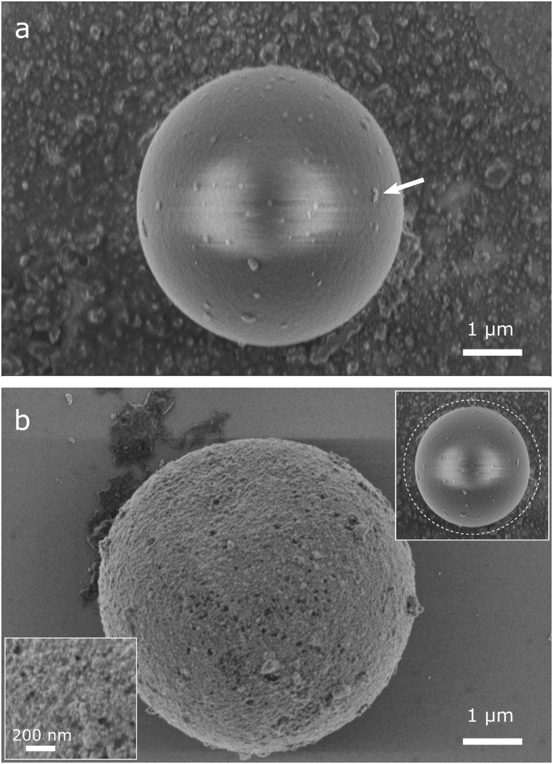
FIG. 2.
(a) SEM image of a particle after one day in gold colloid solution. Larger nanoparticle clusters attached on the surface are clearly visible and indicated by an arrow. (b) SEM image of a gold-coated particle after complete coating procedure. An almost closed layer of about hundreds of nanometers is formed on the particle surface through which the fluorescence still can be detected. Upper inset: illustration of particle size before coating in relation to the size after coating (dashed line). Lower inset: close-up view of the gold surface structure.

