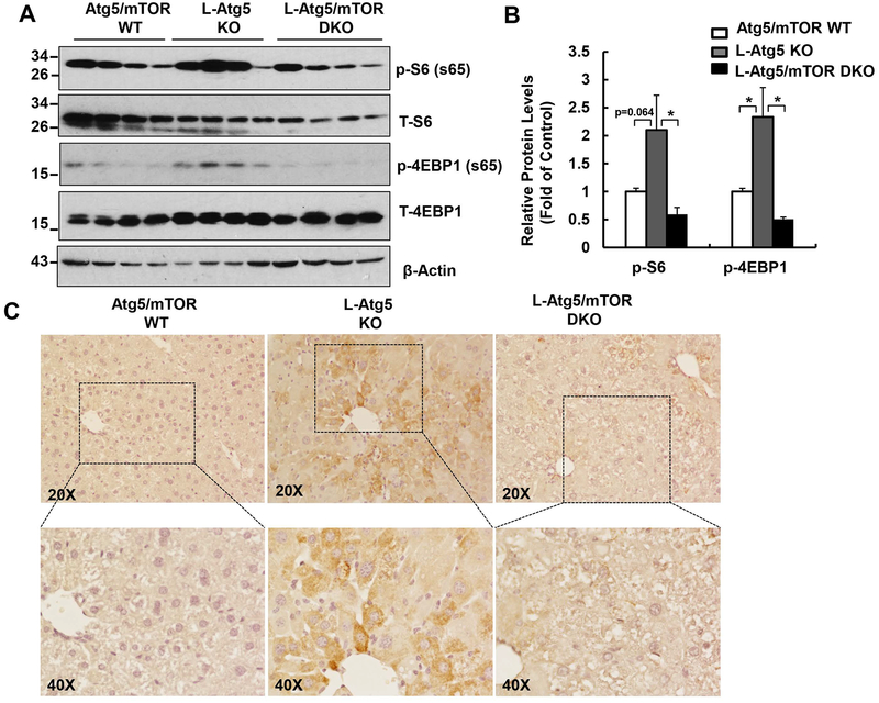
Figure 1. Increased mTOR activation in L-Atg5 KO mice.
(A) Total liver lysates of the indicated genotypes of 2-month-old mice were subjected to western blot analysis. (B) Densitometry analysis of (A). Data are means± SEM (n=4). (C) Representative images of immunostaining of phosphorylated S6 (p-S6) of Atg5/mTOR WT, L-Atg5 KO and L-Atg5/mTOR DKO liver sections.

