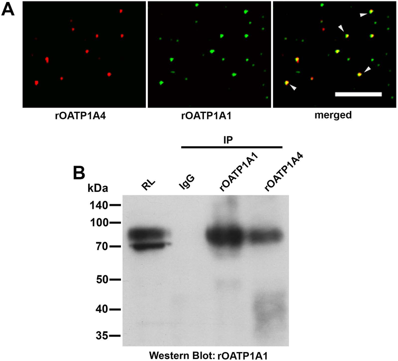
Figure 1: Interaction of rOATP1A1 and rOATP1A4 in rat liver.
A. Endocytic vesicles isolated from rat liver were immunostained for rOATP1A1 and rOATP1A4 using the two primary antibody method. Images from a representative study are shown in which rOATP1A4 is in red, rOATP1A1 is in green, and vesicles in which rOATP1A4 and rOATP1A1 are colocalized are in yellow (arrowheads) in the merged view. rOATP1A1 and rOATP1A4 were colocalized in 74% of the 12,044 vesicles that were examined. Scale bar = 10 μm. B. Colocalization in vesicles does not imply that rOATP1A1 and rOATP1A4 are bound to each other. This was examined by immunoprecipitation. Rat liver homogenate was prepared as in Materials and Methods, solubilized in 1% CHAPS containing protease inhibitors, and subjected to immunoprecipitation with non-immune IgG or antibody to rOATP1A1 or rOATP1A4. The immunoprecipitates were subjected to Western blot with antibody to rOATP1A1 in the absence of reduction. The starting rat liver lysate (RL) in CHAPS was used to show antibody reactivity and specificity.

