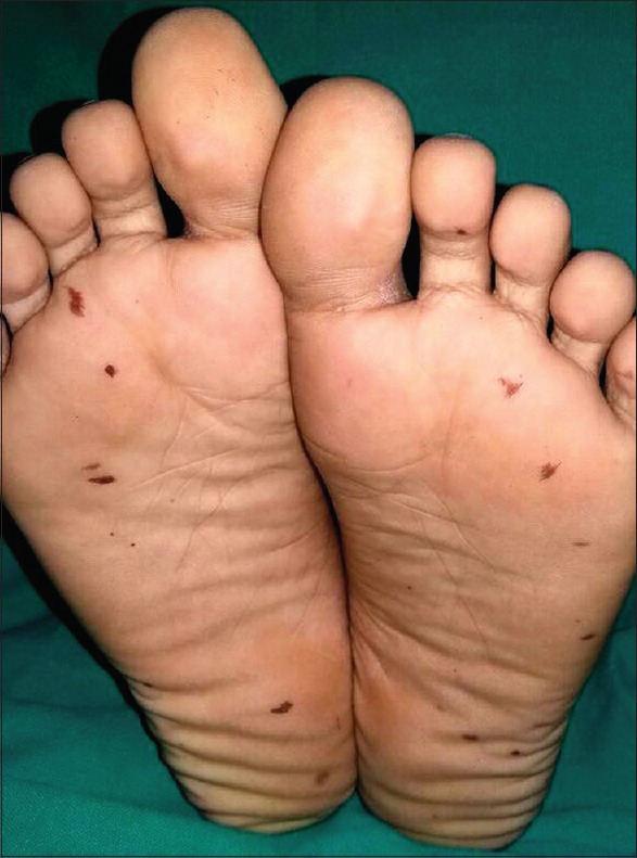
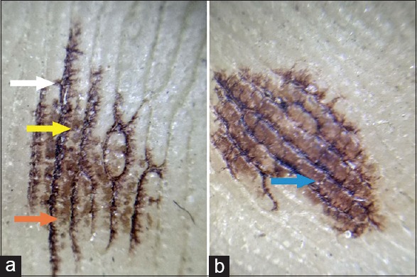
A 7-year-old boy presented with multiple asymptomatic reddish brown spots on the soles of both feet for a duration of 2 days. The lesions had developed after a sudden shower of rain. Physical examination revealed irregularly shaped, reddish brown macules with tapering and streaky ends [Figure 1]. Wet contact dermoscopy revealed dark brownish linear pigmentation along the dermatoglyphic lines (parallel furrows) with feathering (resembling a frayed rope) [Figure 2a]. There were blotches of light brown pigmentation interspersed between the lines which tended to coalesce in the darker lesions [Figure 2a and b]. Pigmentation was accentuated around sweat pores [Figure 2a]. Both grandparents and mother also had similar lesions with similar dermoscopic images. All three family members gave history of contact with numerous small live and dead winged insects on the rainy day. However, no such lesions were noticed in the father, who was at his workplace on that day. Pigmentation persisted despite washing with soap water or spirit. Thus, diagnosis of cydnidae induced pigmentation was established.
Figure 1.

Well-defined reddish-brown macules on soles with streaky and tapering ends on plantar aspect of both feet
Figure 2.

Dermoscopy from light brown (a) and dark brown (b) macule: Wet contact dermoscopy of the lesion revealed dark brownish linear pigmentation along the dermatoglyphic lines (parallel furrows) with feathering (White arrow). In addition, there were blotches of light brown pigmentation interspersed between the lines (Orange arrow), which tended to coalesce giving a diffuse appearance (Blue arrow) in the darker lesions (b). Pigmentation was also accentuated around sweat pores (Yellow arrow) (Mode– non-polarized hand held dermoscope, magnification – 10×)
Cydnidae insects (burrowing bugs) are usually found in rural areas in the fields during monsoon and feed on the roots of plants.[1] A hydrocarbonate, odorous substance is produced from special glands located in its thorax (in adult) and abdomen (in nymph). It acts as a repellant and as a chemoattractant.[2] Red-brown pigmented macules of varying size and shape appear rapidly within minutes at the site of contact with this fluid on accidental crushing or pressure on these insects and gradually resolve in 10–15 days. The differential diagnosis of cydnidae pigmentation includes volar melanotic macules, junctional melanocytic nevi, acral lentigines, and petechiae of dengue fever.[1] Volar melanotic macules are asymptomatic light-brown macules involving palms and/or soles and can occasionally be associated with systemic disorders such as Peutz-Jeghers syndrome or Laugier-Hunziker syndrome. Sudden onset of acral lentigines has been described with underlying malignancies.[3] Petechiae in dengue fever represent the hemorrhagic manifestations and usually appear 4–5 days after the onset of fever. In our patient, the sudden development of macules was restricted to the soles with no other cutaneous or systemic findings suggestive of dengue or malignancy. In addition, the macules were typically irregular in shape with tapering ends. Dermoscopic features of volar melanotic lesions and junctional nevi include parallel furrows, lattice like pigmentation; following the dermatoglyphics and occasionally fibrillar or filamentous pattern.[4,5] Acral lentigines show homogenous, globular, and reticular patterns with occasional irregular dots and the so called “moth eaten appearance.” Petechiae show reddish to violaceous dots/globules in early lesions and brown dots in late lesions. The unique dermoscopic features of cydnidae pigmentation were frayed parallel furrows and accentuation of pigmentation around the sweat glands.
Declaration of patient consent
The authors certify that they have obtained all appropriate patient consent forms. In the form the patient(s) has/have given his/her/their consent for his/her/their images and other clinical information to be reported in the journal. The patients understand that their names and initials will not be published and due efforts will be made to conceal their identity, but anonymity cannot be guaranteed.
Financial support and sponsorship
Nil.
Conflicts of interest
There are no conflicts of interest.
References
- 1.Malhotra AK, Lis JA, Ramam M. Cydnidae (burrowing bug) pigmentation: A novel arthropod dermatosis. JAMA Dermatol. 2015;151:232–3. doi: 10.1001/jamadermatol.2014.2715. [DOI] [PubMed] [Google Scholar]
- 2.Hosokawa T, Kikuchi Y, Fukatsu T. Polyphyly of gut symbionts in stinking bugs of the family cydnidae. Appl Environ Microbiol. 2012;78:4758–61. doi: 10.1128/AEM.00867-12. [DOI] [PMC free article] [PubMed] [Google Scholar]
- 3.Wolf R, Orion E, Davidovici B. Acrallentigines: A new paraneoplastic syndrome. Int J Dermatol. 2008;47:168–70. doi: 10.1111/j.1365-4632.2008.03223.x. [DOI] [PubMed] [Google Scholar]
- 4.Elwan NM, Eltatawy RA, Elfar NN, Elsakka OM. Dermoscopic features of acralpigmented lesions in Egyptian patients: A descriptive study. Int J Dermatol. 2016;55:187–92. doi: 10.1111/ijd.12882. [DOI] [PubMed] [Google Scholar]
- 5.González-Ramírez RA, Guerra-Segovia C, Garza-Rodríguez V, Garza-Báez P, Gómez-Flores M, Ocampo-Candiani J. Dermoscopic features of acral melanocytic nevi in a case series from Mexico. Ann Bras Dermatol. 2018;93:665–70. doi: 10.1590/abd1806-4841.20186695. [DOI] [PMC free article] [PubMed] [Google Scholar]


