Abstract
In July 2017, twenty cattle of a free-grazing herd were found to be infested with leeches in the mouth. Main signs were bloody sialorrhea and/or a purple-red colour of the lower lip. Leeches, in a variable number (1 to 3) per animal, were found at the lingual frenulum or on the sublingual vestibular mucosa and were morphologically identified as Limnatis nilotica. To the best of our knowledge, this is the first report of cattle infestation by L. nilotica in Italy. Besides recalling the attention to leech infestation and suggesting its inclusion in the differential diagnosis of animals with suggestive signs, this short report also provides practitioners with easy-going morphological keys for proper diagnosis and discrimination among species.
Keywords: cattle, leech infestation, Limnatis nilotica, oral cavity, Sicily
Leeches belong to the Annelida phylum and Hirudinea class [14], and are world-wide distributed. Leeches are segmented hermaphroditic blood feeding ectoparasites of man, domestic and wild animals, as well as invertebrate creatures [3]. Leeches are featured by two prominent suckers (i.e., anterior and posterior) that allow them to grip the surface of the prey while they suck the host’s blood. The leech’s mouth located within the anterior sucker, is equipped with a set of razor-sharp jaws that are used to open wound in the prey, while the muscular pharynx pumps the blood into the leech’s digestive tract. A salivary gland secretes the hirudin, an anticoagulant substance that prevents coagulation of the blood [11, 12].
Leeches have a dual significance, indeed, beside the primary role as blood-feeder parasites, some species may also act as vectors of other pathogens. Noteworthily, leeches are acknowledged vectors of protozoans, including Trypanosoma spp. [30] causing disease in humans and/or livestock [18, 19]. The host range of leeches is fairly broad and is not restricted to a single vertebrate class. Therefore, these invertebrates would ingest a range of unrelated trypanosomes with their blood meals, and, in turn, may expose a wide range of vertebrates to the trypanosomes they carry [18, 19].
Several species of leeches have been proven to be beneficial to humans, being Hirudo medicinalis, known as the European medicinal leech, the best-known species used for bloodletting in humans [20], though there are other medicinal leech species such as Hirudo troctina and Haementeria officinalis [15]. Other leech species can lead to damages of different severity in the hosts. In this regard, one of the most important species is Limnatis nilotica, commonly known as horse leech or Nile leech, this species occurs in lakes and streams in southern Europe, Middle-East countries and North Africa [2, 5, 25, 26].
Limnatis nilotica may enter the animal body through drinking from contamined water and usually attaches to the oral cavity or respiratory passages [14, 17, 37]. Indeed, this species is not able to pierce the skin of mammals but it sucks blood from the mouth or nasal mucosa causing inflammation and other damages [24]. Though several animal species (e.g. caw, sheep, goat, horse, dog and pig) and human can be infested [8, 13], hirudiniasis by L. nilotica is considered negligible and only few reports are available in literature (see Table 1). The rareness of L. nilotica reports in European countries may be related to the reclamation of wetlands as well as to the good quality of breeding standards which mostly use clean drinking water. However, one might speculate that the paucity of reports claiming the finding of L. nilotica could be due because unnoticed or misidentified.
Table 1. Reports of Limnatis nilotica infestation in several animal species.
| Animal species | Country | Reference |
|---|---|---|
| Human | Turkey | Ağin et al. 2008 [1] |
| Cattle | Iran | Bahmani et al. 2012 [3] |
| Libya | Negm-Eldin et al. 2013 [23] | |
| Dog | Iran | Rajaei et al. 2014 [28] |
| Italy | Raele et al. 2015 [27] | |
| Goat | Libya | Negm-Eldin et al. 2013 [23] |
| Iran | Bahmani et al. 2013 [6] | |
| Sheep | Libya | Negm-Eldin et al. 2013 [23] |
| Iran | Bahmani et al. 2013 [6] | |
| Donkey | Iran | Bahmani et al. 2014 [4] |
| Camel | Iraq | Al-Ani and Al-Shareefi 1995 [2] |
Here we report the first signalling of oral hirudiniasis by L. nilotica in cattle in Italy; additionally, we also provide easy identification keys that would allow proper and easy diagnosis in case of leech infestation.
In July 2017, cattle, of a cattle-goat mixed herd located in the province of Messina (southern Italy, 38.0239N; 14.7298E; 400 m a.s.l.) were referred for veterinary consultation.
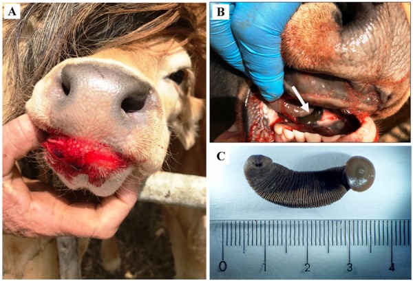
Upon physical examination cattle showed bloody sialorrhea and/or reddish colouring of the lower lip (Fig. 1A), weight loss and restlessness when the oral cavity was touched from the outside. At the opening of the oral cavity, a variable number of live leeches, one to three per cattle, were found attached at the lingual frenulum and/or on the sublingual vestibular mucosa (Fig. 1B–C). The leeches were detached and removed by a gentle traction with forceps, stored in plastic vials containing 70% alcohol and, thereafter, sent to the Unit of Parasitology at the Department of Veterinary Sciences (University of Messina, Messina, Italy) for identification. Once in laboratory, the leeches were observed under a stereomicroscope and photographed with the aid of a digital camera (AxioCam MRc, Carl Zeiss, Göttingen, Germany) mounted directly on the microscope. Additionally, some specimens were also routinely processed for scanning electron microscopy (SEM) as described elsewhere [38] and observed with an SEM Zeiss EVO 10 MA (Carl Zeiss Microscopy GmbH, Jena, Germany).
Fig. 1.

A cow with bloody sialorrhea due to Limnatis nilotica infestation (A); L. nilotica (arrow) attached to oral cavity of a cattle (B); specimen of L. nilotica detached from a cattle (C).
Twenty out of 60 cattle were found leech infested; the infestation was not associated with age of animals as either young (5–6 months of age) and adults (5–6 years) were found parasitized.
The leeches herein investigated measured 2.5–3.0 cm in length and 0.8–1.0 cm in width with dark green-black colour on the dorsal side and a lighter colour on the ventral side.
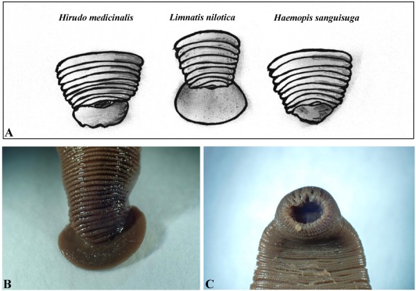
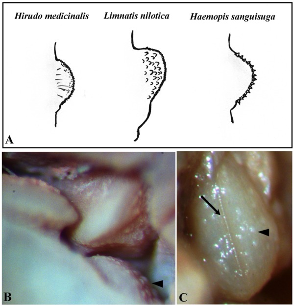
The well-developed posterior sucker (Fig. 2B) had a diameter equal to the maximum width of the body (1 cm), while the anterior sucker with three lobes was featured by a median ventral furrow (Fig. 2C). The anterior sucker showed 10 eye spots arranged in lateral crescents. Triple jaws, consisting of one dorso-medial and asymmetrical pair of ventro-lateral jaws were observed inside the oral cavity and surrounded the oral opening (Fig. 3B). The jaws appeared rounded, soft and light grey with clearer spots. A number of papillae were observed on both sides of jaws (Fig. 3C) and a row of tooth craters, irregular in size, shape and spacing, was observed on their edge (Fig. 3C). Specifically, some specimens herein analysed showed 48 tooth craters for each jaw whereas other ones showed jaws owned 57 tooth craters each.
Fig. 2.

Graphical illustration of species-specific posterior sucker (dorsal view) of some leech species (adapted from Minelli 1979) (A); posterior sucker (B) and anterior sucker (C) of a specimen of Limnatis nilotica.
Fig. 3.

Graphical illustration of species-specific single jaw of some leech (adapted from Minelli 1979) (A); jaw complex of specimens of Limnatis nilotica obtained from the case (B) with papillae on sides of jaw (arrowhead); detail of the tooth craters (black arrow) irregular in size, shape and spacing, and papillae (arrowhead) (C).
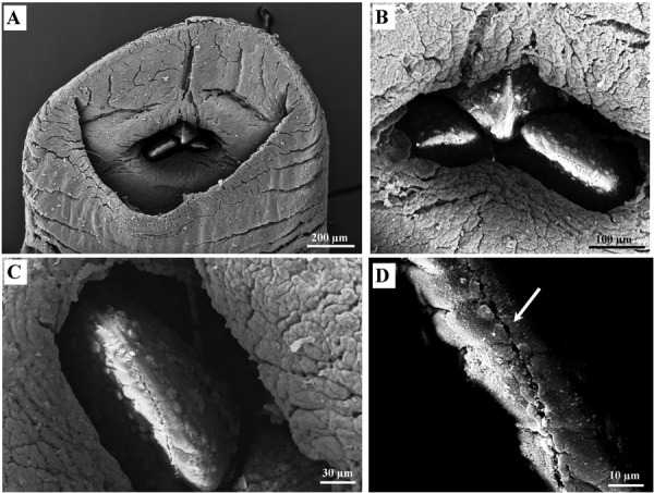
The morphological structures of the anterior extremity observed at the stereomicroscope were also described with SEM (Fig. 4).
Fig. 4.

Scanning Electron Microscope images of anterior sucker of a Limnatis nilotica specimen. Oral sucker and the three jaws in the bottom (A), The jaws are in form of a triangle (B) bearing a number of rounded papillae on both sides (C) and a line of tooth craters in the apex (white arrow) (D).
All the collected leech specimens were identified as L. nilotica according to morphological keys reported by Minelli [24], Nakano et al. [28], Negm-Eldin et al. [29] and Orevi et al. [31].
Macro- and microscopic differences among the species H. medicinalis, L. nilotica and Haemopis sanguisuga are shown in Figs. 2A and 3A, in order to give a basic recognition guide to medicinal and non-medicinal leech species commonly found in Italian territory.
The genus Limnatis includes three species, namely L. nilotica, L. bacescui, and L. paluda [36]. The leech taxonomy, at the species level and higher, is in a revolutionary phase so far. Little care has been devoted till now to represent the genera in molecular studies by their type species, and the species by their topo-type populations, though this approach remains the most reliable tool for assuring the taxonomical identity of specimens. Molecular evidences are available for L. nilotica and for L. paluda [28], whereas no molecular studies are available for L. bacescui. Despite the species in the genus Limnatis are well-known as nasal leeches, the taxonomic status of each is far from being clarified. Also, each nominal species of the genus Limnatis should be reviewed based on specimens collected from the type locality. The type locality of L. nilotica is Egypt, while L. paluda has been reported to occur mainly in the Middle-East [4, 10, 20]. The species L. bacescui was signalled only from its type locality, Romania (Dobruja) [22].
To the best of our knowledge, the present article, for the first-time, reports cattle infestation by the leech L. nilotica in Italy. The morphological structures of the anterior extremity of the examined specimens, particularly the jaws and the number of tooth craters for each jaw allowed the identification to species level. Effectively, it has been stated that L. paluda possesses fewer tooth craters on each jaw (no. 30–47) compared to L. nilotica whose jaws own no. 45–60 tooth craters [21, 27]. In addition, L. bacescui possessed 50–54 teeth on each jaw [22]. Several surveys carried out in the Middle-East countries reported L. nilotica in a large number of animal species such as cattle, donkey, sheep, goat [5, 6], camel [2], dog [34] and humans [1]. Together all these findings suggest that L. nilotica is a generalist blood feeder rather than a host-specific parasite.
In Italy, this leech species has been reported in aquatic environment of Tuscany, Campania, Apulia, Basilicata, Sicily and Sardinia [24]. A case of L. nilotica infestation was described in human from Naples [24] and, more recently, in a dog from Apulia [33]. Although, respiratory disorders, anaemia and topical acute infection have been reported as dominant signs connected with L. nilotica infestation [7, 16], the parasitized animals herein investigated showed bloody sialorrhea and weight loss. The cattle also showed restlessness when the oral cavity was touched from the outside probably due to damage of tissues caused by the leech. In the current study from one to three leeches per cattle was found in the mouth; the low leech burden as well as their anatomical location could explain the moderate severity of clinical signs herein observed.
The variations in leech burden and location among infested animals may reflect the variation in the severity of signs [23]. A total of 19 leeches (4 leeches removed from the nostrils, 15 at necropsy) was reported as a cause of fatal hirudiniasis for cattle in Iran [25]. Bahmani et al. [9] isolated 6–7 leeches from sheep oesophagus as a cause of hemoptysis, anemia, chest pain, fever, vomiting and bloody sialorrhea. Three blood sucking leeches from nose were reported in a children patient as a cause of epistaxis [35].
All the cattle examined in the present report were infested in the oral cavity. This finding is in agreement to what reported in previous surveys [8, 16] on livestock, and not surprising since L. nilotica is an aquatic species that inhabits lakes and streams. Particularly, the juvenile form of this species is smaller than the adult, and it swims on the water surface and can be easily ingested by humans or animals. Thus, it may enter the animal body through drinking from infested waters and most of them attach to the oral cavity or respiratory passages [14, 17, 37]. When infested cattle drink at pools, the fully gorged leech may detach itself into the water and perpetuate the life cycle. This leech species may stay inside the host for several weeks and it feeds on soft internal tissues, and has never been observed on external skin. The feeding habit of L. nilotica is related to the anatomy of its jaws which are soft and rounded with teeth flat-topped, rough-surfaced and smaller and fewer in number than those of other external feeder leeches that are able to pierce the skin’s host [31]. Noteworthy, the attachment of this leech to host has been proven to be facilitated by the enlarged caudal sucker of L. nilotica that represents an adaptation phenomenon of the species that mediates the binding to moist mucous membranes [32]. The animals of the current report, in addition to common drinking troughs, also exploit puddles of rainwater or streams of drying rivers. It is well recognized that L. nilotica can survive two years floating on the water surface like small pieces of aquatic plants [33]. It is for this reason that leech infestations are frequently associated to unsafe habits, as drinking contaminated water. According to previous studies, the leech infestation herein reported concurs with the increased water temperatures of summer season (July). Effectively, an increased prevalence and intensity of infestation by L. nilotica on farm animals was reported in Libya during hot months probably due to the increased proportion of the total leech population (particularly the immature) with the increase of water temperature during summer [29]. In this regard, the first oral hirudiniasis in a stray dog was reported in Italy during summer as well [33].
The current article reports the finding of L. nilotica in cattle in Sicily, a species of leech with a certain pathogenic degree and not previously reported in the area so far.
A straightforward description of the main morphological features of the leech is herein provided in order to propose simple keys that would allow proper diagnosis and easy identification. Further molecular studies and phylogenetical analyses are, however, advocated to corroborate the morphological identification and to better characterize the taxonomic position.
Veterinarians and farmers should take in consideration the possible livestock infestation with L. nilotica during the summer season and a timely recognition of the early clinical signs is mandatory to prevent the impairment of the animal’s health and well-being.
REFERENCES
- 1.Ağin H., Ayhan F. Y., Gülfidan G., Cevik D., Derebaşi H.2008. Severe anemia due to the pharyngeal leech Limnatis nilotica in a child. Turkiye Parazitol. Derg. 32: 247–248. [PubMed] [Google Scholar]
- 2.Al-Ani F. K., Al-Shareefi M. R.1995. Observation on medical leech (Limnatis nilotica) in a camel in Iraq. J. Camel Pract. Res. 2: 145. [Google Scholar]
- 3.Aloto D., Eticha E.2018. Leeches: A review on their pathogenic and beneficial effects. J. Vet. Sci. Technol. 9: 511. doi: 10.4172/2157-7579.1000511 [DOI] [Google Scholar]
- 4.Al-Safadi M. M., El-Shimy N. A.1993. Freshwater leeches from Yemen. Hydrobiologia 263: 185–190. doi: 10.1007/BF00006269 [DOI] [Google Scholar]
- 5.Bahmani M., Eftekhari Z., Mohsezadeghan A., Ghotbian F., Alighazi N.2012. Leech (Limnatis nilotica) causing respiratory distress in a pregnant cow in Ilam province in Iran. Comp. Clin. Pathol. 21: 501–503. doi: 10.1007/s00580-011-1236-1 [DOI] [Google Scholar]
- 6.Bahmani M., Karamati S. A., Anari M., Rahimirad A., Asadzadeh J., Kheiri A., Hajiglolizadeh G., Ghotbian F., Bahmani F.2014. Case report of oral cavity infestation in a 3-year old jackass with Limnatis nilotica from Ilam province, west of Iran. Asian Pac. J. Trop. Dis. 4: 210–212. doi: 10.1016/S2222-1808(14)60508-9 [DOI] [Google Scholar]
- 7.Bahmani M., Nekouei S. H., Parsaei P., Saki K., Banihabib E. K.2011. Case report infestation with Limnatis nilotica in a Persian male dog from Shahrekord. J. Food Microbiol. 2: 29–32. [Google Scholar]
- 8.Bahmani M., Rasouli M., Parsaei P., Bahnihabib E., Saki K., Ghotbian F.2013. Limnatis nilotica infestation in ram and kid in Dehloran city, Ilam province, west of Iran. Asian Pac. J. Trop. Dis. 3: 155–157. doi: 10.1016/S2222-1808(13)60061-4 [DOI] [Google Scholar]
- 9.Bahmani M., Zamani P., Meshgi B., Zadeh A. R.2006. The first reported infestation in sheep at imnatisnilotica in Iran. p. 384. Sixth National Conference and the First Regional Congress of Parasitology and Parasitic Diseases. Karaj.
- 10.Boye E. S., Joshi D. C.1994. Occurrence of the leech Limnatis paluda as a respiratory parasite in man: case report from Saudi Arabia. J. Trop. Med. Hyg. 97: 18–20. [PubMed] [Google Scholar]
- 11.Brum G., Larry M., Gerry K.1993. Biology (Exploring Life), John Wiley and Sons Inc., New York. [Google Scholar]
- 12.Campbell N., Jane B., Reece L. G.1996. Mitchell. Biology: Concepts and Connections (Longman, A. W. ed.), San Francisco. [Google Scholar]
- 13.Cheikh-Rouhou F., Besbes M., Makni F., Chaabouni M., Ayadi A.2000. [Limnatis nilotica, cause of severe anemia in an infant]. Med. Trop. (Mars.) 60: 100–101 (in French). [PubMed] [Google Scholar]
- 14.Fooanant S., Puntasri W., Manorot M., Niwasabutra S.2006. A leech in the nasal cavity: case report. Chiang. Mai. Med. Bull. 45: 27–30. [Google Scholar]
- 15.Geoffrey P., West M. R.1975. Blacks Veterinary Dictionary, Adam and Charles Black, London. [Google Scholar]
- 16.Gharedaghi Y., Elmirazelighi V., Yeganehzad M.2010. Clinical report a rare case of stillbirth goat due to leech. J. Vet. Med. Islamic Azad. Univ. Tabriz. 2: 817–820. [Google Scholar]
- 17.Guerrant R. L., Walker D. H., Weller P. F.2001. Essentials of Tropical Infectious Diseases, Churchill Livingstone, Philadelphia. [Google Scholar]
- 18.Hamilton P. B., Gibson W. C., Stevens J. R.2007. Patterns of co-evolution between trypanosomes and their hosts deduced from ribosomal RNA and protein-coding gene phylogenies. Mol. Phylogenet. Evol. 44: 15–25. doi: 10.1016/j.ympev.2007.03.023 [DOI] [PubMed] [Google Scholar]
- 19.Hamilton P. B., Stevens J. R., Gidley J., Holz P., Gibson W. C.2005. A new lineage of trypanosomes from Australian vertebrates and terrestrial bloodsucking leeches (Haemadipsidae). Int. J. Parasitol. 35: 431–443. doi: 10.1016/j.ijpara.2004.12.005 [DOI] [PubMed] [Google Scholar]
- 20.Henderix M. C.1998. Diagnostic Veterinary Parasitology, Mosby Elsevier, St. Louis. [Google Scholar]
- 21.Kinzelbach R., Rückert F.1985. Annelida of Saudi Arabia, the leeches (Hirudinea) of Saudi Arabia. In: Fauna of Saudi Arabia (Büttiker, W. and Krupp, F. eds.), Pro Entomologia c/o Natural History Museum, Basle. [Google Scholar]
- 22.Manoleli D.1972. A new species of leech Limnatis bacescui sp. nov. (Hirudinoidea: Hirudinidae). Rev. Roum. Biol. Zool. 17: 237–239. [Google Scholar]
- 23.Marquardt L., William R., Demaree S., Robert B. G.2000. Parasitology and Vector Biology, Harcourt Academic Press, London. [Google Scholar]
- 24.Minelli A.1979. Hirudinea (Fauna d’Italia, 16), Calderini, Bologna. [Google Scholar]
- 25.Moghaddar N.2011. Fatal hirudiniasis in a cow. Comp. Clin. Pathol. 20: 205–207. doi: 10.1007/s00580-010-0969-6 [DOI] [Google Scholar]
- 26.Mohammad Y., Rostum M., Dubaybo B. A.2002. Laryngeal hirudiniasis: an unusual cause of airway obstruction and hemoptysis. Pediatr. Pulmonol. 33: 224–226. doi: 10.1002/ppul.10065 [DOI] [PubMed] [Google Scholar]
- 27.Moore J. P.1927. Arhynchobdellæ. In: The Fauna of British India, including Ceylon and Burma. Hirudinea. (Harding, W. A. and Moore, J. P. eds.), Taylor & Francis, London. [Google Scholar]
- 28.Nakano T., Dujsebayeva T., Nishikawa K.2015. First record of Limnatispaluda (Hirudinida, Arhynchobdellida, Praobdellidae) from Kazakhstan, with comments on genetic diversity of Limnatis leeches. Biodivers. Data J. 3: e5004. doi: 10.3897/BDJ.3.e5004 [DOI] [PMC free article] [PubMed] [Google Scholar]
- 29.Negm-Eldin M. M., Abdraba A. M., Benamer H. E.2013. First record, population ecology and biology of the leech Limnatis nilotica in the Green Mountain, Libya. Travaux de l’Institut Scientifique 49: 37–42. [Google Scholar]
- 30.Nehili M., Ilk C., Mehlhorn H., Ruhnau K., Dick W., Njayou M.1994. Experiments on the possible role of leeches as vectors of animal and human pathogens: a light and electron microscopy study. Parasitol. Res. 80: 277–290. doi: 10.1007/BF02351867 [DOI] [PubMed] [Google Scholar]
- 31.Orevi M., Eldor A., Giguzin I., Rigbi M.2000. Jaw anatomy of the blood-sucking leeches, Hirudinea Limnatis nilotica and Hirudo medicinalis, and its relationship to their feeding habits. J. Zool. (Lond.) 250: 121–127. doi: 10.1111/j.1469-7998.2000.tb00583.x [DOI] [Google Scholar]
- 32.Phillips A. J., Arauco-Brown R., Oceguera-Figueroa A., Gomez G. P., Beltrán M., Lai Y. T., Siddall M. E.2010. Tyrannobdella rex n. gen. n. sp. and the evolutionary origins of mucosal leech infestations. PLoS One 5: e10057. doi: 10.1371/journal.pone.0010057 [DOI] [PMC free article] [PubMed] [Google Scholar]
- 33.Raele D. A., Galante D., Cafiero M. A.2015. Oral hirudiniasis in a stray dog, first report in Italy. J. Vet. Med. Sci. 77: 1315–1317. doi: 10.1292/jvms.15-0145 [DOI] [PMC free article] [PubMed] [Google Scholar]
- 34.Rajaei S. M., Khorram H., Ansari Mood M., Mashhadi Rafie S., Williams D. L.2014. Oral infestation with leech Limnatis nilotica in two mixed-breed dogs. J. Small Anim. Pract. 55: 648–651. doi: 10.1111/jsap.12166 [DOI] [PubMed] [Google Scholar]
- 35.Satyawati S. K. S., Singhal S. K., Dass A.2002. Multiple live leeches from nose in a single patient- a rare entity. Indian J. Otolaryngol. Head Neck Surg. 54: 154–155. [DOI] [PMC free article] [PubMed] [Google Scholar]
- 36.Sawyer R. T.1986. Leech Biology and Behaviour, Clarendon Press, Oxford. [Google Scholar]
- 37.Stickland G.2000. Hunter’s Tropical Medicine and Emerging Infectious Disease, W.B. Saunders Company, Philadelphia. [Google Scholar]
- 38.Varcasia A., Scala A., Zidda A., Cabras P. A., Gaglio G., Tamponi C., Pipia A. P., Setsuda A., Sato H.2017. First record of Gongylonema nepalensis in domestic and wild ruminants in Europe. Vet. Parasitol. 246: 11–18. doi: 10.1016/j.vetpar.2017.08.022 [DOI] [PubMed] [Google Scholar]


