

Abstract
Background:
Dietary high fat possibly causes oxidative stress. Also, it alters the pathophysiology of metabolically active myocardial tissues and vascular architecture. Emblica officinalis contains a potential antioxidant that counteracts oxidative stress and possibly maintains vascular integrity.
Objectives:
To assess the effect of ethanolic extract of Emblica officinalis (EEO) on High Fat Diet (HFD) induced changes in vascular chemistry and histopathology of the cardiovascular system in male albino rats.
Materials and Methods:
Ethanolic extract of Emblica Officinalis (EEO) was prepared and phytochemical analysis was done. Rats were divided into four groups, having six rats in each group as follows: group 1- Control (20% fat); group 2 (20% fat+ EEO 100 mg/kg/b w); group 3 (30% fat) and group 4 (30% fat + EEO 100 mg/kg/b w). Dietary and EEO supplementation was continued for 21 days. Gravimetric and oxidative stress markers like MDA, NO, antioxidants like Vitamin C and E, and molecular marker (NOS3) were evaluated. Histopathological analysis was done on the myocardium and elastic artery along with measurement of coronary arterial wall thickness and lumen diameter. One way ANOVA was done for analysis of data.
Results:
High fat diet showed a significant increase in MDA, decrease of NO with unaltered NOS3 protein in rats fed with high fat diet, which indicate possible alteration of vascular pathophysiology. Supplementation of EEO showed an ameliorating effect on high fat diet induced oxidative stress. These results were further corroborated with findings of a histopathological study on the myocardium, elastic artery and coronary arterial architecture.
Conclusion:
Ethanolic extract of Emblica officinalis (EEO) indicates its cardioprotective efficacy against rats fed with high fat diet.
Keywords: Emblica officinalis, high fat diet, histopathology, pathophysiology, oxidative stress, vascular integrity
1. INTRODUCTION
High dietary fat is the major cause of the spread and expansion of atherosclerosis and coronary heart diseases [1]. Specifically, the intake of a diet with high saturated fat has been proved to be the predominant factor in the progress of atherosclerosis with decreasing lumen diameter and elasticity [2]. Atherosclerosis being multi-factorial disease is the prime cause of mortality and morbidity worldwide [3]. Coronary arterial atherosclerosis has been presumed a serious disease.
In addition, high-fat diet has been shown to have acute effects on vascular tone, by reducing endothelial-dependent vasodilation [2]. Subsequently, there is increasing evidence that high-fat diet induced oxidative stress is related to increased risk of cardiovascular diseases and vascular damages [4].
Mainly such conditions are characterized by increased production of ROS, endothelial dysfunction and decreased NO bioavailability [5]. Such oxidative stress enhances the susceptibility of increased lipid pools to lipid oxidation by eliciting lipid Peroxidation [6].
Lipid-lowering drugs like statins and/or fibric acid derivatives have been used mainly to treat elevated levels of lipids and their associated adverse effects. It is likely that modern medicinal system is curing on one hand and causing side effects on the other hand [7]. Nowadays, the development of lipid-lowering drug or formulation from a natural source has gained importance. Hence, much attention has been focused on the use of natural products that have very few side effects [8].
Emblica officinalis (Amla) is considered to have such medicinal values. Recently there has been renewed interest in Emblica Officinalis because of its multimode cardio protective activities. Emblica officinalis (Amla), has a strong antioxidant activity and found to have influences on the regulation of lipid metabolism [9].
This study explains about the influence of supplementation of ethanolic extract of Emblica officinalis to rats fed with high-fat diet on cardiovascular pathophysiology and vascular chemistry including histopathology of myocardium histopathology and morphometry of elastic and the coronary artery.
2. MATERIALS AND METHODS
2.1. Collection and Authentication of Fruits
Fresh and good qualities of healthy fruits of Emblica officinalis (Amla) were procured from local market mainly in the month of November and December 2017. These fruits were identified and authenticated in the Department of Botany, K.C.P. Science College, Vijayapur, and Karnataka, India before further processing.
2.1.1. Process of Fruit Extraction
Fruits of Emblica officinalis were allowed to dry and dried fruits were coarsely powdered. Four hundred and eighty grams of dried, coarsely powdered fruit material was extracted with 99% ethanol using Soxhlet apparatus at a temperature below 60°C for 24 hours. The solvent was evaporated under vacuum which gave semisolid mass with (percentage yield 26%) respect to the dried powder [10]. This extract was stored as a stock solution in the refrigerator and diluted with distilled water when required. Voucher specimen No. BMPP/03 is deposited in our research laboratory for further reference.
2.1.2. Phytochemical Analysis
Preliminary phytochemical analysis of freshly prepared fruit extract was carried out by using standard procedures [11].
2.2. Study Design
2.2.1. Animals
Healthy albino Wistar rats (n=24) of weight 180-220 gm were selected for the study. All animals were allowed to acclimatize for 7 days to the laboratory atmosphere at 22-24°C and were maintained at 12 hr light/dark cycle. Animal care was taken during the experiments as per ‘Committee for the Purpose of Control and Supervision of Experiments on Animals’ (CPSCEA) guidelines, Ministry of Social Justice and Empowerment, Government of India. Institutional Animal Ethics Clearance (IAEC ref. No. 664/15) was procured.
2.2.2. Diet
A control diet was prepared with protein (casein 18%), carbohydrate (Amylum 60%), fat (vegetable oil 20%), vitamin and minerals 2%. Subsequently, high fat diet was prepared by keeping protein (casein18%), carbohydrate (Amylum 50%), fat (Vegetable oil 30%), vitamin and minerals 2% [12].
2.2.3. Experimental Protocol
All experimental rats were randomly divided into 4 groups, 6 rats in each group (Table 1). Ethanolic extracts of Emblica Officinalis (EEO) was diluted in distilled water and dose of 100 mg/Kg b. wt of rats was administered orally for 21 days using force-feeding needle with a syringe [13].
Table 1. Experimental groups.
| Group (n=6) | Group Name | Supplementation |
|---|---|---|
| Group I | Control group | Control diet (fat 20%) for 21 days |
| Group II | Fed with control diet + supplemented with EEO | Control diet (fat 20%) + EEO |
| Group III | Fed with high fat diet | High fat diet (fat 30%) for 21 days |
| Group IV | Fed with high fat diet+ supplemented with EEO | High fat diet (fat 30%) +EEO |
EEO, Ethanolic Extract of Emblica officinalis
2.3. Gravimetry
Body weight of each rat was measured at the beginning of the experiment (day 1) and on the day of sacrifice (day 22nd) by using a digital weighing machine (Practum1102-10IN). Further percent changes of weight gain of all rats were calculated.
2.4. Blood Collection
All animals were kept for an overnight fast on the 21st day. Blood was collected in 10% EDTA tubes by doing retro-orbital puncture. Blood samples were centrifuged at x 2300 G for 10 min and serum was separated.
2.5. Vascular-Biochemical Parameters
2.5.1. Estimation of Serum MDA (Melondialdehyde) Level
MDA as a potent oxidative stress marker is an indicator of the end product of Lipid Peroxidation. It was evaluated by TBARS method [14].
2.5.2. Molecular Marker Analysis
2.5.2.1. Estimation of NO Levels: (by Griess Method, Kinetic Cadmium Reduction)
Principle: Nitrate, the stable product of nitric oxide is reduced to nitrite by cadmium reduction method. The nitrite produced is determined by diazotization with sulphanilamide and coupling to N-naphthylethylenediamine. The intensity of the colored complex is measured at 540 nm.
2.5.2.2. Estimation eNOS/NOS3: (By ELISA Kit Method, YH ELISA Kit)
This kit is based on biotin double antibody sandwich technology to assay rat eNOS and readings were taken by Microplate reader (Merilyzer EIAquant).
2.6. Histopathology of Myocardium, Elastic Artery and Coronary Artery
After collecting the blood sample, all rats were sacrificed carefully by cervical dislocation. The anterior wall of the thoracic cage was opened by taking midline incision. Heart and elastic artery were carefully collected and isolated immediately and fixed in 10% neutral buffered formalin solution. The fixed tissues were processed routinely, and then embedded in paraffin, sectioned to 3-5 μm thickness, deparaffinized, and rehydrated by standard techniques. The impact of high fat diet and amla treatment was evaluated by microscopic changes in the microscopic architecture of myocardium elastic and coronary arteries.
2.7. Vascular Integrity Based on Histological Profile
2.7.1. Estimation of Elastic Artery Thickness
We measured the tunica intima, and tunica media both the layers and also we have estimated elastic artery lumen diameter like transverse, antero-posterior and lumen diameter (Fig. 1).
Fig. (1).
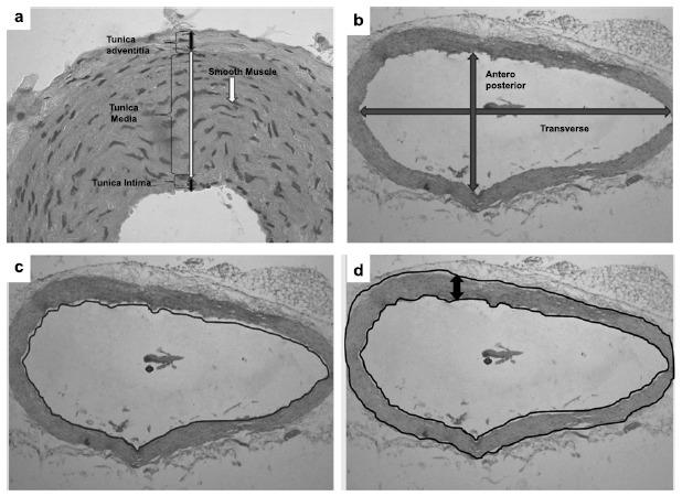
Histopathological architecture of blood vessels in (40X) stained with H&E a) All three layers of the arterial wall and its thickness b) morphometry of lumen with anteroposterior and transverse diameter c) morphometry of lumen diameter & d) morphometry of total wall thickness.
2.7.2. Estimation of Coronary Artery Thickness
We measured the total wall thickness and coronary artery lumen diameter like transverse, antero-posterior and lumen diameter.
2.7.3. Normalized Wall Index
The outer and inner vessel wall counters were manually traced for the coronary artery using the Digimizer Image Analyzer software. The wall area, lumen area, and total vessel area were automatically calculated based on the counters drawn by the software program.
The normalized wall index was calculated by dividing the wall area by the total Vessel area.
Normalized wall Index [15].
Microscopic image of the artery was calibrated with Digimizer Image Analyzer at 40X [16].
2.8. Statistical Analysis
Values were expressed in mean ± SD. Intergroup significance was determined by One Way ANOVA followed by ‘post hoc t tests’ were done by using SPSS software Version 16. P ≤0.05 was considered statistically significant.
2.9. % Change Difference Calculation for Nonstatistical Analysis
Similarly, we calculated E2 and E3 for group III and group IV in comparison tothe group I, respectively.
3. Results
3.1. Phytochemical Screening
Phytochemical analysis in the present study has shown that Emblica officinalis (Amla) does not consist of any toxic ingredients. The major groups of phytochemicals like alkaloids, glycosides, reducing sugars, tannins and flavonoids were identified in the extraction.
3.2. Gravimetry
Table 2 shows a significant increase in body weights of rats in group III (high fat fed rats, 30% fats) as compared to group I (control, 20% fats) on the 21st day. Group IV rats (high fat fed rats, 30%+EEO) showed a significant decrease in final body weight of rats compared to group III rats. However, % body weight gain of rats of group IV was reduced compared to group III even though it was statistically insignificant.
Table 2. Effect of ethanolic extract of Emblica officinalis on body weight, % of body weight gain.
| Parameter | Group I | Group II | Group III | Group IV | ANOVA | |
|---|---|---|---|---|---|---|
| F Value | p Value | |||||
| Initial body weight (1st day) (gms) | 202.3 ± 21 | 191 ± 6.1 | 220.3 ± 6.6 | 212.3 ± 13 | 8.85 | 0.006 |
| Final body weight (22nd day) (gms) | 220.3 ± 20 | 202 ± 11 | 260 ± 21 a, b | 232 ± 8.7 c | 8.2 | 0.003* |
| % of body weight gain | 9.5 ± 1.4 | 16.8 ± 1.7 | 13.7 ± 0.8 | 8.5 ± 0.7 | 3.9 | 0.053 |
Values are expressed as mean ± SD. ANOVA followed by ‘Post hoc t’ test. Group I: control, group II: supplemented with ethanolic extract of Emblica officinalis, group III: high fat fed rats, group and IV: high fat diet + ethanolic extract of Emblica officinalis. Superscript a, b, c, express a significant difference between groups. ‘a’ depicts a comparison with group I, ‘b’ depicts a comparison with group II, ‘c’ depicts a comparison with group III (*p ≤0.05).
Fig. (2) depicts the percentage difference in initial body weight, final body weight and % change of body weight gain among groups. It shows a 39% difference in % of body weight gain between group I (control) vs. group III (high fat fed rat). After supplementation with EEO, high fat fed rats showed 6.8% of decrease in % body weight gain (E-3; group I vs. group IV).
Fig. (2).

% Change of final body weight as compared to initial body weight in terms of % change of body weight gain at the end of 21 days treatment. E-1: group I vs. group II, E-2: group I vs. group III, E-3: group I vs. group IV.
3.3. Vascular Chemistry
MDA levels showed significantly higher values in group III rats compared to control rats which indicate increased lipid Peroxidation and oxidative stress. Levels of antioxidants (Vitamin C and E) were higher in group II rats compared to control. We observed decreased levels of these antioxidants in group III rats (rats fed with high fat diet) Unaltered eNOS/NOS3 protein with decreased levels of NO in high fat fed rats indicates a possible alteration of vascular pathophysiology than oxidative stress (Table 3).
Table 3. Effect of ethanolic extract of Emblica officinalis on vascular chemistry (MDA, Vitamin C and E, NO and NOS3).
| Parameter | Group I | Group II | Group III | Group IV | ANOVA | |
|---|---|---|---|---|---|---|
| F Value | p Value | |||||
| MDA (µM/L) | 0.47 ± 0.6 | 1.03 ± 0.13 | 3.05 ± 0.5a,b | 1.3 ± 0.2c | 30.4 | 0.000* |
| Vitamin C (mg/dl) | 5.2 ± 0.4 | 7.08 ± 0.3a | 5.8 ± 0.32-b | 6.4 ± 0.4 | 4.4 | 0.025* |
| Vitamin E (µg/ml) | 5.8 ± 0.5 | 6 ± 0.4 | 5 ± 0.9b | 5.5 ± 0.8 | 10.8 | 0.04* |
| NO (µM/L) | 3.8 ± 0.49 | 7.2 ± 0.69a | 2.9 ± 0.6b | 4.9 ± 0.8b,c | 22.29 | 0.000* |
| NOS3 (ng/ml) | 30.7 ± 1.7 | 29.5 ± 1.07 | 31.1 ± 1.9 | 31.3 ± 0.9 | 0.855 | 0.489 |
Values are expressed as mean ± SD. ANOVA followed by ‘Post hoc t’ test. Group I: control, group II: supplemented with ethanolic extract of Emblica officinalis, group III: high fat fed rats, group IV: high fat diet + ethanolic extract of Emblica officinalis. Superscript a, b, c express significant difference between groups. ‘a’ depicts comparison with group I, ‘b’ depicts a comparison with group II, ‘c’ depicts a comparison with group III.(*p ≤0.05).
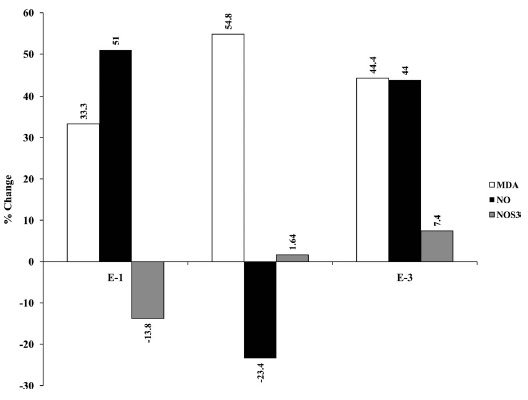
Fig. (3) depicts the percentage difference of oxidative stress markers between groups. Percentage change for MDA between control (group I) and rats fed with high fat diet group (group III) was 54.8% (E-2; group I vs. group III). After supplementation with EEO to high fat diet fed rats, there was a significant decrease in percentage difference i.e. 44.4% (E-3; group I vs. group IV). Percentage change for vitamin C between control (group 1) and rats fed with high fat diet (group 3) was -4.33% (E-2; group1 vs. group 3). After supplementation with EEO to high fat fed rats, there was a significant increase in percentage difference upto 8.01% (E-3; group 1 vs. group 4). There was -12.7% decrease in percentage change for vitamin E between control and rats fed with high fat diet (E-2; group 1 vs. group 3). After supplementation with EEO, this percentage difference was significantly increased to -6.17% between group 1 and group 4 (E-3; group1 vs. group 4). Percentage difference for NO was -23.4% between the control group and rats fed with high fat diet (E-2; group I vs. group III). After supplementation with EEO to rats fed with high fat diet, percentage difference was increased to 44% (E-3; group I vs. group IV). There was 1.64% change for NOS3 between control and rats fed with high fat diet (E-2; group I vs. group III). After treatment with EEO this percentage difference was increased to 7.4% between group I and group IV (E-3; group I vs. group IV).
Fig. (3).
% Change of levels of MDA, Vitamin C and E, NO and NOS3: E-1: group I vs. group II, E-2: group I vs. group III, E-3: group I vs. group IV.
3.4. Histopathology
3.4.1. Myocardium
Myocardium, elastic artery and coronary arteries were stained with H & E and these sections were studied under the compound microscope for all the four groups of rats. Groups I, II and IV showed the healthy morphological architecture of basal part of myocardial tissue within normal limits. High fat diet fed rats (group III) showed no myocardial damage like edema, leukocyte infiltration and necrosis. Subsequently, it wasobserved that group IV rats showed normal, healthy morphology of ventricular musculature (Myocardium) (Fig. 4).
Fig. (4).
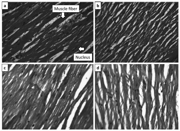
Histopathological architecture of myocardium (40X) stained with H&E. a) Normal architecture of myocardium in control group b) Normal architecture of myocardium in rats supplemented with Emblica officinalis (Amla) c) No pathological changes in rats fed with high fat diet (group III) and d) Normal architecture of myocardium in rats fed with high fat diet supplemented with Emblica officinalis (group IV).
3.4.2. Elastic Artery
The H&E stained 40X section of the group I and II rats showed typical 3 layers i.e. the tunica intima layer with endothelial cells lining, the tunica media layer with the normal arrangement of elastic lamellae and the horizontally oriented spindle-shaped nuclei of the smooth muscle cells and finally the layer of tunica adventitia. Group III rats presented morphological alterations in the cell nuclei of smooth muscle in the tunica media. The tunica media showed the degeneration, round shape and the hyperplasia of the smooth muscle cell nuclei. We have also observed subintimal atheromatous plaques in group III rats (Fig. 5). The microscopic structure of all 3 layers of the elastic artery (tunica intima, media and adventitia) of group IV rats showed the healthy architecture of elastic artery and significantly normal compared with group III.
Fig. (5).

Histopathological architecture of elastic artery (40X) stained with H&E a) Normal architecture of elastic artery in control group b) Normal architecture of elastic artery in rats supplemented with Emblica officinalis (Amla); group II c) elastic artery revealing the subintimal deposition of fat in tunica intima layer in rats fed with high fat diet (group III) and d) elastic artery revealing no pathological changes in rats fed with high fat diet supplemented with Emblica officinalis (group IV).
3.4.3. Histomorphometry of Elastic Artery
We observed a significant decrease in thickness of tunica intima layer of the elastic artery in group III rats compared to control group rats whereas (Table 4), Emblica officinalis (Amla) supplemented groups show normal elastic artery thickness. Interestingly tunica media thicknesses in group III rats have not shown any significant alteration in thickness.
Table 4. Effect of ethanolic extract of Emblica officinalis (Amla) on elastic artery thickness.
| Parameters | Group I | Group II | Group III | Group IV | ANOVA | |
|---|---|---|---|---|---|---|
| F Value | p Value | |||||
| Tunica Intima (µm) | 35.8 ± 2.1 | 37.8 ± 3.8 | 29 ± 4.2a | 37.5 ± 4.2c | 7.28 | 0.002 |
| Tunica Media (µm) | 196 ± 47 | 211 ± 41 | 191 ± 32 | 193 ± 48 | 0.26 | 0.89 |
Values are expressed as mean ± SD. ANOVA followed by ‘Post hoc t’ test. Group I: control, group II: supplemented with ethanolic extract of Emblica officinalis, group III: high fat fed rats, group IV: high fat diet + ethanolic extract of Emblica officinali. Superscript a, b, c, express significant differences between groups. ‘a’ depicts comparison with group I, ‘b’ depicts a comparison with group II, ‘c’ depicts a comparison with group III.(*p ≤0.05).
The results shown in Table 5 clearly indicate a significant decrease in anteroposterior lumen diameter, transverse diameter and area of arterial lumen in group III rats. Group IV rats have shown significant improvement in antero-posterior, transverse diameter and lumen area compared to group III rats.
Table 5. Effect of ethanolic extract of Emblica officinalis (Amla) on morphometry of elastic arterial lumen.
| Parameters | Group I | Group II | Group III | Group IV | ANOVA | |
|---|---|---|---|---|---|---|
| F Value | p Value | |||||
| Antero-post (µm) | 765.9 ± 76 | 777.3 ± 37 | 571.9 ± 42a,b | 777.5 ± 27c | 26.6 | 0.000 |
| Transverse (µm) | 212.5 ± 109 | 217.6 ± 40 | 168.2 ± 60a,b | 221.8 ± 40c | 53.7 | 0.000 |
| Arterial Lumen (µm) | 722.7 ± 28 | 788.7 ± 15 | 577.3 ± 27a,b | 775.3 ± 36c | 59.4 | 0.000 |
Values are expressed as mean ± SD. ANOVA followed by ‘Post hoc t’ test. Group I: control, group II: supplemented with ethanolic extract of Emblica officinalis, group III: high fat fed rats, and group IV: high fat diet + ethanolic extract of Emblica officinalis. Superscript a, b, c, express a significant difference between groups. ‘a’ depicts a comparison with group I, ‘b’ depicts a comparison with group II, ‘c’ depicts a comparison with group III.(*p ≤0.05).
3.4.4. Coronary Artery
The H & E stained section of myocardium containing coronary artery of group I, II and IV are showing normal and healthy microscopic architecture. The tunica intima layer is signifying with intact endothelial cell lining, tunica media with normal arrangement of internal elastic lamellae and well-defined spindle-shaped smooth muscle cells. Coronary artery in group III rats represented morphological alterations in the lumen and arterial wall. We have observed an early change of atherosclerotic plaques in the coronary arterial lumen in group III rats. The atherosclerotic plaques are in the form of fatty changes in the subendothelial layer as well as in the arterial wall. Hence, coronary arterial wall showed mild degeneration, round shaped hyperplasia of smooth muscle cell nuclei (Fig. 6). We even observed that the coronary arterial wall thickness is significantly increased in group III rats compared to group I, whereas coronary artery wall thickness in group IV is significantly decreased and is showing healthy normal architecture when compared to group II. High fat fed rats supplemented with Emblica Officinalis (Group IV) have shown a remarkable improvement of architecture of coronary arterial wall as compared to group III (Fig. 6d).
Fig. (6).
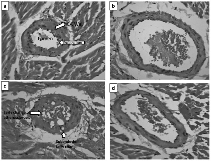
Histopathological architecture of coronary artery (40X) stained with H&E. a) Normal architecture of coronary artery in control group b) Normal architecture of coronary artery in rats supplemented with Emblica officinalis (Amla); group II c) Coronary artery revealing the subintimal deposition of fat (Microvesicular and macrovesicular) in tunica intima and media in rats fed with high fat diet (group III) and d) Coronary artery revealing no pathological changes in rats fed with high fat diet supplemented with Emblica officinalis (group IV).
3.4.5. Histomorphometry of Coronary Artery
Table 6 depicts the significant increase of coronary arterial wall thickness in group III rats compared to the control group. Supplementation of Emblica officinalis (Amla) to high fat fed rats has shown remarkable improvement in coronary arterial wall thickness compared to group III rats.
Table 6. Effect of ethanolic extract of Emblica Officinalis (Amla) on Coronary arterial wall thickness.
| Parameters | Group I | Group II | Group III | Group IV | ANOVA | |
|---|---|---|---|---|---|---|
| F Value | p Value | |||||
| Coronary Artery Thickness (µm) | 192.4 ± 6.7 | 199.4 ± 16.6b | 251.9 ± 18.4 a,b | 193.8 ± 9.8c | 23.32 | 0.000 |
Values are expressed as mean ± SD. ANOVA followed by ‘Post hoc t’ test. Group I: control, group II: supplemented with ethanolic extract of Emblica officinalis, group III: high fat fed rats, group IV: high fat diet + ethanolic extract of Emblica officinalis. Superscript a, b, c, express a significant difference between groups. ‘a’ depicts comparison with group I, ‘b’ depicts a comparison with group II, ‘c’ depicts a comparison with group III.(*p ≤0.05).
The results shown in Table 7 depict a significant narrowing of vascular integrity by reducing anteroposterior diameter, transverse diameter and compromising area of the coronary arterial lumen. Interestingly, group IV rats showed ameliorating effect of Emblica officinalis on lipid-induced impact on coronary arterial vascular integrity.
Table 7. Effect of ethanolic extract of Emblica officinalis (Amla) on Coronary arterial lumen.
| Parameters | Group I | Group II | Group III | Group IV | ANOVA | |
|---|---|---|---|---|---|---|
| F Value | p Value | |||||
| Antiro-Post (µm) | 793.9 ± 57.7 | 638.3 ± 194 | 569 ± 117 a | 726.7 ± 136 | 7.76 | 0.004 |
| Transverse (µm) | 591.8 ± 35 | 1208.4 ± 98.1 a,c | 499.1 ± 45 | 1027.7 ± 166 a,c | 27.8 | 0.000 |
| Arterial Lumen (µm) | 366.9 ± 45 | 496 ± 29.4 c | 268.1 ± 46 a | 653 ± 124 a,c | 24.08 | 0.000 |
Values are expressed as mean ± SD. ANOVA followed by ‘Post hoc t’ test. Group I: control, group II: supplemented with ethanolic extract of Emblica officinalis, group III: high fat fed rats, group and IV: high fat diet + ethanolic extract of Emblica officinalis fed rats. Superscript a, b, c, express a significant difference between groups. ‘a’ depicts comparison with group I, ‘b’ depicts a comparison with group II, ‘c’ depicts a comparison with group III.(*p ≤0.05).
The results shown in Table 8 clearly indicate a significant increase in NWI in group III rats compared with group I rats. In the case of group IV rats, NWI was found significantly decreased compared to group III rats. Increase in the thickness of arterial wall and a decrease in the arterial lumen in group III were due to early changes of atherosclerosis in the form of foam cells macrovesicular and microvesicular. The mean wall area and mean lumen area slightly decreased in the elastic and coronary arteries of group III when compared to group I.
Table 8. Effect of ethanolic extract of Emblica officinalis (Amla) on Normalized Wall Index (NWI).
| Parameters | Group I | Group II | Group III | Group IV | ANOVA | |
|---|---|---|---|---|---|---|
| F Value | p Value | |||||
| Normalized wall Index (NWI) |
0.30 ± 0.01 | 0.30 ± 0.02a | 0.44 ± 0.03a,b | 0.31 ± 0.02c | 37.4 | 0.000 |
Values are expressed as mean ± SD. ANOVA followed by ‘Post hoc t’ test. Group I: control, group II: supplemented with ethanolic extract of Emblica Officinalis, group III: high fat fed rats, group and IV: high fat diet + ethanolic extract of Emblica officinalis fed rats. Superscripts a, b, c, express a significant difference between the groups. ‘a’ depicts comparison with group I, ‘b’ depicts a comparison with group II, ‘c’ depicts a comparison with group III.(*p ≤0.05).
4. DISCUSSION
4.1. Vascular Chemistry
Results of this study imply that the development of Cardiovascular Diseases (CVD) is multifactorial but among all, dietary fat is having a detrimental effect on cardiovascular health. High fat diet stimulates metabolic and vascular alterations [17]. Our results further indicate the cardioprotective actions of ethanolic extract of Emblica officinalis (Amla) in high fat fed rats. It is due to an increase in cardiac glycogen and myocardial adaptation by augmenting endogenous antioxidants and protects rat heart from oxidative stress [18].
Mainly saturated fats produce positive energy balance and lead to increase fat deposition specifically in the vasculature and around visceral organs. High-fat diet may affect the cardiovascular system through a direct, endothelial dependent pathway and/or an indirect, cholesterol-dependent pathway [2].
Our results show vascular abnormalities with an alteration in endothelial L-arginine/NO pathway. The production and/or release of Nitric Oxide (NO) is an important endothelial factor involved in the regulation of vascular tone [19].
Eventually, endothelial NOS (eNOS) is a potent regulator for numerous essential cardiovascular functions. Endothelial NOS-derived NO dilates all types of blood vessels by stimulating soluble guanylyl cyclase and increasing cyclic GMP in smooth muscle cells [20]. Cardiovascular and vascular diseases represent with endothelial dysfunction, i.e. the inability of the endothelial to generate sufficient amounts of bioactive NO (and to produce NO-mediated vasodilation) [21]. These cardiovascular risk factors and vascular diseases are linked with increased production of ROS. Oxidative stress converts eNOS from NO producing enzyme to an enzyme which generates O2 [22]. The main adverse effect of ROS on endothelial cells is that it shows decreased bioavailability of NO, as a result of eNOS uncoupling [23].
Recently it has been reported that high fat diet induced rats have shown endothelial dysfunction through increased NADPH oxidase derived oxidative stress and production of pro-inflammatory cytokines. The altered angiogenic process occurring due to change in NO- was observed upon fat expansion, giving rise to hypoxia [24]. Hence, ROS-induced endothelial dysfunction will not only impair blood flow regulation but also restrict capillary network formation. Such alterations will result ultimately in the attenuation of microcirculatory network in metabolic active tissues [25]. Unaltered eNOS protein with decreased NO in high fat fed rats of the present study indicate a possible alteration of vascular pathophysiology probably through oxygen sensing cell signaling pathway [26].
It has been well-documented in various studies that Amla is a powerful antioxidant. It has also been noted that phenolic and flavonoid contents in ethanolic extracts of Amla have shown an antioxidant potential by inhibiting auto-oxidation via free radical scavenging, singlet oxygen quenching and hydrogen donating mechanisms [27, 28]. Subsequently, results also show that ellagic acid and ascorbic acid present in extracts of amla may accelerate antioxidant property by the increase in nitric oxide and decrease in hydroxyl radicals through its free radical scavenging property and by preventing LDL oxidation [29].
4.2. Cardiovascular Histopathology
4.2.1. Myocardium
Normally, the excess lipid may stimulate mitochondria overload and activate myocardial molecular intimal cardiac remodeling. In the present study, ventricular histology did not show any significant change in group III rats, except in few rat’s myocardium containing coronary artery is showing an early change of atherosclerosis which indicates minimal cardiac metabolic disturbances by high dietary fat.
4.3. Vascular Histopathology
4.3.1. Elastic Artery
The present study indicates that the endothelial layer of elastic artery shows early changes of atherosclerotic plaque and also there is a mild alteration in the arterial wall histology. These alterations may include arterial wall modification with component changes in the arterial wall and same in the stiffer aorta [30]. It has already been reported that alterations in layers of elastic artery (tunica intima and tunica media) with respect to high fat diet may further lead to increased arterial stiffness from small arteries to large arteries [31]. It was reported that aortic intima and media thickness was an earlier marker of clinical atherosclerosis, which had been observed in group III rats in the present study [32]. The function of elastic fibers in the arterial wall was to maintain the tension without the constant expenditure of energy. According to Burton, the arterial tension has a correlation to the amount of elastic tissue present in the vessel wall. Since coronary arteries arise from the root of the aorta, they are subjected to maximum pressure during each cardiac cycle and hence have abundant elastic fibers to maintain arterial tension [33].
The result indicates loss of arterial compliance with possible stiffening accompanied by histological modification of arterial wall due to high fat diet. Perhaps the internal elastic lamina or media component might be enriching fiber components such as collagen and elastin. The high fat diet induces changes in this vascular integrity and induces loss of elasticity. This increase in collagen was partly an addition to the bulk of the media but in later life, it was partly at the expense of smooth muscle [34]. Thus, alteration in mechanical priority may lead to severe cardiovascular dysfunction [35].
In the present study, the supplementation of Emblica officinalis (Amla) shows a significant improvement in the diameter of lumen accompanied by a significant decrease of arterial wall thickness in rats fed with high lipid diet. These results clearly show improvement of the elastic arterial property with supplementation of Emblica officinalis (Amla) in group IV rats.
4.3.2. Coronary Artery
In our observation of coronary arterial wall and lumen integrity, changes shown in the lumen area indicate an early change of atherosclerosis in tunica intima as well as tunica media in high fat fed rats. Atherosclerosis is a disease of the Tunica intima and which is separated from the Tunica media layer by the internal elastic lamina [36]. The tunica media consists of up to 40 layers of circumferential or helical oriented smooth muscles. The normal tunica media ranges in thickness from 125-350 pm (average 200 pm). Tunica media thickness underlying diseased intima (atherosclerotic plaque) is considerably thinner, ranging from 16 to 190 pm (mean 80 pm) [37]. The smooth muscle cells are embedded in a glycoprotein mix that stains heavily with the periodic acid-Schiff reactions (being PAS positive). One of the most important initial events in the development of atherosclerosis is the accumulation of cells containing excess lipid within the arterial wall which are mostly macrophages and transformed monocytes, which engulf oxidized LDL to become foam cell of fat-laden macrophages [38].
In the present study, abnormal increase in intimal thickening due to high fat diet probably induces vascular derangement resulting in insufficient oxygen tension in tissues of arterial wall [33]. Further, coronary arterial histopathology in high fat diet showed internal elastic lamina splitting, fraying, fragmentation and reduplication [33]. The present study also reported increased coronary arterial wall thickness with concomitant reduction of the coronary arterial wall area (change of anteroposterior and transverse diameter) in group III rats. This is attributed to happen series of pathology such as of high oxidized LDL oxidation, ROS generated oxidative stress, transformation of monocytes to macrophages and further develop foam cell, which fills subintimal layer and forms fatty streak in the coronary artery [39]. The fat induced injury on a subintimal layer may also initiate various cytokines and growth factors which stimulate migration and proliferation of smooth muscle cell that became intermix with the area of inflammation to form intermediary lesions and reduces lumen diameter. Such responses continue further, may cause an increase in the thickness of the coronary arterial wall with compensatory slow dilation [40]. Thickening of coronary arterial wall definitely compromises coronary arterial lumen diameter and surface area which we have noticed in our observation in high fat fed rats.
The antioxidant effect of Embilica officinalis (Amla) observed in the present study may be mediated by protecting LDL oxidation [41]. The substantial improvement of coronary arterial wall thickness, lumen diameter and lumen area after supplementation with Embilica officinalis (Amla) might be due to their potential impact of HMG CoA reductase pathways in lipid metabolism [42].
4.3.3. Normalized Wall Index
It has been reported that the normalized wall index as an indicator of cardiovascular diseases and mean wall index might be useful to assess the atherosclerotic disease burden [43]. The present study reported significant changes in mean lumen area in high fat fed rats, although many cardiovascular diseases do not show any changes in lumen area. Hence, lumen area is considered to be a less sensitive marker than normalized wall index in assessing the atherosclerotic disease burden. The decrease in normalized wall index in rats fed with high fat diet indicate negative remodeling and supplementation of Emblica officinalis (Amla) shows a remarkable improvement in normalized wall index which may be considered passive indicator for coronary arterial structural integrity [43]. This may be one of the first attempts to evaluate the normalized wall index of coronary artery in an experimental animal by histopathology.
CONCLUSION
10% of the extra fat for subchronic period albino Wister rats develops alterations in vascular chemistry possibly through oxidant-antioxidant imbalance in albino rats. High fat diet (30% fat) also induces altered pathophysiology of metabolically active tissues and vascular architecture. These observations were further corroborated with the histopathological study on the myocardium, elastic artery and coronary artery. Ethanolic extracts of Emblica Officinalis supplementation were found to be beneficial against high fat induced vascular alterations in terms of functions and architecture. It is mainly due to the fact that Emblica Officinalis contains many biological compounds like tannins, gallic acids, flavonoids, etc. which possess medicinal properties. Probably polyphenolic compounds and flavonoids of EEO might have the protective role by various means like antioxidant activities. EEO might have cardioprotective actions in high fat fed rats by modulating cardiac functions. Supplementation of EEO as an antioxidant ameliorates fat induced oxidative stress in metabolically active tissues which further protects cardiovascular health. The supportive possible mechanisms are depicted in Fig. (7) to elaborate the beneficial effect of EEO in high fat fed rats.
Fig. (7).
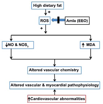
Postulated mechanisms of action of ethanolic extract of Emblica officinalis on high fat fed albino rats in cardiovascular abnormalities.
ACKNOWLEDGEMENTS
Declared none.
LIST OF ABBREVIATIONS
- EEO
Ethanolic Extract of Emblica officinalis
- NO
Nitric Oxide
- NOS3/eNOS
Endothelial Nitric Oxide Synthase
- TBARS
Thiobarbituric Acid Reactive Substances
ETHICS APPROVAL AND CONSENT TO PARTICIPATE
The study protocol was approved by the Institutional Animal Ethics Committee (IAEC) of BLDE University’s Shri B.M. Patil Medical College, India. IAEC number is 664/15.
HUMAN AND ANIMAL RIGHTS
No humans were used in this study. The reported experiments on animal were in accordance with the guideline of Committee for the Purpose of Control and Supervision of Experiments on Animals (CPCSEA, Government of India).
CONSENT FOR PUBLICATION
Not applicable.
AVAILABILITY OF DATA AND MATERIALS
Not applicable.
FUNDING
This work was supported by VGST (Vision Group of Science and Technology), Karnataka Science and Technology Promotion Society, Government of Karnataka, India [Ref. No:KSTePS/05/K-FIST/2015-16. Dtd. 22-06-2016].
CONFLICT OF INTEREST
The authors declare no conflict of interest, financial or otherwise.
REFERENCES
- 1.Choudhary M.I., Naheed S., Jalil S., Alam J.M. Effects of ethanolic extract of Iris germanica on lipid profile of rats fed on a high-fat diet. J. Ethnopharmacol. 2005;98(1-2):217–220. doi: 10.1016/j.jep.2005.01.013. [DOI] [PubMed] [Google Scholar]
- 2.Jakulj F., Zernicke K., Bacon S.L., Van Wielingen L.E., Key B.L., West S.G., Campbell T.S. A high-fat meal increases cardiovascular reactivity to psychological stress in healthy young adults. J. Nutr. 2007;137(4):935–939. doi: 10.1093/jn/137.4.935. [DOI] [PubMed] [Google Scholar]
- 3.Kabiri N., Asgary S., Madani H., Mahzouni P. Effects of Amaranthuscaudatusl. Extract and lovastatin on atherosclerosis in hypercholesterolemic rabbits. J. Med. Plants Res. 2010;4(3):55–61. [Google Scholar]
- 4.Hopps E., Noto D., Caimi G., Averna M.R. A novel component of the metabolic syndrome: The oxidative stress. Nutr. Metab. Cardiovasc. Dis. 2010;20(1):72–77. doi: 10.1016/j.numecd.2009.06.002. [DOI] [PubMed] [Google Scholar]
- 5.Galili O., Versari D., Sattler K.J., Olson M.L., Mannheim D., McConnell J.P., Chade A.R., Lerman L.O., Lerman A. Early experimental obesity is associated with coronary endothelial dysfunction and oxidative stress. Am. J. Physiol. Heart Circ. Physiol. 2007;292(2):H904–H911. doi: 10.1152/ajpheart.00628.2006. [DOI] [PubMed] [Google Scholar]
- 6.Chakraborti D., Verma R. Ameliorative effect of Emblica officinalis aqueous extract on ochratoxin-induced lipid peroxidation n the kidney and liver of mice. Int. J. Occup. Med. Environ. Health. 2010;23(1):63–73. doi: 10.2478/v10001-010-0009-4. [DOI] [PubMed] [Google Scholar]
- 7.Lankin V.Z., Tikhaze A.K., Kukharchuk V.V., Konovalova G.G., Pisarenko O.I., Kaminnyi A.I., Shumaev K.B., Belenkov Y.N. Antioxidants decreases the intensification of low density lipoprotein in vivo peroxidation during therapy with statins. Mol. Cell. Biochem. 2003;249(1-2):129–140. [PubMed] [Google Scholar]
- 8.Kanthe P.S., Patil B.S., Bagali S.C., Reddy R.C., Aithala M.R., Das K.K. Protective effects of ethanolic extract of Emblica officinalis (amla) on cardiovascular pathophysiology of rats, fed with high fat diet. J. Clin. Diagn. Res. 2017;11(9):CC05. doi: 10.7860/JCDR/2017/28474.10628. [DOI] [PMC free article] [PubMed] [Google Scholar]
- 9.Antony B., Merina B., Sheeba V., Mukkadan J. Effcet of standardized Amla extract on atherosclerosis and dyslipidemia. Indian J. Pharm. Sci. 2006;68(4):437–441. [Google Scholar]
- 10.Kanthe P.S., Patil B.S., Aithala M.R., Das K.K. Effect of ethanolic extract of Emblica officinalis (amla) on glucose homeostasis in rats fed with high fat diet. J. Krishna Inst. Med. Sci. Univ. 2017;6(3):31–37. [Google Scholar]
- 11.Tiwari P., Kumar B., Kaur M., Kaur G., Kaur H. Phytochemical screening and extraction: A review. Int. Pharm. Sci. 2011;1:103–104. [Google Scholar]
- 12.Das K.K., Dasgupta S. Effect of nickel sulfate on testicular steroidogenesis in rats during protein restriction. Environ. Health Perspect. 2002;110(9):923–926. doi: 10.1289/ehp.02110923. [DOI] [PMC free article] [PubMed] [Google Scholar]
- 13.Pingali U., Fatima N., Murlidhar N. Effects of phyllanthus Emblica extract on endothelial dysfunction and biomarkers of oxidative stress in patients with type 2 diabetes mellitus: A randomized, double-blind, controlled study. Diab. Metab. Syndrome Obesity: Target Ther. 2013;6:275–284. doi: 10.2147/DMSO.S46341. [DOI] [PMC free article] [PubMed] [Google Scholar]
- 14.Okhawa H., Ohishi N., Yagi K. Assay for lipid peroxides in animal tissues by thiobarbituric acid reaction. Anal. Biochem. 1979;95(2):351–358. doi: 10.1016/0003-2697(79)90738-3. [DOI] [PubMed] [Google Scholar]
- 15.Hayashi K., Mani V., Nemade A., Aguiar S., Postley J.E., Fuster V., Fayad Z.A. Variations in atherosclerosis and remodeling patterns in aorta and carotids. J. Cardiovasc. Magn. Reson. 2010;12(1):10. doi: 10.1186/1532-429X-12-10. [DOI] [PMC free article] [PubMed] [Google Scholar]
- 16. https://www.micro-shop.zeiss.com/?l=en&p=hr&f=e&i=10290& (Accessed February 26, 2015).
- 17.Collins P. Risk factors for cardiovascular disease and hormone therapy in women. Heart. 2006;92(3):24–28. doi: 10.1136/hrt.2005.071787. [DOI] [PMC free article] [PubMed] [Google Scholar]
- 18.Rajak S., Banerjee S.K., Sood S., Dinda A.K., Gupta Y.K., Gupta S.K., Maulik S.K. Emblica officinalis causes myocardial adaptation and protects against oxidative stress in ischemic‐reperfusion injury in rats. Phytother. Res. Int. J. Pharmacol. Toxicol. Eval. Nat. Prod. Derivatives. 2004;18(1):54–60. doi: 10.1002/ptr.1367. [DOI] [PubMed] [Google Scholar]
- 19.Nascimento T.B., Baptista R.D., Pereira P.C., Campos D.H., Leopoldo A.S., Leopoldo A.P., Oliveira S.A., Junior, Padovani C.R., Cicogna A.C., Cordellini S. Vascular alterations in high fat diet obese rats: Role of endothelial L-arginine/NO pathway. Arq. Bras. Cardiol. 2011;97(1):40–45. doi: 10.1590/s0066-782x2011005000063. [DOI] [PubMed] [Google Scholar]
- 20.Radomski M.W., Palmer R.M., Moncada S. The anti-aggregating properties of vascular endothelium: Interactions between prostacyclin and nitric oxide. Br. J. Pharmacol. 1987;92:639–646. doi: 10.1111/j.1476-5381.1987.tb11367.x. [DOI] [PMC free article] [PubMed] [Google Scholar]
- 21.Nakaki T., Nakayama M., Kato R. Inhibition by nitric oxide and nitric oxide producing vasodilators of DNA synthesis in vascular smooth muscle cells. Eur. J. Pharmacol. Mol. Pharmacol. 1990;189:347–353. doi: 10.1016/0922-4106(90)90031-r. [DOI] [PubMed] [Google Scholar]
- 22.Mueller C.F., Laude K., McNally J.S., Harrison D.G. Redox mechanism in blood vessels. Arterioscler. Thromb. Vasc. Biol. 2005;25:274–278. doi: 10.1161/01.ATV.0000149143.04821.eb. [DOI] [PubMed] [Google Scholar]
- 23.Payne G.A., Bohlen H.G., Dincer U.D., Borbouse L., Tune J.D. Periadventitial adipose tissue impairs coronary endothelial function via PKC ß-dependent phosphorylation of nitric oxide synthase. Am. J. Physiol. Heart Circ. Physiol. 2009;297(1):460–465. doi: 10.1152/ajpheart.00116.2009. [DOI] [PMC free article] [PubMed] [Google Scholar]
- 24.Hosogai N., Fukuhara A., Oshima K., Miyata Y., Tanaka S., Segawa K., Furukawa S., Tochino Y., Komuro R., Matsuda M., Shimomura I. Adipose tissue hypoxia in obesity and its impact on adipocytokine dysregulation. Diabetes. 2007;56(4):901–911. doi: 10.2337/db06-0911. [DOI] [PubMed] [Google Scholar]
- 25.Vásquez V.J., Kalyanaraman B., Martásek P., Hogg N., Masters B.S., Karoui H., Tordo P., Pritchard K.A. Superoxide generation by endothelial nitric oxide synthase: The influence of cofactors. Proc. Natl. Acad. Sci. USA. 1998;95(16):9220–9225. doi: 10.1073/pnas.95.16.9220. [DOI] [PMC free article] [PubMed] [Google Scholar]
- 26.Das K.K., Chadchan K.S., Reddy R.C., Biradar M.S., Kanthe P.S., Patil B.S., Ambekar J.G., Bagoji I.B., Das S.N. Effects of some indigenous plants of North Karnataka (India) on cardiovascular and glucose regulatory systems in alloxan-induced diabetic rats. Cardiovasc. Hematol. Agents Med. Chem. 2017;15(1):49–61. doi: 10.2174/1871525715666170712121347. [DOI] [PubMed] [Google Scholar]
- 27.Sultana S., Ahmad S., Khan N., Jahangir T. Effects of Emblica officinalis (Gaertn) on CCl4-induced hepatic toxicity and DNA synthesis of Wistar rats. Indian J. Exp. Biol. 2005;43:430–436. [PubMed] [Google Scholar]
- 28.Kaur J., Kaur D., Singh H., Khan M.U. Emblica officinalis Emblica officinalis: A meritocratic drug for treating various disorders. Indo Am. J. Pharm. Res. 2013;3(6) [Google Scholar]
- 29.Nampoothiri S.V., Prathapan A., Cherian O.L., Raghu K.G., Venugopalan V.V., Sundaresan A. In vitro antioxidant and inhibitory potential of Terminalia bellerica and Emblica officinalis fruits against LDL oxidation and key enzymes linked to type 2 diabetes. Food Chem. Toxicol. 2011;49(1):125–131. doi: 10.1016/j.fct.2010.10.006. [DOI] [PubMed] [Google Scholar]
- 30.Townsend R.R., Wilkinson I.B., Schiffrin E.L., Avolio A.P., Chirinos J.A., Cockcroft J.R., Heffernan K.S., Lakatta E.G., McEniery C.M., Mitchell G.F., Najjar S.S. Recommendations for improving and standardizing vascular research on arterial stiffness: A scientific statement from the American Heart Association. Hypertension. 2015;66(3):698–722. doi: 10.1161/HYP.0000000000000033. [DOI] [PMC free article] [PubMed] [Google Scholar]
- 31.Sentelices L.C., Rutaman S.J., Ertart J.C., Pranti R.L., Hanay J.N., Vorp D.A., Aharan J.M. Experimental system for ex vivo measurement of Murine aortic stiffness. Physiol. Meas. 2007;28(8):39–49. doi: 10.1088/0967-3334/28/8/N01. [DOI] [PubMed] [Google Scholar]
- 32.Haurington J., Pana A.S., Gent R., Hirtec C.J. Aortic intima media thickness is an early marker of atherosclerosis in children with type 1 diabetes mellitus. J. Pedator. 2010;156:237–241. doi: 10.1016/j.jpeds.2009.08.036. [DOI] [PubMed] [Google Scholar]
- 33.Deopujari R., Dixit A. The study of age related changes in coronary arteries and its relevance to the atherosclerosis. J. Anat. Soc. India. 2010;59(2):192–196. [Google Scholar]
- 34.Billaud M., Johnstone S.R., Isakson B.E. Loss of compliance in small arteries, but not in conduit arteries, after 6 weeks exposure to high fat diet. J. Cardiovasc. Transl. Res. 2012;5(3):256–263. doi: 10.1007/s12265-012-9354-y. [DOI] [PubMed] [Google Scholar]
- 35.Rizzoni D.D., Ciuceis C., Porteri E., Semerarao F., Rosri E.A. Structural alterations in small resistance arteries in obesity. Basic Clin. Pharmacol. Toxicol. 2011:1742–1843. doi: 10.1111/j.1742-7843.2011.00786.x. [DOI] [PubMed] [Google Scholar]
- 36.Waller B.F., Orr C.M., Slack J.D., Pinkerton C.A., Van T.J., Peters T. Anatomy, histology, and pathology of coronary arteries: A review relevant to new interventional and imaging techniques-Part I. Clin. Cardiol. 1992;15(6):451–457. doi: 10.1002/clc.4960150613. [DOI] [PubMed] [Google Scholar]
- 37.Benditt E.P., Schwartz S.M. Blood vessels. In: Rubin E., Farber J.L., editors. Pathology. Philadelphia: JB Lippincott; 1988. pp. 454–465. [Google Scholar]
- 38.Waller B.F. The eccentric coronary atherosclerotic plaque morphologic observations and clinical relevance. Clin. Cardiol. 1989;12:14–20. doi: 10.1002/clc.4960120103. [DOI] [PubMed] [Google Scholar]
- 39.Leopold J.A., Loscalzo J. Oxidative mechanisms and athero thrombotic cardiovascular disease. Drug Discov. Today Ther. Strateg. 2008;5(1):5–13. doi: 10.1016/j.ddstr.2008.02.001. [DOI] [PMC free article] [PubMed] [Google Scholar]
- 40.Ross R. Atherosclerosis-an inflammatory disease. N. Engl. J. Med. 1999;340(2):115–126. doi: 10.1056/NEJM199901143400207. [DOI] [PubMed] [Google Scholar]
- 41.Oubina M.P., De Las Heras N., Cediel E., Sanz-Rosa D., Aragoncillo P., D’iaz C., Hernandez G., Lahera V., Cachofeiro V. Synergetic effect of angiotensin converting enzyme ACE 3 hydroxy 3 mythelyne CoA HMG CoA reductase inhibition on inflammatory in atherosclerosis Rabbits. Clin. Sci. (Lond.) 2003;105:655–662. doi: 10.1042/CS20030127. [DOI] [PubMed] [Google Scholar]
- 42.Stocker R., Keaney J.F. Role oxidative modifications in atherosclerosis. Physiol. Rev. 2004;84:1381–1478. doi: 10.1152/physrev.00047.2003. [DOI] [PubMed] [Google Scholar]
- 43.Saam T., Raya J.G., Cyran C.C., Bochmann K., Meimarakis G., Dietrich O., Clevert D.A., Frey U., Yuan C., Hatsukami T.S., Werf A. High resolution carotid black-blood 3TMR with parallel imaging and dedicated 4-channel surface coils. J. Cardiovasc. Magn. Reson. 2009;4(1):11–41. doi: 10.1186/1532-429X-11-41. [DOI] [PMC free article] [PubMed] [Google Scholar]
Associated Data
This section collects any data citations, data availability statements, or supplementary materials included in this article.
Data Availability Statement
Not applicable.