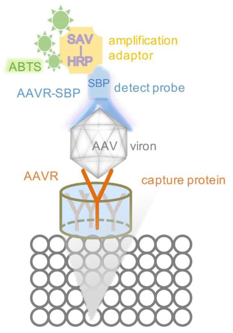
Figure 1.
The principle of VIRELISA for schematic demonstration. The soluble AAVR was immobilized in the 96-well plate as the captured protein. AAV samples were loaded into each well. AAVR-SBP was used as the detection probe, equivalent to a primary antibody. Then SAV (streptavidin) conjugated with HRP (horseradish peroxidase) was applied as the amplification adaptor, as a secondary antibody. The ABTS (2,2′-azino-di-(3-ethylbenzthiazoline sulfonic acid)) was finally added to develop visual signals.

