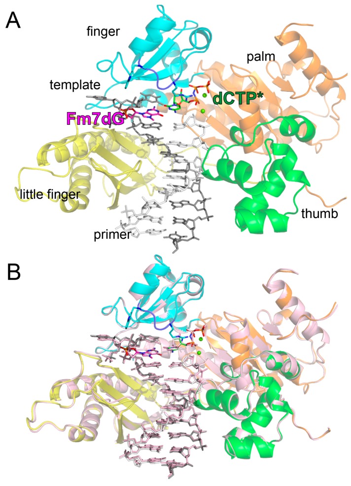
Figure 3.
Overall conformation of the polη-Fm7dG:dCTP* complex structure. (A) The ternary complex structure of polη bound to Fm7dG and the incoming nonhydrolyzable dCTP analog. The palm, thumb, finger, and little finger domains are shown in orange, green, cyan, and yellow, respectively. The flexible Arg61–Trp64 loop is shown in blue. The primer strand is colored in white and the template strand in gray. The active site Mg2+ ions are shown in green spheres. (Β) Superposition of the polη-Fm7dG:dCTP* and the polη-dG:dCTP* (light pink, PDB ID: 4O3N [41]) structures.

