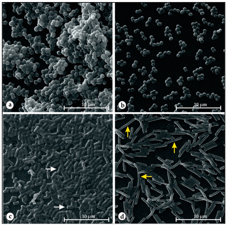
Figure 4.
Scanning electron microscopy images of preformed biofilms of S. aureus (5B) and E. coli (P12) treated with the EO of Ocimum gratissimum L. (EOOG) at MIC doses. Untreated 5B (a) and P12, (c) and EOOG-treated 5B, (b) and P12 (d) cells. In the control condition, E. coli presented rod-shaped (white arrow) cells, and after EOOG treatment, the P12 cells displayed an elongated (yellow arrow) morphology status.

