



Abstract
Memory impairment after stroke in young adults is poorly understood. In elderly stroke survivors memory impairments and the concomitant loss of hippocampal volume are usually explained by coexisting neurodegenerative disease (e.g., amyloid pathology) in interaction with stroke. However, neurodegenerative disease, such as amyloid pathology, is generally absent at young age. Accumulating evidence suggests that infarction itself may cause secondary neurodegeneration in remote areas. Therefore, we investigated the relation between long‐term memory performance and hippocampal volume in young patients with first‐ever ischemic stroke. We studied all consecutive first‐ever ischemic stroke patients, aged 18–50 years, admitted to our academic hospital center between 1980 and 2010. Episodic memory of 173 patients was assessed using the Rey Auditory Verbal Learning Test and the Rey Complex Figure and compared with 87 stroke‐free controls. Hippocampal volume was determined using FSL‐FIRST, with manual correction. On average 10 years after stroke, patients had smaller ipsilateral hippocampal volumes compared with controls after left‐hemispheric stroke (5.4%) and right‐hemispheric stroke (7.7%), with most apparent memory dysfunctioning after left‐hemispheric stroke. A larger hemispheric stroke was associated with a smaller ipsilateral hippocampal volume (b=−0.003, P<0.0001). Longer follow‐up duration was associated with smaller ipsilateral hippocampal volume after left‐hemispheric stroke (b=−0.028 ml, P=0.002) and right‐hemispheric stroke (b=−0.015 ml, P=0.03). Our results suggest that infarction is associated with remote injury to the hippocampus, which may lower or expedite the threshold for cognitive impairment or even dementia later in life. Hum Brain Mapp 36:2432–2442, 2015. © 2015 Wiley Periodicals, Inc.
Keywords: episodic memory, hippocampal volume, ischemic stroke, young adults
INTRODUCTION
Episodic memory deficits are common after ischemic stroke with high risk of conversion to poststroke dementia over time [Allan et al., 2011; Henon et al., 2001; Ivan et al., 2004; Leys et al., 2005]. These memory deficits in elderly stroke survivors are often explained by coexisting neurodegenerative pathological changes like for example amyloid pathology that directly affects hippocampal volume, in interaction with the infarction (e.g., due to poststroke Wallerian degeneration) as hippocampal strokes are rare [Blum et al., 2012; Henon et al., 1998, 2001; Wu et al., 2008].
Also, up to 37.1% of young stroke patients (18–50 years) suffer from episodic memory impairments independent of site and severity of the infarction, even up to 10 years after stroke [Schaapsmeerders et al., 2013]. However, these memory impairments in young stroke patients are poorly understood, as the presence of coexisting neurodegenerative pathology observed in elderly stroke survivors (e.g., presence of amyloid pathology) is very unlikely to occur at such a young age [Lesne et al., 2013].
Infarctions located anywhere in the brain can induce widespread effects causing disruption of functional networks of the cortical regions [Snaphaan et al., 2009; Tuladhar et al., 2013]. Furthermore, apart from these functional changes, atrophy of nonischemic remote brain regions, presumably due to neuropathological consequences of ischemic stroke, has been observed [Chen et al., 2014; Xie et al., 2011; Zhang et al., 2012].
Hippocampal lesion studies have shown that the hippocampus is an important brain structure for episodic memory formation, that is, encoding and long‐term consolidation of new information [Squire and Wixted, 2011]. These processes can be assessed using the Rey‐Auditory Verbal Learning Test (RAVLT) [Vakil and Blachstein, 1993] and the Rey‐Osterrieth Complex Figure Test (ROCF) [Osterrieth, 1944].
One smaller study of 36 younger patients with medial cerebral artery (MCA) occlusion found a smaller ipsilateral hippocampal volume compared with the contralateral hippocampus and concomitant impairment in episodic verbal memory dysfunction, up to 2 years after stroke [Xie et al., 2011].
Memory performance is of utmost importance in young stroke patients as they are in a demanding time of life and usually have a life expectancy of decades ahead. Therefore, understanding the long‐term underlying structural correlates of decline in memory functioning after stroke is of key importance and may provide opportunities to tailor poststroke treatment and rehabilitation. However, currently these data are lacking. Therefore, we investigated the relation between long‐term memory performance and hippocampal volume in young patients with first‐ever ischemic stroke. We hypothesized that infarction in these young adults is associated with long‐term ipsilateral hippocampal atrophy and longer follow‐up duration to be associated with a smaller ipsilateral hippocampal volume. We further hypothesized that stroke has a generalized effect on the ipsilateral hemisphere, not only restricted to the hippocampus. To investigate this, we determined thalamic volume after stroke, because the thalamus, apart from the hippocampus, has also been found to be associated with memory function [Exner and Weniger, 2001] and previous studies have shown that ipsilateral thalamic volume is reduced after MCA occlusion [Herve et al., 2005; Tamura et al., 1991].
MATERIALS AND METHODS
Study Design
This study is part of the “Follow‐Up of Transient ischemic attack and stroke patients and Unelucidated Risk factor Evaluation”‐study (FUTURE study), a prospective cohort study of prognosis after young stroke or transient ischemic attack (TIA) in adults aged 18 through 50 years admitted to the Radboud university medical centre, the Netherlands, between January 1, 1980 and November 1, 2010 [Rutten‐Jacobs et al., 2011, 2013; Schaapsmeerders et al., 2013]. The Medical Review Ethics Committee region Arnhem‐Nijmegen approved the study and written informed consent was obtained from all participants. Patients were identified through a prospective registry of all consecutive young stroke/TIA patients that has been kept at the department since the 1970s with a standardized collection of baseline and clinical characteristics (including demographics, stroke subtype, vascular risk factors, and a history of epilepsy). The present magnetic resonance imaging (MRI) study comprises all consecutive patients with a first‐ever ischemic stroke. Ischemic stroke was defined as focal neurologic deficit persisting more than 24 h. Primary exclusion criteria for ischemic stroke in the FUTURE study were previous stroke or TIA, cerebral venous sinus thrombosis, and retinal infarction. Additional exclusion criteria for the present MRI study were recurrent stroke/TIA and hippocampal stroke.
Participants had the opportunity to undergo an extensive neuropsychological examination after long follow‐up which was administered between November 2009 and December 2011.
Lesion location (left hemisphere, right hemisphere, and infratentorial) was based on medical records and radiological findings. The exact lesion location (frontal lobe, parietal lobe, temporal lobe, occipital lobe, basal ganglia, thalamus, brainstem, and cerebellum) was determined using the T1‐weighted images and fluid attenuated inversion recovery (FLAIR) sequence.
Stroke‐free control participants were recruited among the patients' spouses, relatives, or social environment. Controls had to be at least 18‐years‐old without a history of TIA or stroke. Controls were all living independently, none fulfilled the clinical criteria of dementia.
Episodic Memory
Verbal episodic memory was assessed using the three‐trial version of the RAVLT [Vakil and Blachstein, 1993; van Norden et al., 2008] and visuospatial episodic memory was assessed using the ROCF [Osterrieth, 1944]. The verbal and visual memory indices derived from these tests are considered to be associated with hippocampal functioning [Babiloni et al., 2009; Snaphaan et al., 2009], integrity [Carlesimo et al., 2010; den Heijer et al., 2012; van Norden et al., 2012], and volume [den Heijer et al., 2010, 2012; McConley et al., 2008; Vyhnalek et al., 2014]. The encoding phase of memory functioning was assessed using the “Immediate verbal recall” (total number of correctly recalled words over the three consecutive learning trials of the RAVLT), and “immediate visuospatial recall” (immediate recall score/copy trial score of the ROCF). Storage of previously acquired information is reflected by “delayed verbal recall” (number correctly reproduced words 30 min after Trial 3) and “delayed visuospatial recall” (delayed recall score after 30 min/copy trial). However, as delayed recall performance might be confounded by a retrieval deficit, that is, the inability to access previously stored information, the RAVLT also incorporates “delayed verbal recognition” (number of hits and correct rejections on a list of 30 items consisting of 15 target items among 15 distractor items) that bypasses retrieval and reflects successful storage [Vakil and Blachstein, 1993].
Other Measurements
Age, sex, level of education, depressive symptoms, and fatigue, were considered possible confounders for memory performance. Level of education was scored with a widely‐used Dutch scoring system (1=less than primary school; 7=university degree). Depressive symptoms and fatigue were assessed using the depression subscale of the Hospital Anxiety and Depression Scale (HADS) [Zigmond and Snaith, 1983] and the subscale Subjective Fatigue of the revised Checklist Individual Strength (CIS‐20R) [Vercoulen et al., 1994]. Functional outcome during follow‐up visit was evaluated using the modified Rankin Scale (mRS) [Leifer et al., 2011]. The mRS is a measure for degree of disability or dependence in daily activities of patients with stroke (0=no symptoms; 6=death).
Furthermore, assessment of stroke etiology (modified Trial of Org 10172 in Acute Stroke Treatment [TOAST] classification) [Adams et al., 1993] and severity (National Institutes of Health Stroke Scale [NIHSS]) [Brott et al., 1989] was done retrospectively for all cases by a validated approach [Kasner et al., 1999; Williams et al., 2000], as these scales did not exist at the time when a substantial proportion of the patients experienced their qualifying event. The TOAST is a system to categorize subtypes of ischemic stroke based on etiology. It includes six categories: (1) large‐artery atherosclerosis, (2) cardioembolism, (3) small artery occlusion (lacune), (4) rare causes, (5) multiple causes, and (6) unknown cause. The NIHSS is a 15‐item neurologic examination stroke scale used to evaluate the effect of acute cerebral infarction on the levels of consciousness, language, neglect, visual‐field loss, extraocular movement, motor strength, ataxia, dysarthria, and sensory loss.
Neuroimaging Data Acquisition
Participants underwent 1.5‐T MRI scanning on the Siemens, Magnetom Avanto. The scanning protocol included a T1‐weighted MPRAGE whole‐brain scan (TI /TR/TE 1,000 ms / 2,730 ms / 2.95 ms; flip angle 7°; field of view [FOV] = 256 mm, voxel size 1.0 × 1.0 × 1.0 mm3) and a FLAIR pulse sequence (TI /TR/TE 2,200 ms / 12,220 ms/85 ms; interslice gap: 0.6 mm; voxel size 1.2 × 1.0 × 3.0 mm3).
Neuroimaging Data Processing
Matlab 7 was used to perform all MRI data analyses. We used optimized Voxel‐Based Morphometry toolbox (VBM8) (http://dbm.neuro.uni-jena.de/vbm.html) within SPM8 (Wellcome Trust Centre for Neuroimaging, University College London, London, UK; http://www.fil.ion.ucl.ac.uk/spm) for each T1‐weighted image to determine the relative proportion of grey matter, white matter, and cerebrospinal fluid (CSF) per voxel. We used the thorough clean up option, turned the final masking and skull‐striping with graph‐cut off to have better segmentation of CSF in case of large cortical infarction. Still, in patients with large cortical infarcts the CSF was underestimated and manual correction was needed based on visual inspection. FSLview (FMRIB Software Library release 5.0, http://www.fmrib.ox.ac.uk/fsl) was used to draw a mask using the dura mater as the boundary for intracranial space [Jenkinson et al., 2012]. Total brain volume was taken as the sum of total grey and white matter. Intracranial volume (ICV) was taken as the sum of total grey and white matter and CSF volume. T1‐weighted images were used to segment the hippocampus using FSL‐FIRST [Patenaude et al., 2011]. Each mask was manually corrected using FSLview [Jenkinson et al., 2012]. One experienced investigator, blinded for memory performance scores and baseline characteristics, corrected all masks. Anatomical boundaries were determined in the coronal section, actual segmentation was performed using a previously published standardized protocol in which segmentation correction was performed from posterior to anterior [van Norden et al., 2008]. We used a standard neuroanatomical atlas as a guide [Duvernoy, 1997]. The slice before the level in which the crurae fornices appeared in full view was defined as the posterior border of the hippocampus. The anterior border of the hippocampus was defined as the slice in which the hippocampus was no longer present, and the amygdala fully covered the hippocampus. The inferior horn of the lateral ventricle was used as the superior border. The inferior border was determined by the white matter. The lateral border was defined by the temporal horn of the lateral ventricle and the white matter adjacent to the hippocampus. To correct for differences in head size, normalized total brain volumes and normalized hippocampal volumes (ml) were calculated with the following formula: (average ICV of the total population * (total brain or hippocampal volume of the participant/ICV of the participant)) [van Norden et al., 2008]. Inter‐rater reliability on a random sample of 10% of all cases showed an intraclass correlation coefficient for the left hippocampus of 0.81, and for the right hippocampus of 0.83. The intrarater reliability for hippocampal volumes yielded an intraclass correlation coefficient for the left hippocampus of 0.93 and for the right hippocampus 0.96.
Lesion Volume and Lesion Probability Maps
Cerebral infarctions were defined as hypointense areas on a T1‐weighted MPRAGE whole‐brain scan with corresponding gliotic rim on FLAIR. The T1‐weighted image from each patient was used to manually trace lesions, with the aid of corresponding slices on the FLAIR image. One experienced investigator traced all the lesions and was blinded for baseline characteristics and outcome measures (hippocampal volume and memory performance). Normalized lesion volumes (ml) were calculated with the same formula used to normalize hippocampal volume. Next, the T1‐weighted images were brain‐extracted (FSL‐BET: Brain Extraction Tool)[Smith, 2002] and subsequently, along with the lesion mask, registered to the Montreal Neurological Institute (MNI) standard space by an affine transformation with 12 degrees of freedom using FSL‐FLIRT (FMRIB's Linear Image Registration Tool; Software Library release 5.0, http://www.fmrib.ox.ac.uk/fsl), followed by nonlinear registration using FNIRT (FMRIB's Nonlinear Image Registration Tool)[Andersson et al., 2007]. Next, for each patient group all lesion masks were merged and averaged, which resulted in a lesion probability map for left‐hemispheric stroke, right‐hemispheric stroke, and infratentorial stroke patients.
Thalamic Volume
To determine whether ipsilateral stroke specifically affects the hippocampus we additionally investigated thalamic volumes using FSL‐FIRST [Patenaude et al., 2011]. Patients with a thalamic stroke were excluded from the analysis. Normalized thalamic volumes were calculated with the same formula used to normalize hippocampal volumes.
Statistical Analyses
Baseline characteristics were presented as mean (±SD), median (Q1–Q3), or number of cases (%) and group differences were tested with a Pearson's chi‐square test, Mann‐Whitney U test, or Student's t‐test when appropriate. Two‐sided P‐values<0.05 were considered statistically significant. We compared lesion volumes between the three patient groups (left‐hemispheric stroke, right‐hemispheric stroke, and infratentorial stroke) with analysis of co‐variance (ANCOVA), adjusted for age at follow‐up, sex, and follow‐up duration.
The mean memory performance for patients with left‐hemispheric stroke, right‐hemispheric stroke, and infratentorial stroke were compared with controls by means of ANCOVA, adjusted for age at follow‐up, sex, education, depressive symptoms, and fatigue. Aphasia may negatively influence verbal memory performance and, therefore, we examined the effect of excluding aphasic patients from the memory analyses.
Mean hippocampal volumes were calculated for each of the three lesion locations and were compared with controls using an ANCOVA model, adjusted for age at follow‐up and sex.
We investigated whether ipsilateral hippocampal volume was associated with immediate verbal recall, delayed verbal recall, immediate visuospatial recall, and delayed visuospatial recall (in Z‐scores) after left‐hemispheric stroke and right‐hemispheric stroke. Linear regression was used for this purpose (b‐weights; 95%CI), adjusted for age, sex, level of education, follow‐up duration, normalized lesion volume, and ipsilateral thalamic volume. Patients with thalamic stroke were excluded from this analysis to investigate whether ipsilateral hippocampal volume was independently associated with memory performance.
To investigate whether lesion volume was related to ipsilateral and contralateral hippocampal volume after hemispheric stroke linear regression was used (b‐weights; 95%CI), adjusted for age at follow‐up, sex, lesion location (left or right), and follow‐up duration.
The association between follow‐up duration and left and right hippocampal volume was analyzed by left‐hemispheric stroke, right‐hemispheric stroke, and infratentorial stroke separately by means of linear regression, corrected for age at follow‐up, sex, and normalized lesion volume.
Finally, mean thalamic volumes were calculated for each of the three lesion locations and compared with controls using an ANCOVA model, adjusted for age at follow‐up and sex. Patients with thalamic stroke were excluded. For left and right‐hemispheric stroke patients separately, we were also interested whether ipsilateral thalamic volume was associated with immediate verbal recall, delayed verbal recall, immediate visuospatial recall, and delayed visuospatial recall (in Z‐scores). The same linear regression model as described in the section on hippocampal volume and memory functioning was used for this purpose and we subsequently reported whether there was an independent association between thalamic volume (b‐weights; 95%CI) and memory performance.
RESULTS
The study population consisted of 176 ischemic stroke participants and 87 controls. No differences were observed in baseline characteristics between participants (n=176) and those who refused (n=96) or were lost follow‐up (n=63). Basic demographical and clinical characteristics of those who did participate in the FUTURE study, but did not participate in the present sub study are presented in Table 1.
Table 1.
Demographic and clinical characteristics of participants, controls, and nonparticipants in present MRI study
| Characteristics | Total Ischemic stroke population | Hemispheric stroke | Infratentorial stroke | Controls | No memory assessment /no MRI | |
|---|---|---|---|---|---|---|
| Left | Right | |||||
| No. | 176 | 73 | 60 | 40 | 87 | 84 |
| Mean age at event (SD) | 39.6 (8.0) | 39.9 (7.7) | 39.1 (8.5) | 39.9 (7.8) | 41.8 (7.0)* | |
| Mean follow‐up duration (SD) | 10.1 (7.9) | 10.0 (7.8) | 9.9 (7.7) | 10.7 (8.5) | 10.5 (9.7) | |
| Follow‐up ≥ 15 yr, No | 47 (26.7%) | 18 (24.7%) | 14 (23.3%) | 14 (35.0%) | ||
| Mean age at follow‐up (SD) | 49.7 (9.7) | 49.9 (9.0) | 49.1 (9.5) | 50.6 (11.0) | 49.0 (11.7) | 52.3 (11.1) |
| Men, No (%) | 78 (44.3%) | 26 (35.6%) | 25 (41.7%) | 26 (65.0%) | 39 (44.8%) | 30 (35.7%) |
| NIHSS at stroke onset, median (Q1–Q3) | 4 (2–8) | 3 (1–5) | 6 (2.25–10) | 5 (3–8) | 5 (2–12) | |
| Education, median (Q1–Q3) | 5 (4–6) | 5 (4–6) | 5 (4–5) | 5 (5–6) | 5 (5–6) | 5 (4–5)* |
| MRS at follow‐up, median (Q1–Q3) | 1 (0–2) | 1 (0–2) | 1 (0–2) | 1 (0–1) | 0 (0–0) | 1 (1–2)* |
| HADS—depressive symptoms | 3.6 (3.5) | 4.4 (3.8) | 3.1 (3.1) | 2.8 (3.2) | 2.5 (2.8) | |
| CIS‐20R—subjective fatigue | 29.3 (14.0) | 32.6 (14.1) | 27.5 (11.9) | 25.7 (15.9) | 22.9 (12.6) | |
| Mean normalized lesion volume, ml (SD) | 16.5 (32.4) | 36.9 (54.4) | 2.8 (5.5) | |||
| Mean normalized total brain volume, ml (SD) | 1133.0 (57.3) | 1110.4 (62.4) | 1149.1 (32.5) | 1159.4 (32.8) | ||
| Mean intracranial volume (ICV), ml (SD) | 1401.5 (159.4) | 1385.7 (147.5) | 1450.9 (130.5) | 1387.2 (119.7) | ||
| TOAST, No (%) | ||||||
| Atherothrombotic stroke | 39 (22.2%) | 13 (17.8%) | 16 (26.7%) | 9 (22.5%) | 22 (26.2%) | |
| Cardioembolic stroke | 12 (6.8%) | 9 (12.3%) | 1 (1.7%) | 2 (5.0%) | 12 (14.3%) | |
| Lacunar stroke | 21 (11.9%) | 12 (16.4%) | 5 (8.3%) | 4 (10.0%) | 13 (15.5%) | |
| Rare causes | 34 (19.3%) | 12 (16.4%) | 13 (21.7%) | 9 (22.5%) | 19 (22.6%) | |
| Multiple causes | 4 (2.3%) | 1 (1.4%) | 1 (1.7%) | 1 (2.5%) | 3 (3.6%) | |
| Unknown cause | 66 (37.5%) | 26 (35.6%) | 24 (40.0%) | 15 (37.5%) | 15 (17.9%)* | |
| Lesion location, No (%) | ||||||
| Left hemispheric stroke | 73 (41.5%) | 34 (40.5%) | ||||
| Right hemispheric stroke | 60 (34.1%) | 35 (41.7%) | ||||
| Infratentorial stroke | 40 (22.7%) | 13 (15.5%) | ||||
| Bilateral stroke | 3 (1.7%) | 2 (2.4%) | ||||
| Cortical strokea | ||||||
| Frontal lobe | 33 (45.2%) | 37 (61.7%) | ||||
| Parietal lobe | 30 (41.1%) | 30 (50.0%) | ||||
| Temporal lobe | 25 (34.2%) | 31 (51.7%) | ||||
| Occipital lobe | 19 (26.0%) | 8 (13.3%) | ||||
| Subcortical strokea | ||||||
| Basal ganglia | 28 (38.4%) | 30 (50.0%) | ||||
| Thalamus | 8 (11.0%) | 16 (26.7%) | ||||
| Brainstem strokea | 27 (67.5%) | |||||
| Cerebellar strokea | 19 (47.5%) | |||||
| Language, No (%) | ||||||
| Aphasia at stroke onset | 35 (20.1%) | 29 (40.3%) | 3 (5.0%) | 2 (5.3%) | ||
| Aphasia at discharge | 20 (11.4%) | 17 (23.6%) | 2 (3.3%) | 0 (0.0%) | ||
Data are expressed as mean (SD), number (%), or median (Q1–Q3). NIHSS, National Institutes of Health Stroke Scale; mRS, modified Rankin scale; HADS, Hospital Anxiety and Depression Scale; CIS‐20R, Checklist Individual Strength; TOAST, Trial of Org 10172 in Acute Stroke Treatment. First‐ever ischemic stroke, no recurrent events with no memory assessment/MRI: No RAVLT + No ROCF: n=4, no MRI: n=47, participated in the FUTURE study, but refused to visit the research center: n=33. Missing data in ischemic stroke participants: education=0.6%, NIHSS at admission=1.1%, HADS‐depressive symptoms= 0.6%, CIS‐20R=0.6%, aphasia at discharge= 0.6%. Missing data in nonparticipants: education=3.6%. *P <0.05, denotes a significant difference between ischemic stroke participants in present MRI study and patients with no memory assessment/no MRI. a Stroke could be located in more than one region in a patient.
The mean follow‐up duration was 10.1 (SD 7.9) years and the mean age at follow‐up was 49.7 years (SD 9.7). Eighty‐seven controls (mean age: 49.0, SD 11.7) with memory assessment and T1‐weighted whole‐brain image were available. The number of patients with bilateral infarction (n=3) was too small for subsequent analyses, therefore, they were excluded and 173 patients remained for subsequent analyses.
Table 1 shows the exact lesion location of patients. Left‐hemispheric stroke patients and right‐hemispheric stroke patients showed the highest lesion probability in the MCA territory (Fig. 1). Infratentorial stroke patients had the highest lesion probability in right cerebellum and pons (Fig. 1).
Figure 1.
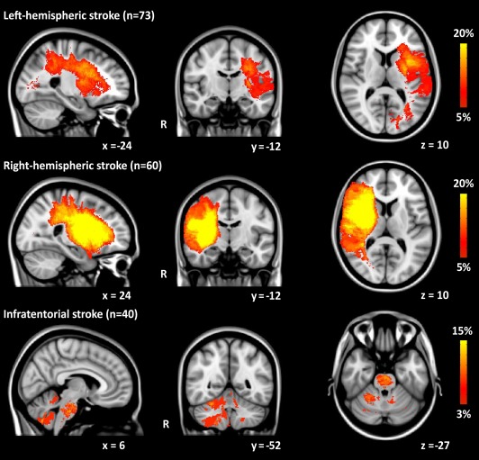
Lesion probability maps in patients with left‐hemispheric stroke, right‐hemispheric stroke, and infratentorial stroke. The color overlay created on top of the MNI standard brain template shows the probability of each voxel containing a lesion in each patient group. The color bar denotes the probability range. [Color figure can be viewed in the online issue, which is available at http://wileyonlinelibrary.com.]
Strokes in the right hemisphere were on average larger than strokes in the left hemisphere (F(1,128)=8.03, P=0.005) and than infratentorial strokes (F(1,95)=14.09, P=0.0003) (Table 1 and Fig. 1). Left‐hemispheric stroke patients had larger strokes compared with infratentorial stroke patients (F(1,108)=3.93, P=0.049). Patients with a right‐hemispheric stroke had a more severe stroke (higher NIHSS score at stroke onset) compared with left‐hemispheric stroke patients (Mann‐Whitney U test, P=0.002), but not compared with infratentorial stroke patients (P=0.7) (Table 1). Infratentorial stroke patients had a more severe stroke compared with left‐hemispheric stroke patients (P=0.009).
Memory Performance
Table 2 shows the mean memory performance of patients (expressed in Z‐scores) compared with controls. Left‐hemispheric stroke patients performed significantly worse on immediate verbal recall (unadjusted mean Z‐score:−0.67, 95% CI: −0.88 to −0.46) and delayed verbal recall (mean:−0.56, 95% CI: −0.80 to −0.32), delayed visuospatial recall (mean=−0.49, 95% CI: −0.78 to −0.19), and delayed verbal recognition (mean:−0.73, 95% CI: −1.02 to −0.45) compared with controls (mean Z‐score for every measure=0.00, 95% CI: −0.21 to 0.21) (Table 2). Right‐hemispheric stroke patients only performed significantly worse than controls on delayed verbal recognition (mean Z‐score=−0.54, 95% CI: −0.82 to −0.27). Infratentorial stroke patients performed significantly worse on immediate verbal recall (mean=−0.44, 95% CI: −0.74 to −0.15) and delayed verbal recognition (mean=−0.63, 95% CI: −1.01 to −0.25) compared with controls.
Table 2.
10‐year follow‐up of mean memory performance (Z‐scores) for patients compared with controls
| Hemispheric stroke | Infratentorial stroke | ||
|---|---|---|---|
| Left | Right | ||
| No. | 73 | 60 | 40 |
| Verbal memory (RAVLT), Mean (SD) | |||
| Immediate verbal recall | −0.70(0.9)c | −0.25(0.9) | −0.44(0.9)a |
| Delayed verbal recall | −0.57(1.0)b | −0.28(0.9) | −0.46(0.9) |
| Delayed verbal recognition (hits + correct negatives) | −0.73(1.2)c | −0.53(1.1)b | −0.60(1.2)a |
| Visuospatial memory (ROCF), Mean (SD) | |||
| Immediate visuospatial recall | −0.42(1.1) | 0.13(1.1) | 0.18(1.1) |
| Delayed visuospatial recall | −0.51(1.2)a | 0.13(1.2) | 0.00(1.1) |
Data are expressed as unadjusted mean Z‐score (±SD). RAVLT, Rey Auditory Verbal Learning Test; ROCF, Rey‐Osterrieth Complex Figure. Missing data in patients: no ROCF: 1.7%, no RAVLT: 1.2%. Controls (n=87) are not displayed since their mean Z‐score is zero (SD=1) for each measure. A star denotes a significant worse performance for patients compared with controls after adjustments for age at follow‐up, sex, education, depressive symptoms, and fatigue.
P <0.05
P <0.01
P <0.001.
Exclusion of patients with aphasia (n=19) did not affect our results, except for a change into a trend toward significance for the delayed visuospatial recall (P=0.058) in left‐hemispheric stroke patients.
Hippocampal Volume After Stroke
After a mean follow‐up duration of 10 years patients with left‐hemispheric stroke showed a smaller left hippocampal volume (2.98 ml, SD 0.4) compared with controls (3.15 ml, SD 0.3)(F(1,156)=8.28, P=0.005), which was an unadjusted mean difference of 5.4% (adjustment for age and sex did not affect this difference) (Fig. 2). Right‐hemispheric stroke was associated with smaller right hippocampal volume (3.11 ml, SD 0.4) compared with controls (3.36 ml, SD 0.4) (F(1,143)=16.79, P<0.0001), which was a difference of 7.4% (after adjustment for age and sex: 7.7%)(Fig. 2). The mean contralateral hippocampal volume of both left and right‐hemispheric stroke patients did not significantly differ from controls (Fig. 2). There were no differences between mean hippocampal volume of patients with infratentorial stroke and controls (Fig. 2).
Figure 2.
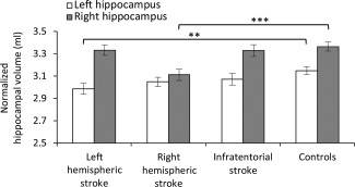
Unadjusted mean hippocampal volume (±SEM) 10 years after ischemic stroke in young adults compared with stroke‐free controls. Significant differences are shown after adjusting for age at follow‐up and sex. *P<0.05, **P<0.01, ***P<0.001.
After adjusting for age, sex, level of education, follow‐up duration, normalized lesion volume, and left thalamic volume, there was a significant positive association between left hippocampal volume and immediate visuospatial recall (b=0.90, 95%CI= 0.20–1.60, t=2.58, P=0.012) and delayed visuospatial recall (b=1.01, 95%CI= 0.27–1.75, t=2.74, P=0.008) in left‐hemispheric stroke patients. No association was observed between left hippocampal volume and verbal memory performance after left‐hemispheric stroke. In right‐hemispheric stroke patients no significant associations between right hippocampal and memory performance volume were observed.
Relationship Between Lesion Volume and Hippocampal Volume
Even after adjustment for age at follow‐up, sex, lesion location (left/right), and follow‐up duration, a larger hemispheric stroke was significantly associated with smaller ipsilateral hippocampal volume (b=−0.003, 95%CI= −0.005 to −0.002 per ml increase in lesion volume, t=−4.46, P<0.0001), not contralateral hippocampal volume (b=−0.001, 95%CI= −0.002 to 0.000, t=−1.36, P=0.18).
Relationship Between Follow‐Up Duration and Hippocampal Volume
After controlling for age, sex, and normalized lesion volume, in patients with left‐hemispheric stroke there was a negative association between follow‐up duration and left hippocampal volume (b= −0.028 ml, 95% CI=−0.04 to −0.01, t=−3.25, P=0.002). After each decade the left hippocampus is estimated to be 0.28 ml smaller. In right‐hemispheric stroke patients longer follow‐up duration was associated with a smaller right hippocampal volume (b= −0.015 ml, 95% CI=−0.03 to −0.003, t=−2.54, P=0.03). This is an estimated reduction of 0.15 ml after a decade. We did not observe a relationship between duration of follow‐up and contralateral hippocampal volume after hemispheric stroke. No associations were observed after infratentorial stroke.
Thalamic Volume After Stroke
To investigate whether the results were specific to the hippocampus, thalamic volumes after stroke were also examined. In left‐hemispheric stroke patients a significantly smaller mean left thalamic volume (7.00 ml, SD 0.9) was found compared with controls (7.60 ml, SD 0.6) (F(1,148)=24.66, P<0.0001). Right‐hemispheric stroke patients had a smaller mean right thalamic volume (6.38 ml, SD 1.1) compared with controls (7.39 ml, SD 0.6) (F(1,127)=49.5, P<0.0001). Hemispheric stroke was not associated with smaller contralateral thalamic volume compared with controls. Patients with infratentorial stroke did not have significantly smaller thalamic volumes compared with controls.
After adjustment for age, sex, level of education, follow‐up duration, ipsilateral hippocampal volume, and normalized lesion volume, no independent association between ipsilateral thalamic volume and memory performance was found in hemispheric stroke patients (all P‐values>0.05).
DISCUSSION
We showed that on average 10 years following ischemic stroke in adults 18 through 50 years, hemispheric stroke outside the medial‐temporal lobe is associated with a smaller ipsilateral hippocampal volume compared with a stroke‐free population, accompanied by a worse memory performance.
We showed that patients with a left‐hemispheric stroke after a mean follow‐up of 10 years had a worse memory performance on almost all memory measures with an attendant smaller ipsilateral hippocampal volume compared with controls. The memory performance was only very mildly affected in right‐hemispheric stroke patients compared with controls, although again a smaller ipsilateral hippocampal volume was observed. Possibly, aphasia limits the proper assessment of verbal memory performance in left‐hemispheric stroke patients. However, these patients also had a worse visual memory performance and the exclusion of patients with aphasia did not significantly alter our findings making this a very unlikely explanation. Other studies also found that left hippocampal atrophy is more evidently related to poorer memory function than right hippocampal atrophy [Bigler et al., 1996; Burgess et al., 2002; Kilpatrick et al., 1997; McConley et al., 2008]. This finding is in line with hippocampal lesion data [Alessio et al., 2004; Szabo et al., 2009]. These previous results and our present findings suggest that volumetric changes in the left hippocampus may be more important to memory performance than volumetric changes in the right hippocampus.
In left‐hemispheric stroke patients, our results suggest poorer encoding ability as well as a decay of acquired visuospatial information over time, both of which have been found to correlate with hippocampal volume, integrity, and functioning [Babiloni et al., 2009; McConley et al., 2008; van Norden et al., 2012].
Somewhat surprisingly, the regression analyses yielded no association between verbal memory and ipsilateral hippocampal volume, whereas this relationship has been found in patients with Alzheimer's disease [Estevez‐Gonzalez et al., 2003]. In patients with Alzheimer's disease larger reductions in hippocampal volume have been observed (left: 30% and right: 35%) compared to our young stroke patients (left: 5.4% and right: 7.7%)[Frisoni and Whitwell, 2008]. Possibly, once volume loss exceeds a certain volume, as for example in Alzheimer's disease, the relation between hippocampal volume and verbal episodic memory may become more evident [Frisoni and Whitwell, 2008; Leube et al., 2008].
A previous study by Xie et al. [2011] investigated 36 relatively young patients up to 2 years after middle cerebral artery occlusion. However, life expectancy of most of these young patients exceeds by far 2 years. Our findings, therefore, extend those of Xie et al. [2011] as we found a relation between hippocampal volume and hemispheric stroke, but not infratentorial stroke, with a much longer follow‐up of 10 years. Data on lesion side (left/right) was lacking in the study of Xie et al. [2011], which is important since the asymmetry between left and right hippocampal volume, with larger right than left volumes, is established in healthy adults [Pedraza et al., 2004]. Therefore, we compared volumes of patients with volumes of controls within each hemisphere.
Furthermore, another important finding of our study is the inverse relation between follow‐up duration in years and ipsilateral hippocampal volume, reflecting decreasing hippocampal volume from the early months after stroke up to 30 years after stroke. This relation persisted even after adjusting for age. This might explain the negative association between follow‐up duration and memory performance previously observed in these young adults [Schaapsmeerders et al., 2013].
Finally, our results suggest that hemispheric stroke has a generalized effect on ipsilateral brain structures remote from the infarction, such as a smaller ipsilateral hippocampus and thalamus. However, after adjusting for age, sex, education, follow‐up duration, normalized lesion volume, and ipsilateral hippocampal volume no independent relation was found between thalamic volume and memory performance. Ipsilateral hippocampal volume was still associated with visual memory function in left‐hemispheric stroke patients after excluding patients with thalamic stroke and adjusting for left thalamic volume in the regression analysis, suggesting an independent association between ipsilateral hippocampal volume and memory performance.
Underlying Mechanism of Smaller Ipsilateral Hippocampal Volume
Rodent studies showed loss of pyramidal cells in the CA1 area of the hippocampus after ischemia in other areas of the brain [Wang et al., 2004], possibly caused by spreading depression (SD), causing secondary neuronal damage and infarct expansion. SD is associated with failure of brain ion homeostasis, efflux of excitatory amino acids from neurons, resulting in increased energy metabolism and changes in cerebral blood flow [Lauritzen et al., 2011]. Support for SD as underlying mechanism of hippocampal damage after infarction comes from the finding that inhibition of SD propagation by blocking intercellular communication via gap junction channels after ischemic stroke in rodents, results in less hippocampal damage and fewer deficits in memory [Xie et al., 2011].
The observation of smaller hippocampi dependent on follow‐up duration after stroke may induce a vulnerability of the hippocampus in the affected hemisphere to the effect of ageing, possibly leading to accelerated cognitive decline at an already earlier age. When these patients finally come to an age when neurodegenerative features come into play, their remaining hippocampal volume may already be lower or expedite the threshold for cognitive impairment or even dementia, compared with persons without a stroke at young age. This notion is in agreement with the finding that patients who develop Alzheimer's disease, loss of hippocampal volume seems to be present years before the clinical diagnosis [den Heijer et al., 2010].
Strengths and Limitations
The strengths of our study include its large sample size and the long follow‐up. This single centre study allowed us to collect baseline and follow‐up information according to identical procedures in all patients, with high inter‐rater and intrarater agreement for hippocampal volumes. We used strict protocols and researchers were trained, to reduce the risk of information bias.
Some limitations of our study need to be addressed. Although the FUTURE study has a prospective design, the current analysis is cross‐sectional and we, therefore, can only report on smaller hippocampal volume after stroke. While this may be due to poststroke hippocampal atrophy, a longitudinal design is required to further support causality. However, our data clearly demonstrate a relation between the stroke and future loss of ipsilateral hippocampal volume, since we did not observe smaller contralateral hippocampal volumes and longer follow‐up duration was associated with smaller ipsilateral hippocampal volume. Additional support comes from our finding that a larger hemispheric stroke was associated with smaller ipsilateral hippocampal volume. In contrast with one smaller study (n=36) [Xie et al., 2011], we did observe an inverse relationship between lesion volume and ipsilateral hippocampal volume. Possibly, in the previous study low statistical power due to a small sample size resulted in no association [Xie et al., 2011]. Thus, our findings suggest that it is highly unlikely that these patients already had smaller ipsilateral hippocampal volumes before stroke onset.
Second, selection bias might have occurred, since patients who refused or were not eligible for MRI scanning had a poorer outcome compared with participants. However, bias would only have occurred when the relation between the stroke and hippocampal volume would be selectively different in nonparticipants, which does not seem likely. Although nonparticipants might have had more vascular risk factors, which may accelerate decline of hippocampal volume.
Third, MRI quantification (with manual correction of hippocampal volume and ICV in case of large cortical infarcts) could not have been done blindly for lesion location and size. This might have biased the results. However, automated analyses on thalamic volume showed the same results as compared with manually corrected hippocampal volume, suggesting no bias due to unblinded reading.
Conclusions and Clinical Implications
To conclude, the greater (probable life‐long) memory impairment after stroke may be explained by a smaller ipsilateral hippocampus. At present the mean memory performance of our patients was not within the “impaired” range (i.e., less than 1.5 standard deviations below the mean of the controls). Given their young age, these patients are expected to live on for several decades and the reduced ipsilateral hippocampal volume accompanied by a worse memory score may point toward a lower neural reserve in these patients, making them at risk for further cognitive decline, especially when neurodegenerative disease comes into play. Also, in these young patients even small decrements in memory function may affect vocational and academic achievements, which make our findings highly relevant.
The stronger association between left‐hemispheric stroke, a smaller left hippocampal volume, and worse memory performance has clinical implications, since it seems that especially small baseline left hippocampal volume is associated with higher risk of conversion into dementia [Eckerstrom et al., 2008]. These patients with memory decline and smaller hippocampal volumes, therefore, might be especially at risk for further cognitive decline. It is important to inform patients on these long‐term consequences and provide realistic outlooks given the underlying structural changes. Future studies are needed that prospectively investigate the underlying structural changes of cognitive decline to support causality.
ACKNOWLEDGMENT
The funder had no role in study design, data collection and analysis, decision to publish, or preparation of the manuscript.
REFERENCES
- Adams HP, Jr ., Bendixen BH, Kappelle LJ, Biller J, Love BB, Gordon DL, Marsh EE, 3rd (1993): Classification of subtype of acute ischemic stroke. Definitions for use in a multicenter clinical trial. TOAST. Trial of Org 10172 in Acute Stroke Treatment. Stroke 24:35–41. [DOI] [PubMed] [Google Scholar]
- Alessio A, Damasceno BP, Camargo CH, Kobayashi E, Guerreiro CA, Cendes F (2004): Differences in memory performance and other clinical characteristics in patients with mesial temporal lobe epilepsy with and without hippocampal atrophy. Epilepsy Behav 5:22–27. [DOI] [PubMed] [Google Scholar]
- Allan LM, Rowan EN, Firbank MJ, Thomas AJ, Parry SW, Polvikoski TM, O'Brien JT, Kalaria RN (2011): Long term incidence of dementia, predictors of mortality and pathological diagnosis in older stroke survivors. Brain 134:3716–3727. [DOI] [PMC free article] [PubMed] [Google Scholar]
- Andersson JLR, Jenkinson M, Smith S (2007): Non‐Linear Registration, Aka Spatial Normalisation. Technical Report TR07JA2. Oxford Centre for Functional MRI of the Brain. [Google Scholar]
- Babiloni C, Vecchio F, Mirabella G, Buttiglione M, Sebastiano F, Picardi A, Di Gennaro G, Quarato PP, Grammaldo LG, Buffo P, Esposito V, Manfredi M, Cantore G, Eusebi F (2009): Hippocampal, amygdala, and neocortical synchronization of theta rhythms is related to an immediate recall during rey auditory verbal learning test. Hum Brain Mapp 30:2077–2089. [DOI] [PMC free article] [PubMed] [Google Scholar]
- Bigler ED, Johnson SC, Anderson CV, Blatter DD, Gale SD, Russo AA, Ryser DK, Macnamara SE, Bailey BJ, Hopkins RO, Abildskov TJ (1996): Traumatic brain injury and memory: The role of hippocampal atrophy. Neuropsychology 10:333–342. [DOI] [PubMed] [Google Scholar]
- Blum S, Luchsinger JA, Manly JJ, Schupf N, Stern Y, Brown TR, DeCarli C, Small SA, Mayeux R, Brickman AM (2012): Memory after silent stroke: Hippocampus and infarcts both matter. Neurology 78:38–46. [DOI] [PMC free article] [PubMed] [Google Scholar]
- Brott T, Adams HP, Jr ., Olinger CP, Marler JR, Barsan WG, Biller J, Spilker J, Holleran R, Eberle R, Hertzberg V, M Rorick, CJ Moomaw, M Walker (1989): Measurements of acute cerebral infarction: A clinical examination scale. Stroke 20:864–870. [DOI] [PubMed] [Google Scholar]
- Burgess N, Maguire EA, O'Keefe J (2002): The human hippocampus and spatial and episodic memory. Neuron 35:625–641. [DOI] [PubMed] [Google Scholar]
- Carlesimo GA, Cherubini A, Caltagirone C, Spalletta G (2010): Hippocampal mean diffusivity and memory in healthy elderly individuals: A cross‐sectional study. Neurology 74:194–200. [DOI] [PubMed] [Google Scholar]
- Chen IH, Novak V, Manor B (2014): Infarct hemisphere and noninfarcted brain volumes affect locomotor performance following stroke. Neurology 82:828–834. [DOI] [PMC free article] [PubMed] [Google Scholar]
- den Heijer T, van der Lijn F, Koudstaal PJ, Hofman A, van der Lugt A, Krestin GP, Niessen WJ, Breteler MM (2010): A 10‐year follow‐up of hippocampal volume on magnetic resonance imaging in early dementia and cognitive decline. Brain 133:1163–1172. [DOI] [PubMed] [Google Scholar]
- den Heijer T, der Lijn F, Vernooij MW, de Groot M, Koudstaal PJ, der Lugt A, Krestin GP, Hofman A, Niessen WJ, Breteler MM (2012): Structural and diffusion MRI measures of the hippocampus and memory performance. Neuroimage 63:1782–1789. [DOI] [PubMed] [Google Scholar]
- Duvernoy H (1997): The human hippocampus: Functional anatomy, vascularization and serial sections with MRI. New York: Springer‐Verlag. [Google Scholar]
- Eckerstrom C, Olsson E, Borga M, Ekholm S, Ribbelin S, Rolstad S, Starck G, Edman A, Wallin A, Malmgren H (2008): Small baseline volume of left hippocampus is associated with subsequent conversion of MCI into dementia: The Goteborg MCI study. J Neurol Sci 272:48–59. [DOI] [PubMed] [Google Scholar]
- Estevez‐Gonzalez A, Kulisevsky J, Boltes A, Otermin P, Garcia‐Sanchez C (2003): Rey verbal learning test is a useful tool for differential diagnosis in the preclinical phase of Alzheimer's disease: Comparison with mild cognitive impairment and normal aging. Int J Geriatr Psychiatry 18:1021–1028. [DOI] [PubMed] [Google Scholar]
- Exner C, Weniger G, Irle E (2001): Implicit and explicit memory after focal thalamic lesions. Neurology 57:2054–2063. [DOI] [PubMed] [Google Scholar]
- Frisoni GB, Whitwell JL (2008): How fast will it go, doc? New tools for an old question from patients with Alzheimer disease. Neurology 70:2194–2195. [DOI] [PubMed] [Google Scholar]
- Henon H, Pasquier F, Durieu I, Pruvo JP, Leys D (1998): Medial temporal lobe atrophy in stroke patients: Relation to pre‐existing dementia. J Neurol Neurosurg Psychiatry 65:641–647. [DOI] [PMC free article] [PubMed] [Google Scholar]
- Henon H, Durieu I, Guerouaou D, Lebert F, Pasquier F, Leys D (2001): Poststroke dementia: Incidence and relationship to prestroke cognitive decline. Neurology 57:1216–1222. [DOI] [PubMed] [Google Scholar]
- Herve D, Molko N, Pappata S, Buffon F, LeBihan D, Bousser MG, Chabriat H (2005): Longitudinal thalamic diffusion changes after middle cerebral artery infarcts. J Neurol Neurosurg Psychiatry 76:200–205. [DOI] [PMC free article] [PubMed] [Google Scholar]
- Ivan CS, Seshadri S, Beiser A, Au R, Kase CS, Kelly‐Hayes M, Wolf PA (2004): Dementia after stroke: The Framingham Study. Stroke 35:1264–1268. [DOI] [PubMed] [Google Scholar]
- Jenkinson M, Beckmann CF, Behrens TE, Woolrich MW, Smith SM (2012): Fsl. Neuroimage 62:782–790. [DOI] [PubMed] [Google Scholar]
- Kasner SE, Chalela JA, Luciano JM, Cucchiara BL, Raps EC, McGarvey ML, Conroy MB, Localio AR (1999): Reliability and validity of estimating the NIH stroke scale score from medical records. Stroke 30:1534–1537. [DOI] [PubMed] [Google Scholar]
- Kilpatrick C, Murrie V, Cook M, Andrewes D, Desmond P, Hopper J (1997): Degree of left hippocampal atrophy correlates with severity of neuropsychological deficits. Seizure 6:213–218. [DOI] [PubMed] [Google Scholar]
- Lauritzen M, Dreier JP, Fabricius M, Hartings JA, Graf R, Strong AJ (2011): Clinical relevance of cortical SD in neurological disorders: Migraine, malignant stroke, subarachnoid and intracranial hemorrhage, and traumatic brain injury. J Cereb Blood Flow Metab 31:17–35. [DOI] [PMC free article] [PubMed] [Google Scholar]
- Leifer D, Bravata DM, Connors JJ, 3rd, Hinchey JA, Jauch EC, Johnston SC, Latchaw R, Likosky W, Ogilvy C, Qureshi AI, Summers D, Sung GY, Williams LS, Zorowitz R, American Heart Association Special Writing Group of the Stroke C, Atherosclerotic Peripheral Vascular Disease Working G, Council on Cardiovascular S, Anesthesia, Council on Cardiovascular N (2011): Metrics for measuring quality of care in comprehensive stroke centers: Detailed follow‐up to Brain Attack Coalition comprehensive stroke center recommendations: A statement for healthcare professionals from the American Heart Association/American Stroke Association. Stroke 42:849–877. [DOI] [PubMed] [Google Scholar]
- Lesne SE, Sherman MA, Grant M, Kuskowski M, Schneider JA, Bennett DA, Ashe KH (2013): Brain amyloid‐beta oligomers in ageing and Alzheimer's disease. Brain 136:1383–1398. [DOI] [PMC free article] [PubMed] [Google Scholar]
- Leube DT, Weis S, Freymann K, Erb M, Jessen F, Heun R, Grodd W, Kircher TT (2008): Neural correlates of verbal episodic memory in patients with MCI and Alzheimer's disease—A VBM study. Int J Geriatr Psychiatry 23:1114–1118. [DOI] [PubMed] [Google Scholar]
- Leys D, Henon H, Mackowiak‐Cordoliani MA, Pasquier F (2005): Poststroke dementia. Lancet Neurol 4:752–759. [DOI] [PubMed] [Google Scholar]
- McConley R, Martin R, Palmer CA, Kuzniecky R, Knowlton R, Faught E (2008): Rey Osterrieth complex figure test spatial and figural scoring: relations to seizure focus and hippocampal pathology in patients with temporal lobe epilepsy. Epilepsy Behav 13:174–177. [DOI] [PubMed] [Google Scholar]
- Osterrieth PA (1944): Le test de copie d'une figure complexe: Contribution à l' étude de la perception et de la mémoire. Arch Psychol 30:206–353. [Google Scholar]
- Patenaude B, Smith SM, Kennedy DN, Jenkinson M (2011): A Bayesian model of shape and appearance for subcortical brain segmentation. Neuroimage 56:907–922. [DOI] [PMC free article] [PubMed] [Google Scholar]
- Pedraza O, Bowers D, Gilmore R (2004): Asymmetry of the hippocampus and amygdala in MRI volumetric measurements of normal adults. J Int Neuropsychol Soc 10:664–678. [DOI] [PubMed] [Google Scholar]
- Rutten‐Jacobs LC, Maaijwee NA, Arntz RM, Van Alebeek ME, Schaapsmeerders P, Schoonderwaldt HC, Dorresteijn LD, Overeem S, Drost G, Janssen MC, van Heerde WL, Kessels RP, Zwiers MP, Norris DG, van der Vlugt MJ, van Dijk EJ, de Leeuw FE (2011): Risk factors and prognosis of young stroke. The Future study: A prospective cohort study. Study rationale and protocol. BMC Neurol 11:109. [DOI] [PMC free article] [PubMed] [Google Scholar]
- Rutten‐Jacobs LC, Arntz RM, Maaijwee NA, Schoonderwaldt HC, Dorresteijn LD, van Dijk EJ, de Leeuw FE (2013): Long‐term mortality after stroke among adults aged 18 to 50 years. JAMA 309:1136–1144. [DOI] [PubMed] [Google Scholar]
- Schaapsmeerders P, Maaijwee NA, van Dijk EJ, Rutten‐Jacobs LC, Arntz RM, Schoonderwaldt HC, Dorresteijn LD, Kessels RP, de Leeuw FE (2013): Long‐term cognitive impairment after first‐ever ischemic stroke in young adults. Stroke 44:1621–1628. [DOI] [PubMed] [Google Scholar]
- Smith SM (2002): Fast robust automated brain extraction. Hum Brain Mapp 17:143–155. [DOI] [PMC free article] [PubMed] [Google Scholar]
- Snaphaan L, Rijpkema M, van Uden I, Fernandez G, de Leeuw FE (2009): Reduced medial temporal lobe functionality in stroke patients: A functional magnetic resonance imaging study. Brain 132:1882–1888. [DOI] [PubMed] [Google Scholar]
- Squire LR, Wixted JT (2011): The cognitive neuroscience of human memory since H.M. Annu Rev Neurosci 34:259–288. [DOI] [PMC free article] [PubMed] [Google Scholar]
- Szabo K, Forster A, Jager T, Kern R, Griebe M, Hennerici MG, Gass A (2009): Hippocampal lesion patterns in acute posterior cerebral artery stroke: Clinical and MRI findings. Stroke 40:2042–2045. [DOI] [PubMed] [Google Scholar]
- Tamura A, Tahira Y, Nagashima H, Kirino T, Gotoh O, Hojo S, Sano K (1991): Thalamic atrophy following cerebral infarction in the territory of the middle cerebral artery. Stroke 22:615–618. [DOI] [PubMed] [Google Scholar]
- Tuladhar AM, Snaphaan L, Shumskaya E, Rijpkema M, Fernandez G, Norris DG, de Leeuw FE (2013): Default mode network connectivity in stroke patients. PLoS One 8:e66556. [DOI] [PMC free article] [PubMed] [Google Scholar]
- Vakil E, Blachstein H (1993): Rey auditory‐verbal learning test: structure analysis. J Clin Psychol 49:883–890. [DOI] [PubMed] [Google Scholar]
- van Norden AG, Fick WF, de Laat KF, van Uden IW, van Oudheusden LJ, Tendolkar I, Zwiers MP, de Leeuw FE (2008): Subjective cognitive failures and hippocampal volume in elderly with white matter lesions. Neurology 71:1152–1159. [DOI] [PubMed] [Google Scholar]
- van Norden AG, de Laat KF, Fick I, van Uden IW, van Oudheusden LJ, Gons RA, Norris DG, Zwiers MP, Kessels RP, de Leeuw FE (2012): Diffusion tensor imaging of the hippocampus and verbal memory performance: The RUN DMC study. Hum Brain Mapp 33:542–551. [DOI] [PMC free article] [PubMed] [Google Scholar]
- Vercoulen JH, Swanink CM, Fennis JF, Galama JM, van der Meer JW, Bleijenberg G (1994): Dimensional assessment of chronic fatigue syndrome. J Psychosom Res 38:383–392. [DOI] [PubMed] [Google Scholar]
- Vyhnalek M, Nikolai T, Andel R, Nedelska Z, Rubinova E, Markova H, Laczo J, Bezdicek O, Sheardova K, Hort J (2014): Neuropsychological correlates of hippocampal atrophy in memory testing in nondemented older adults. J Alzheimers Dis 42(Suppl 3):S81–S90. [DOI] [PubMed] [Google Scholar]
- Wang W, Redecker C, Bidmon HJ, Witte OW (2004): Delayed neuronal death and damage of GDNF family receptors in CA1 following focal cerebral ischemia. Brain Res 1023:92–101. [DOI] [PubMed] [Google Scholar]
- Williams LS, Yilmaz EY, Lopez‐Yunez AM (2000): Retrospective assessment of initial stroke severity with the NIH Stroke Scale. Stroke 31:858–862. [DOI] [PubMed] [Google Scholar]
- Wu W, Brickman AM, Luchsinger J, Ferrazzano P, Pichiule P, Yoshita M, Brown T, DeCarli C, Barnes CA, Mayeux R, Vannucci SJ, Small SA (2008): The brain in the age of old: The hippocampal formation is targeted differentially by diseases of late life. Ann Neurol 64:698–706. [DOI] [PMC free article] [PubMed] [Google Scholar]
- Xie M, Yi C, Luo X, Xu S, Yu Z, Tang Y, Zhu W, Du Y, Jia L, Zhang Q, Dong Q, Zhu W, Zhang X, Bu B, Wang W (2011): Glial gap junctional communication involvement in hippocampal damage after middle cerebral artery occlusion. Ann Neurol 70:121–132. [DOI] [PubMed] [Google Scholar]
- Zhang J, Zhang Y, Xing S, Liang Z, Zeng J (2012): Secondary neurodegeneration in remote regions after focal cerebral infarction: A new target for stroke management? Stroke 43:1700–1705. [DOI] [PubMed] [Google Scholar]
- Zigmond AS, Snaith RP (1983): The hospital anxiety and depression scale. Acta Psychiatr Scand 67:361–370. [DOI] [PubMed] [Google Scholar]