

Abstract
Many studies have shown the involvement of the premotor cortex in action observation, recognizing this region as the neural marker of action simulation (i.e., internal modeling on the basis of the observer's own motor repertoire). So far, however, we have remained unaware of how action simulation differs from more general action representation in terms of premotor activation. The present fMRI experiment is the first to demonstrate how premotor structures contribute to action simulation as opposed to other action‐related cognitive tasks, such as maintaining action representations. Using similar stimuli, a prediction condition requiring internal simulation of transiently occluded actions was compared to three different action‐related control tasks differing solely in task instructions. Results showed right pre‐SMA activation as a correlate of maintaining action representations in general. Moreover, the prediction condition was most efficient in activating the left pre‐SMA and left PMd. These results suggest that the conjoint activation of the pre‐SMA and PMd reflects a core neural driver of action simulation. Hum Brain Mapp, 2011. © 2010 Wiley‐Liss, Inc.
Keywords: fMRI, action observation, prediction, simulation, occlusion, presupplementary motor area, pre‐SMA, dorsal premotor cortex, PMd
INTRODUCTION
In monkeys and humans, motor system activation is robustly found in experiments involving the observation of other individuals [Gallese et al., 1996; Grèzes and Decety, 2001; Rizzolatti et al., 2002]. Although there has been considerable theorizing about the gain of motor system involvement during action observation, the purpose of translating perceptual action information into sensorimotor representations is constantly under debate. Evidence suggesting that premotor cortex activation during action observation may have predictive purposes is also constantly accumulating. The present study aimed at elucidating the contribution of the premotor cortex to action prediction in contrast to other action‐related tasks.
It has been suggested that a particularly efficient way of achieving action prediction may be based on exploiting predictive functions of the observer's own motor system in a simulation mode [Blakemore and Decety, 2001; Grush, 2004; Wilson and Knoblich, 2005]. Corresponding neurophysiological evidence in monkeys points to a predictive activation of sensorimotor action representations during observation [Umiltà et al., 2001]: mirror neurons in the macaque premotor area have been shown to exhibit similar firing patterns, irrespective of whether food was grasped behind an occluder or in front of the monkey. As its monkey homolog, the human premotor cortex contains somatotopically organized sensorimotor representations [Buccino et al., 2001; Godschalk et al., 1995; Graziano and Gandhi, 2000; Rizzolatti et al., 1988; Sakreida et al., 2005].
Several behavioral studies provide evidence of predictive use of sensorimotor representations when humans watch actions. For instance, when an attended action was occluded in behavioral experiments [Graf et al., 2007], participants achieved temporally precise action predictions. This was taken as evidence of real‐time action simulation on the basis of sensorimotor representations. Likewise, when humans observed someone stacking wooden blocks, they coordinated their gaze in a predictive way with the observed hand movement [Flanagan and Johansson, 2003]. Anticipatory EEG activation was also recorded over the motor cortex of observers prior to the onset of an observed transitive hand movement [Kilner et al., 2004]. Further, action imagery induces motor system activation in the absence of (dynamic) perceptual input [Gerardin et al., 2000; Hanakawa et al., 2008; Lotze et al., 1999]. Moreover, current research extends the role of the premotor cortex to the prediction of event dynamics in general [Schubotz, 2007; Schubotz and von Cramon, 2003].
The current fMRI experiment was designed to dissociate brain activation specific to action prediction from the background of activation related to more general action representation such as memorizing certain aspects of an action. Therefore, comparison of a prediction task with other action‐related control tasks was required. To pinpoint the impact of task instructions while balancing the influence of perceptual input between conditions, similar stimuli were used in four tasks. Participants watched short video‐clips showing an actress performing everyday actions in a naturalistic environment. The clips were repeatedly occluded for 1 s. After each occlusion, participants indicated whether the action continued with coherent or incoherent timing (Prediction condition). As a second condition, a memory task (Freezing condition) used the same stimuli and required participants to indicate whether the last frame before occlusion was identical to the first frame after occlusion. Two further conditions were used to control for effects of the occluder intervention (Counting condition) and the motor response (Detection condition) (see Methods and Fig. 1 for details).
Figure 1.
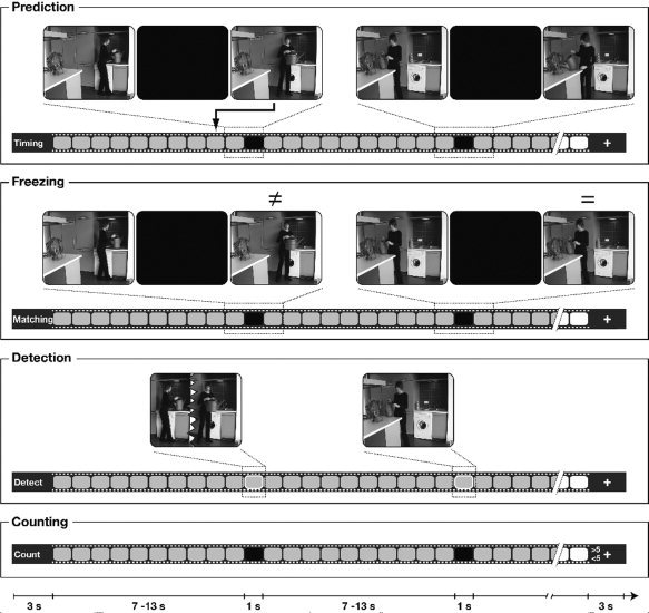
Experimental conditions. Panels show four conditions (Prediction, Freezing, Detection, and Counting) employing the same video‐clips. The bars on the bottom of each panel represent ongoing video presentation during which black boxes repeatedly occluded the whole screen for 1 s in Prediction, Freezing, and Counting conditions. On average, a 60‐s video was occluded five times. In Detection, instead of occlusions, a border surrounding the display changed color for 1 s. With every new video, the task changed and this was announced visually. In the conditions Prediction, Freezing and Detection, the observation of the ongoing course of the action was required. In Prediction, participants indicated whether the action continued with coherent timing after each occluder. In Freezing, they memorized the last frame before occlusion to judge whether the video continued from the same position. When the border was colored in Detection, participants were asked to detect occasional disruptions in the smoothness of the ongoing action. In these three conditions, yes/no response‐buttons were pressed as soon as possible after the offset of occlusions or colored borders. The depicted scenes are taken from a video‐clip to illustrate two single trials for each condition. The pictures on the left show an incoherent continuation in Prediction, a mismatch in Freezing, and a disrupted action in Detection. The examples depicted on the right illustrate accurate trials for each condition. The Counting condition did not draw attention to the action. Instead, participants counted occlusions and indicated whether a clip contained more or fewer than five of these.
The first step of data analysis was to assess activation common to the Prediction and Freezing conditions. Therefore, contrasts with the control conditions Detection and Counting were calculated separately for Prediction and Freezing and subsequently integrated using conjunction analyses. Functionally, the Prediction and Freezing conditions similarly required the maintenance of an internal reference against which the continuation of the video was matched after occlusion. In the second step, prediction‐specific activation was obtained by contrasting the Prediction condition directly with the Freezing condition. This contrast aimed to reveal functional resources that Prediction needed, but Freezing did not. The two conditions differed with respect to characteristics of the maintained internal reference. In Freezing, the internal reference was derived from the last preoccluder frame kept in memory as a static posture. The Prediction condition additionally required a dynamic transformation over time. That is, the formation of the internal reference departed from the last preoccluder frame (that was also crucial in Freezing), but needed to be transformed through mental simulation in Prediction. With the outlined two‐step analysis, we were able to dissociate brain activation specifically related to internal action prediction from effects of rather general maintenance of an action‐related internal reference. As indicated by the literature reviewed, we hypothesized that action prediction elicits stronger premotor activation relative to the other tasks. Although the experiment was designed to reveal differences reflecting prediction task‐induced effects in the premotor cortex rather than stimulus‐driven effects, whole‐brain images were analyzed.
METHODS
Participants
Twenty‐two right‐handed women (mean age 25.7 years; range 23.7–28.8) participated in the experiment. None had a neurological or psychiatric disorder or was on medication at the time of measurement. Four participants were excluded from statistical analysis as their correct response rates were below 65%, resulting in a total of 18 participants in the final analysis. All participants gave written consent and were paid for their participation. They were treated according to the regulations of ethics in the Declaration of Helsinki and fMRI procedures were approved by the local ethics committee.
Stimuli
Four different video‐clips showed a woman performing everyday actions (making coffee, fertilizing a houseplant, setting the table and hanging up laundry). Videos lasted for ∼60 s each (range 54–76 s). Several times during a clip, the whole display was transiently occluded by a black rectangle. Occlusions lasted for 1 s and were pseudo‐randomly distributed with an interval between two occlusions jittered between 7, 9, 11 or 13 s. After an occlusion, the video continued immediately. The videos were presented in full color with a resolution of 720 × 576 pixels and a frame rate of 25 frames per second using a back projection system including a LCD projector which projected onto a screen placed behind the magnet. The screen was reflected on a mirror which was installed above the eyes of the participants. For stimulus presentation and response registration, the software “Presentation” (Neurobehavioral SystemsTM, Albany, CA) was used.
Design and Procedure
The video‐clips were repeatedly presented in four experimental conditions. The number of presentations of each clip was counterbalanced between the conditions. Within one clip, the condition (i.e., task) never changed and thus several successive occlusions (i.e., trials) applied to the same experimental condition. On average, one video‐clip contained five occlusions. A new condition was announced during 3 s prior to the beginning of a new clip with a visually presented instruction. Immediately before the fMRI experiment, participants were familiarized with the video‐clips and trained in the different tasks in a short practice session.
Tasks
The order of task presentation was counterbalanced across participants, ensuring equal distribution of conditions over the experimental session. In the Prediction condition (P), participants had to decide whether the action continued with congruent (67% of the trials) or incongruent (33%) timing after occlusion. Timing was congruent when the action proceeded for 1 s during occlusion, matching with the period of occlusion. In incongruent trials, videos continued at a previous scene (corresponding to congruent trials minus 1,480 ms) or were shifted to the future (congruent plus 2,000 ms). Participants were instructed to respond as soon as possible after occlusion. Responses were given with the right middle and index fingers on a response device equipped with two buttons (“yes” and “no”). In the Freezing condition (F), participants were instructed to keep the last visible action stage before occlusion in memory. After each occluder, participants were required to indicate whether the video continued with the same frame (67% match) or with a different one (33% mismatch). Mismatch trials showed a past action stage (same frame minus 600 ms) or a future stage (same frame plus 3,000 ms). The use of different shifting ranges in incoherent trials of the Freezing condition compared with Prediction was motivated by the aim to match difficulty between the two conditions. In a behavioral pilot experiment, these ranges were carefully adjusted to achieve a correct response rate of 80% in both conditions. Instead of occlusions, the Detection condition (D) required participants to determine disruptions of the action's smoothness. In analogy to occlusions, disruptions were similarly distributed over the clips and occurred during 1‐s intervals that were marked by a color change (from gray to blue) of a border which surrounded the scene at all times. Participants were asked to indicate if the video was displayed correctly or not. Action smoothness was manipulated by cutting the video such that 25 consecutive frames were removed (corresponding to a gap of 1 s). The Counting condition (C) employed the same videos and required silent counting of occlusions during a given video‐clip. Using a button‐press after the clip ended, participants were asked to indicate if they had counted more or fewer than five occlusions. A resting baseline showing a gray screen with a centered white fixation cross was presented 20 times throughout the experimental session for 14 s.
Scanning Procedure
For MRI scanning, participants were provided with earplugs to attenuate scanner noise and were installed supine on the scanner bed with a response device in their right hand. Using a 3 Tesla Bruker Medspec 30/100 system equipped with a standard birdcage head coil, functional images were acquired with a single shot gradient echo‐planar imaging (EPI) sequence with the following parameters: echo time TE = 30 ms, flip angle 90°, repetition time TR = 2,000 ms, acquisition bandwidth 100 kHz. Twenty‐two axial slices were acquired (pixel matrix = 64 × 64, FOV = 19.2 cm, resulting in an in‐plane resolution of 3 mm × 3 mm, slice thickness = 4 mm, interslice gap = 1 mm). Slices were oriented parallel to the bicommissural plane (AC‐PC). In a single fMRI run, a total of 1,120 images were acquired. Prior to the functional run, 22 two‐dimensional anatomical images (256 × 256 pixel matrix, MDEFT sequence) were recorded as well as T1 weighted EPI images with the same parameters as functional scans to be used for registration purposes.
Data Analysis
For data processing, the software package LIPSIA was used [Lohmann et al., 2001]. Motion‐correction was performed with a matching metric based on linear correlation. The temporal offset between the slices was corrected by sinc‐interpolation based on the Nyquist‐Shannon‐Theorem. Low frequency components such as baseline drifts were suppressed by high‐pass filtering with a cut‐off frequency of 1/120 Hz. Spatial smoothing was achieved by a Gaussian kernel with a standard deviation of 0.8. Because of the voxel size of 3 mm, this is equivalent to 5.65‐mm full width at half maximum (FWHM).
To align the functional data slices with a high‐resolution three‐dimensional (3D) reference dataset, a rigid linear registration with 6° of freedom (three rotational, three translational) was performed. The rotational and translational parameters were acquired through registration of the two‐dimensional MDEFT (160 slices with 1 mm thickness) and EPI‐T1 datasets with an individual 3D reference dataset which was acquired during a previous scanning session. The rotational and translational parameters were subsequently transformed to the Talairach stereotactic space [Talairach and Tournoux, 1988] by linear scaling. Resulting parameters were used to align the functional slices with the stereotactic coordinate system using trilinear interpolation. After interpolation of slice gaps, the spatial resolution of output data was 3 × 3 × 3 mm3 (27 mm3).
Statistical analysis was based on a least‐squares estimation using the general linear model for serially autocorrelated observations [Friston, 1994; Friston et al., 1995a, b; Worsley and Friston, 1995]. The design matrix corresponded to an event‐related design and was generated with a synthetic hemodynamic response function [Friston et al., 1998; Glover, 1999; Josephs et al., 1997] and its first derivative. In the conditions Prediction, Freezing and Counting, onsets of occlusions were defined as target events for event‐related analysis. In the Detection condition, the target event was the onset of the detection phase. By performing a general linear regression [using the prewhitening approach proposed by Worsley et al., 2002], a linear model was fitted to fMRI time series. In the first stage, autocorrelation parameters were estimated from the least squares residuals using the Yule‐Walker equations. Then, the autocorrelation parameters were used to “whiten” the data and the design matrix. In a second stage, the linear model was reestimated using least squares on the whitened data to produce estimates of effects and their standard errors. Subsequently, for each participant, five contrast‐images (i.e., estimates of raw‐score differences) were generated between the target events (occlusions/detection phases) in the different conditions: Prediction > Counting, Prediction > Detection, Freezing > Counting, Freezing > Detection, Prediction > Freezing.
For group analysis of each of these contrast images, a one‐sample t‐test was carried out to ascertain whether the observed between condition differences were significantly distinct from zero [Holmes and Friston, 1998]. Subsequently, t‐values were transformed to z‐scores. A correction for multiple comparisons using Monte‐Carlo simulations was performed, combining a single voxel probability threshold with a double threshold comprised of minimum cluster‐size and minimum cluster‐z‐value [see also Forman et al., 1995; Xiong et al., 1995 for the advantages of combining a voxel‐based threshold with a minimum cluster size]. An original voxel probability threshold of Z = 2.576 was used to define clusters in randomly generated maps. Using 1,000 iterations, the true false‐positive rate (proportional to the total number of voxels in the dataset) was then estimated, determining the minimum cluster size and minimum cluster z‐value thresholds for a corresponding P‐value (P = 0.05). Subsequently, these values were used to threshold the statistical maps to mask out all nonsignificant activations.
To compare the percent signal change (PSC) in activated areas between conditions in pairwise comparisons (t‐test, two‐tailed, Bonferroni corrected), timelines were averaged within a 3‐s interval around peak‐time.
Conjunction analyses using the logical combination method as suggested by Joseph et al. [2002] were employed to assess effects specific for the Prediction (P) and Freezing conditions (F). For each of these two conditions, corrected contrasts with the control conditions Counting (C) and Detection (D) were integrated in two separate conjunction analyses ((P > C) ∩ (P > D) and (F > C) ∩ (F > D)). In a third conjunction analysis, all four contrasts were merged to assess similarities between the prediction and the freezing condition ((P > C) ∩ (P > D) ∩ (F > C) ∩ (F > D)).
RESULTS
Behavioral Results
Correct response rates did not differ between Prediction (Mean = 83.85%, SD = 6.08) and Freezing (Mean = 83.91%, SD = 5.75) (t(17) = −0.032, P = 0.975). These two tasks differed from Detection, as indicated by a higher correct response rate for Detection (Mean = 90.31%, SD = 3.69) which was statistically confirmed (Prediction < Detection: t(17) = −4.724, P < 0.001; Freezing < Detection: t(17) = −4.208, P = 0.001). For the Counting condition, a correct response rate of 91.66% was obtained.
A comparison of reaction times (RTs) between Prediction and Freezing using a paired samples t‐test revealed significantly longer RTs for Prediction (Mean = 1069.44 ms, SD = 171.74) as compared to Freezing (Mean = 963.75 ms, SD = 168.04) (t(17) = 5.721, P < 0.001). The Detection condition was not included in this comparison since RTs were markedly faster (Mean = 572.15 ms, SD = 111.71) due to early response selection in this condition.
Imaging Results
Analysis was carried out in two steps. First, activation common to the Prediction and Freezing conditions was assessed in a conjunction approach. This analysis revealed task‐unspecific effects of maintaining an internal reference. By subsequently contrasting Prediction with Freezing, activation specific to the prediction of action dynamics was obtained.
Effects Related to the Maintenance of an Internal Reference
A conjunction approach allowed the assessment of effects specific to both the Prediction and Freezing condition separately, while controlling for stimulus‐related activation (i.e., occluder intervention) in contrasts with the Counting condition and activation associated with the motor response in contrasts with the Detection condition. First, the occluder event in Prediction was contrasted with the same period in Counting and in a second contrast with the detection phase in Detection. Subsequently, a conjunction analysis was calculated including the contrasts Prediction (P) vs. Detection (D) and Prediction (P) vs. Counting (C) ((P > C)∩(P > D)). It resulted in dorsal premotor cortex (PMd) activation, which was larger on the left (x = −27, y = 7, z = 48; z‐score = 4.3) as compared to the right hemisphere (x = 33, y = 4, z = 45; z‐score = 4.2), and activation in the presupplementary motor area (pre‐SMA), which was maximal in the right hemisphere (x = 3, y = 13, z = 48; z‐score = 5.4). Further, activations in the middle frontal gyrus (MFG), the ventral premotor cortex (PMv) and regions in the visual system were obtained (for details see Table I and Fig. 2). Second, equivalent analyses were carried out for Freezing ((F > C)∩(F > D)), revealing activation in the right pre‐SMA (x = 3, y = 13, z = 51; z‐score = 4.9), the left intraparietal lobe (IPL; x = −39, y = −41, z = 42; z‐score = 3.4) and regions in the visual system (see Table I and Fig. 2).
Table I.
Effects of maintaining an internal reference
| Conjunction analyses | |||||||||||||
|---|---|---|---|---|---|---|---|---|---|---|---|---|---|
| (P > D) ∩ (P > C) | (F > D) ∩ (F > C) | (P > D) ∩ (P > C) ∩ (F > D) ∩ (F > C) | |||||||||||
| Anatomy | Hem | x | y | z | z‐score | x | y | z | z‐score | x | y | z | z‐score |
| pre‐SMA | 3 | 13 | 48 | 5.4 | 3 | 13 | 51 | 4.9 | 3 | 13 | 51 | 4.9 | |
| PMd | L | −27 | 7 | 48 | 4.3 | ||||||||
| R | 33 | 4 | 45 | 4.2 | |||||||||
| PMv | L | −39 | 4 | 30 | 4.0 | ||||||||
| R | 33 | 4 | 45 | 4.2 | |||||||||
| MFG | L | −48 | 19 | 27 | 4.1 | ||||||||
| R | 42 | 13 | 24 | 4.1 | |||||||||
| IPL | L | −39 | −41 | 42 | 3.4 | ||||||||
| SOG | L | −36 | −74 | 27 | 3.6 | −33 | −74 | 27 | 3.4 | −33 | −74 | 27 | 3.3 |
| Cingulate G. | L | −18 | −59 | 18 | 4.4 | ||||||||
| R | 15 | −56 | 24 | 3.5 | |||||||||
| Parahipp. G. | L | −21 | −41 | 0 | 4.0 | −18 | −41 | 0 | 3.7 | −18 | −41 | 0 | 3.7 |
| R | 15 | −47 | −3 | 3.4 | 18 | −50 | −3 | 3.2 | 15 | −50 | −3 | 3.1 | |
| Calcarine S. | L | −12 | −80 | 3 | 3.7 | ||||||||
| a‐d INS | R | 27 | 25 | 9 | 4.4 | 30 | 19 | 9 | 4.6 | 27 | 22 | 9 | 4.3 |
| Thalamus dm | 3 | −11 | 15 | 3.2 | |||||||||
| VTA | 3 | −23 | 0 | 4.9 | |||||||||
Brain areas activated in z‐maps resulting from integration of contrasts in three conjunction analyses.
Anatomical specification, hemisphere, Talairach coordinates (x, y, z), and maximal z‐scores of brain areas exhibiting significant activation in each contrast contained in the conjunction. Anatomical abbreviations: pre‐SMA, pre‐supplementary motor area; PMd, dorsal premotor cortex; PMv, ventral premotor cortex; MFG, medial frontal gyrus; IPL, intraparietal lobe; SOG, superior occipital gyrus; G, Gyrus; S, Sulcus; Parahipp., parahippocampal; a‐d INS, anterior‐dorsal Insula; Thalamus dm, dorsomedian nucleus; VTA, ventral tegmental area. L, left hemisphere; R, right hemisphere.
Figure 2.

Effects related to the maintenance of an internal reference. Depicted statistical maps (group‐averaged, n = 18) show results obtained in conjunction analyses using the logical combination method [Joseph et al., 2002] to integrate (A) contrasts of the Prediction condition with Counting (P > C) and Detection (P > D), (B) contrasts of the Freezing condition with Counting (F > C) and Detection (F > D) and (C) all contrasts of the Prediction condition and the Freezing condition with control conditions ((P > C) ∩ (P > D) ∩ (F > C) ∩ (F > D)). Z‐maps were thresholded at z = 2.58 (P < 0.05 corrected). The pre‐SMA was activated during maintenance of an internal reference in the Prediction condition (A) and also in the Freezing condition (B) which explains its activation in the conjunction analysis (C). Lateral premotor areas (PMd and PMv) were only activated when contrasting the Prediction condition with control conditions (A). See Table I for detailed information on coordinates and z‐scores and for anatomical abbreviations.
With the purpose of obtaining activation related to maintenance of an action‐related internal reference which was required due to occlusions in both Prediction and Freezing, all four contrasts were finally merged in a third conjunction analysis. The intersection between these contrasts ((P > C)∩(P > D)∩(F > C)∩(F > D)) revealed activation in the right pre‐SMA (x = 3, y = 13, z = 51; z‐score = 4.9) (Fig. 2) as the premotor area which is similarly required in both conditions, Prediction and Freezing (for additional areas in the visual system see Table I).
Prediction‐Specific Effects
To obtain activation specific to action prediction as opposed to memorizing action information, the occluder event in the Prediction condition was contrasted with that of Freezing. Action prediction activated a left lateralized cluster in the pre‐SMA (x = −9, y = 16, z = 42; z‐score = 4.1) and one in the PMd (x = −24, y = 1, z = 51; z‐score = 4.0; results are depicted in Fig. 3). Additionally, activation was obtained in the area of the rostral cingulate zone/parahippocampal gyrus (x = −9, y = −44, z = 12; z‐score = 3.5).
Figure 3.

Brain correlates of active, internal action prediction. Group‐averaged (n = 18) statistical maps show significant activation in the left pre‐SMA (x = −9, y = 16, z = 42) and left PMd (x = −24, y = 1, z = 51) in the Prediction condition as opposed to the Freezing condition. Z‐maps were thresholded at z = 2.58 (P < 0.05 corrected). The bar diagrams show percent signal changes (PSC) in the activated clusters. Separate bars represent the experimental conditions. The crossing of the category axis is aligned to null‐events. In addition to the differences between Prediction and Freezing, as revealed in statistical maps, Freezing differed significantly from Detection and Counting with regard to pre‐SMA activation. In the PMd, a significant difference was found between Freezing and Counting. Significant differences are indicated by asterisks; ** P < 0.01; * P < 0.05). Anatomical abbreviations: pre‐SMA, presupplementary motor area; PMd, dorsal premotor cortex.
Subsequently, percent signal changes (PSC) were obtained for the four conditions to assess differences between the control conditions providing a more detailed description of the specificity of these activations. Therefore, PSC were pooled within a spherical region of interest of 7 voxels centered on the maximally activated voxel of each the left pre‐SMA and left PMd. Amplitudes were averaged over a time window of 3 s around peak time for each participant. Plotting PSC values for all conditions yielded maximal activation in the Prediction condition for both the left pre‐SMA and left PMd which makes up the effects in the statistical maps (Fig. 3). Activation increased gradually over the conditions in both regions; with the Counting condition exhibiting lowest activation, followed by Detection and Freezing. This was in part statistically confirmed by means of a paired samples t‐test. In the left pre‐SMA, activation in Freezing was greater than in Detection (t(17) = 3.16, P < 0.05) and in Counting (t(17) = 3.16, P < 0.05). In the left PMd, Freezing differed significantly from Counting (t(17) = 3.16, P < 0.05).
Although the reaction time difference between Prediction and Freezing amounted to not more than 100 ms, we ensured that this behavioral effect did not explain BOLD activation in pre‐SMA and PMd. For each of the three conditions in which participants gave responses (Prediction, Freezing, and Detection), percent signal changes (as used in the above described comparisons) were correlated (Pearson, two‐tailed) with reaction times, separately for the two regions of interest. No significant correlations were found for any condition and region of interest (Prediction: pre‐SMA—RT: r = 0.015, P = 0.952; PMd—RT: r = 0.347, P = 0.158; Freezing: pre‐SMA—RT: r = 0.191, P = 0.447; PMd—RT: r = 0.375, P = 0.125; Detection: pre‐SMA—RT: r = 0.230; P = 0.259, PMd—RT: r = 0.268, P = 0.282). Likewise a parallel analysis using β‐values (preprocessed raw data convoluted with the design file) did not reveal significant correlations.
DISCUSSION
The present fMRI study addressed neural correlates of action prediction. Everyday actions were presented in short videos and were repeatedly occluded. As occlusions transiently deprived participants from ongoing action perception, we used them as a means of enhancing top‐down control while attending to an observed action. Depending on the task, participants were asked to judge after occlusion if the action continued coherently in time (Prediction condition) or at the memorized last frame (Freezing condition). Two further conditions employed the same videos and required participants to detect disruptions in the smoothness of the non‐occluded action (Detection condition) or to count occlusions instead of attending to the action (Counting condition). First, activation was obtained that relates to the maintenance of an internal reference (i.e., a requirement common to Prediction and Freezing) but not Detection and Counting. The right pre‐SMA was found as a correlate of this functional overlap. Contrasting the Prediction condition with the Freezing condition in a second step revealed activation specific to action prediction in the left PMd and the left pre‐SMA. This finding suggests that both areas are conjointly involved in internally driving the prediction of transiently occluded actions. Thus, different parts of the premotor network were differentially involved, depending on the cognitive operations carried out on action information. These findings are in line with the literature associating the neural substrate of action planning with that of action observation and imagery [Cross et al., 2006; Filimon et al., 2007; Gallese et al., 1996; Grèzes and Decety, 2001]. The present study extends this to include a comparison of premotor involvement in different aspects of action observation (i.e., maintenance of action representations vs. action prediction), thus allowing us to draw conclusions about the neural signature of action simulation.
Effects Related to the Maintenance of an Internal Reference
Both Prediction and Freezing similarly required the evaluation of perceptual action information after occlusion according to an internal reference. At the time of matching, this reference is internal in the sense that it is not provided by current stimulus information, but rather is maintained in working memory. No such maintenance of an internal reference was required in the two control conditions Detection and Counting. Contrasts with the Detection condition allowed the subtraction of activation related to accessing sensorimotor representations while detecting disruptions in ongoing, nonoccluded actions. In addition, it served to control for the preparation of a motor response. The Counting task did not require attentive action observation but the counting of occlusions in a clip. It was used to control for effects of the occluder intervention. Integrating contrasts with these control conditions (as obtained for Prediction and Freezing separately) in a conjunction analysis revealed brain areas reflecting task‐unspecific maintenance of an internal reference. Right pre‐SMA activation was obtained as a correlate of this functional overlap.
Properties of pre‐SMA neurons flesh out the core role of this region in action planning [Clower and Alexander, 1998; Isoda and Tanji, 2004; Shima and Tanji, 2000]. Instead of being directly associated with movement execution, sequence‐ and rank‐order selective neurons in the pre‐SMA are involved in organizing the ordinal structure of motor events during action planning. This results in a complex interplay between inhibition and facilitation in sensorimotor areas [e.g., Nachev et al., 2007]. However, pre‐SMA activation is not restricted to the motor domain, but seems to be quite generally associated with delay intervals when external stimuli are transiently not available. In motor tasks, these intervals are associated with internally guided action planning, that is, when actions are planned without referring to external cues, on the basis of memory [Cunnington et al., 2006; Deiber et al., 1999; Gowen and Miall, 2007; Ogawa et al., 2006; Sakai et al., 2002]. Cognitive tasks inducing pre‐SMA activation share the requirement of maintained access to mnemonic representations, as during anticipation [Ikeda et al., 1999, Schubotz and von Cramon, 2004], time estimation [Coull et al., 2004; Pouthas et al., 2005] and in working memory tasks [Gazzaley et al., 2004; Pollmann and von Cramon, 2000; Smith and Jonides, 1999].
Action occlusions are comparable to intervals of internal guidance, since external stimuli are transiently not available to determine which sensorimotor representations need to be accessed and when. Thus, pre‐SMA functions are required to guide the access to sensorimotor representations to maintain an internal reference of upcoming, expected, or currently unavailable action information. Notice that pre‐SMA activation correlating with maintenance in both Prediction and Freezing was located in the right hemisphere. Activation in the right pre‐SMA has repeatedly been found in studies of cognitive timing. It increases with the amount of attention drawn to the time domain instead of stimulus color [Coull et al., 2004] or location in space [Beudel et al., 2009] and parametrically with the duration of the estimated intervals [Pouthas et al., 2005]. Accordingly, activation in the right pre‐SMA seems to be modulated by the load on internal guidance and maintenance, which may point to more general conceptions of hemispheric involvement in attention, associating the right hemisphere with sustained attention [Sarter et al., 2001] (which includes the maintenance of task‐relevant information by definition) or suggesting enhanced interhemispheric exchange under high task demands [Banich, 1998].
Prediction‐Specific Effects
To obtain activation associated with active, internal action prediction, the occluder phase in the Prediction condition was contrasted with that in the Freezing condition. As expected, this contrast revealed premotor activation. In particular, activation specific to Prediction was found in the left pre‐SMA and in the left PMd. Prediction and Freezing were highly similar conditions and differed only with respect to the task instructions while stimuli and task difficulty were matched. However, differences in task instructions required participants to attend to different aspects of the action. Crucially, the task‐set determined the internal reference to which the continuation was matched after occlusion. While in the Freezing condition, the internal reference consisted of the configuration of the memorized last frame (as unchanged as possible), the Prediction condition required an additional dynamic transformation departing from this last frame. Importantly, the prediction of occluded actions needed mental simulation in order to achieve such transformation of the internal reference over time but none of the other tasks did. We accordingly take differences in pre‐SMA and PMd activation between Prediction and Freezing to reflect the difference between simulation proper and the maintenance of an internal reference, which is more generally required to substitute or evaluate perceptual action information.
The pre‐SMA and PMd are assumed to work in close exchange during cognitive motor control [Picard and Strick, 2001]. The PMd is described as integrating the arm being used and the spatial target location during hand/arm movements such as reaching and pointing [Beurze et al., 2007; Filimon et al., 2007; Hoshi and Tanji, 2000; Raos et al., 2003; Sakreida et al., 2005; for a review see Schubotz and von Cramon, 2003]. The functional description of the PMd corresponds to the characteristics of the actions that were predicted in the present study: most occlusions covered right hand/arm movements like reaching for an object or object transport in space. Specifically, in the Prediction condition, participants had to rely on rough postural configurations (e.g., distances or angles) between the hand/body of the actress and the manipulated object(s). Activation found during the prediction of occluded actions in macaque monkeys [Umiltà et al., 2001] was located in an area which is suggested the homolog of a more ventral part of the human premotor cortex. Missing correspondence in localization along the ventral to dorsal axis between these and the present results is most likely due to a different action type that was observed by the monkeys, namely finger coordination during grasping actions. According to the somatotopic organization of the premotor cortex [Buccino et al., 2001], coordinated finger actions are represented in a more ventral part of the premotor cortex.
The conjoint activation of the left PMd and left pre‐SMA suggests that to internally transform spatially defined actions during prediction, sensorimotor representations in the PMd were accessed under the guidance of the pre‐SMA. While right pre‐SMA activation was associated with the maintenance of the internal reference (as a function required by both Prediction and Freezing), prediction‐specific activation was found in the pre‐SMA and PMd of the left hemisphere. Left lateralization may have resulted from right‐handed participants predicting a right‐hander's actions and therefore accessing sensorimotor representations in the contralateral premotor structures. Alternatively, lateralization could reflect more abstract functional preferences of the hemispheres. Previous studies reporting lateralization in premotor areas during motor sequence learning suggest effector‐unspecific involvement of the left PMd in the generation of action sequences, since its activation increases with sequence complexity regardless of the performing hand [Grafton et al., 2002; Haaland et al., 2004; Sakai et al., 2002]. The association of the left PMd with sequence generation has recently been extended to non‐motor cognitive tasks by studies finding that mainly the rostral portion of the left PMd is involved in the reorganization of memorized digit sequences [Abe and Hanakawa, 2009; Abe et al., 2007].
Thus, while the right pre‐SMA provided sustained access to task‐relevant representations, the left pre‐SMA could have guided the access to sensorimotor representations on a narrower time scale. Functional involvement of the two hemispheres may reflect biases for the activation of two different types of pre‐SMA neurons that are described in electrophysiological studies in monkeys [e.g., Ohbayashi et al., 2003]. According to the authors, the generation of a motor program from maintained information needs activation of “transient neurons” involved in determining the order of single motor acts, together with “sustained neurons” that hold task‐relevant information. The additional left PMd activation found in the present study may either concretely reflect the sensorimotor representations that were accessed under the guidance of the pre‐SMA in correspondence with characteristics of the observed actions. Alternatively (drawing on the sequence learning literature), it may result from a more abstract cognitive function of the left PMd that overlaps with pre‐SMA functions, that is, the generation of ordinally structured sequences from mnemonic representations.
Comparing signal changes in the left pre‐SMA and in the left PMd between the Freezing and the other two control conditions suggests a gradual activation increase towards those conditions that employed occlusions (cf. Fig. 3). The same areas were also activated, but to a lesser extent, in the Detection condition which indicates that sensorimotor representations were exploited in order to determine disruptions in the ongoing action. However, premotor involvement became stronger when perceptual information was transiently absent and had to be internally substituted. The gradual increase in pre‐SMA and PMd activation thus reflects the increasing demand to internally organize the access to sensorimotor representations when perceptual cues were absent.
It has to be kept in mind, however, that due to temporal smoothing of the hemodynamic response function, brain activation induced by the occluder event of one second cannot be dissociated from the period after the offset of occlusion when the internal reference was matched with the actual perceptual information. Nevertheless, the difference between Prediction and Freezing persisted during matching, as the action continuation after occlusion was matched to different types of internal references depending on the task. Studies using temporally more precise electrophysiological measures provide evidence for premotor involvement during anticipation or the transient absence of stimuli [e.g., Ikeda et al., 1999; Umiltà et al., 2001; described above]. In fMRI experiments, premotor areas have been repeatedly shown to be activated in the absence of sensory input during periods of action imagery [Filimon et al., 2007; Gerardin et al., 2000; Hanakawa et al., 2008].
To sum up, action observation occasionally involves both maintenance and prediction of upcoming actions. The current results indicate that different portions of the premotor cortex play different roles in both of these aspects. The right pre‐SMA was involved in maintaining an internal reference of transiently occluded actions. In contrast, dynamic transformation of an action‐related internal reference over time was needed to predict upcoming parts of an action and was reflected in the pre‐SMA and PMd in the left hemisphere. This suggests that pre‐SMA and PMd are conjointly required to drive action simulation due to their subfunctions.
Acknowledgements
The authors thank Sarah Schräder for acting, Jasmin Sadat Shaffai for helping to create the stimulus material, Andrea Gast‐Sandmann and Stephan Liebig for their aid with the illustrations, Joseph King and Rosie Wallis for proof reading, Michael Welt and Simone Brandstädter for experimental assistance, and two anonymous reviewers who's comments on a previous version of the manuscript helped to increase the quality of the present one.
REFERENCES
- Abe M,Hanakawa T ( 2009): Functional coupling underlying motor and cognitive functions of the dorsal premotor cortex. Behav Brain Res 198: 13–23. [DOI] [PubMed] [Google Scholar]
- Abe M,Hanakawa T,Takayama Y,Kuroki C,Ogawa S,Fukuyama H ( 2007): Functional coupling of human prefrontal and premotor areas during cognitive manipulation. J Neurosci 27: 3429–3438. [DOI] [PMC free article] [PubMed] [Google Scholar]
- Banich MT ( 1998): The missing link: The role of interhemispheric interaction in attentional processing. Brain Cogn 36: 128–157. [DOI] [PubMed] [Google Scholar]
- Beudel M,Renken R,Leenders KL,de Jong BM ( 2009): Cerebral representations of space and time. Neuroimage 44: 1032–1040. [DOI] [PubMed] [Google Scholar]
- Beurze SM,de Lange FP,Toni I,Medendorp WP ( 2007): Integration of target and effector information in the human brain during reach planning. J Neurophysiol 97: 188–199. [DOI] [PubMed] [Google Scholar]
- Blakemore SJ,Decety J ( 2001): From the perception of action to the understanding of intention. Nat Rev Neurosci 2: 561–567. [DOI] [PubMed] [Google Scholar]
- Buccino G,Binkofski F,Fink GR,Fadiga L,Fogassi L,Gallese V,Seitz RJ,Zilles K,Rizzolatti G,Freund HJ ( 2001): Action observation activates premotor and parietal areas in a somatotopic manner: An fMRI study. Eur J Neurosci 13: 400–404. [PubMed] [Google Scholar]
- Clower WT,Alexander GE ( 1998): Movement sequence‐related activity reflecting numerical order of components in supplementary and presupplementary motor areas. J Neurophysiol 80: 1562–1566. [DOI] [PubMed] [Google Scholar]
- Coull JT,Vidal F,Nazarian B,Macar F ( 2004): Functional anatomy of the attentional modulation of time estimation. Science 303: 1506–1508. [DOI] [PubMed] [Google Scholar]
- Cross ES,Hamilton AF,Grafton ST ( 2006): Building a motor simulation de novo: Observation of dance by dancers. Neuroimage 31: 1257–1267. [DOI] [PMC free article] [PubMed] [Google Scholar]
- Cunnington R,Windischberger C,Robinson S,Moser E ( 2006): The selection of intended actions and the observation of others' actions: A time‐resolved fMRI study. Neuroimage 29: 1294–1302. [DOI] [PubMed] [Google Scholar]
- Deiber MP,Honda M,Ibanez V,Sadato N,Hallett M ( 1999): Mesial motor areas in self‐initiated versus externally triggered movements examined with fMRI: Effect of movement type and rate. J Neurophysiol 81: 3065–3077. [DOI] [PubMed] [Google Scholar]
- Filimon F,Nelson JD,Hagler DJ,Sereno MI ( 2007): Human cortical representations for reaching: Mirror neurons for execution, observation, and imagery. Neuroimage 37: 1315–1328. [DOI] [PMC free article] [PubMed] [Google Scholar]
- Flanagan JR,Johansson RS ( 2003): Action plans used in action observation. Nature 424: 769–771. [DOI] [PubMed] [Google Scholar]
- Forman SD,Cohen JD,Fitzgerald M,Eddy WF,Mintun MA,Noll DC ( 1995): Improved assessment of significant activation in functional magnetic resonance imaging (f MRI): Use of a cluster‐size threshold. Magn Res Med 33: 636–647. [DOI] [PubMed] [Google Scholar]
- Friston KJ ( 1994): Statistical parametric mapping In: Thatcher RW, Hallet M, Zeffiro T, John ER, Huerta M, editors. Functional Neuroimaging: Technical Foundations. San Diego: Academic Press; pp 79–93. [Google Scholar]
- Friston KJ,Holmes AP,Poline JB,Grasby PJ,Williams SCR,Frackowiak RSJ,Turner R ( 1995a): Analysis of fMRI time‐series revisited. Neuroimage 2: 45–53. [DOI] [PubMed] [Google Scholar]
- Friston KJ,Holmes AP,Worsley KJ,Poline JP,Frith CD,Frackowiak RSJ ( 1995b): Statistical parametric maps in functional imaging: A general linear approach. Hum Brain Mapp 2: 189–210. [Google Scholar]
- Friston KJ,Fletcher P,Josephs O,Holmes A,Rugg MD,Turner R ( 1998): Event‐related fMRI: Characterizing differential responses. Neuroimage 7: 30–40. [DOI] [PubMed] [Google Scholar]
- Gallese V,Fadiga L,Fogassi L,Rizzolatti G ( 1996): Action recognition in the premotor cortex. Brain 119: 593–609. [DOI] [PubMed] [Google Scholar]
- Gazzaley A,Rissman J,Desposito M ( 2004): Functional connectivity during working memory maintenance. Cogn Affect Behav Neurosci 4: 580–599. [DOI] [PubMed] [Google Scholar]
- Gerardin E,Sirigu A,Lehericy S,Poline JB,Gaymard B,Marsault C,Agid Y,Le Bihan D ( 2000): Partially overlapping neural networks for real and imagined hand movements. Cereb Cortex 10: 1093–1104. [DOI] [PubMed] [Google Scholar]
- Glover GH ( 1999): Deconvolution of impulse response in event‐related BOLD fMRI. NeuroImage 9: 416–429. [DOI] [PubMed] [Google Scholar]
- Godschalk M,Mitz AR,van Duin B,van der Burg H ( 1995): Somatotopy of monkey premotor cortex examined with microstimulation. Neurosci Res 23: 269–279. [DOI] [PubMed] [Google Scholar]
- Gowen E,Miall RC ( 2007): Differentiation between external and internal cuing: An fMRI study comparing tracing with drawing. Neuroimage 36: 396–410. [DOI] [PMC free article] [PubMed] [Google Scholar]
- Graf M,Reitzner B,Corves C,Casile A,Giese M,Prinz W ( 2007): Predicting point‐light actions in real‐time. Neuroimage 36 ( Suppl 2): T22–T32. [DOI] [PubMed] [Google Scholar]
- Grafton ST,Hazeltine E,Ivry RB ( 2002): Motor sequence learning with the nondominant left hand. A PET functional imaging study. Exp Brain Res 146: 369–378. [DOI] [PubMed] [Google Scholar]
- Graziano MS,Gandhi S ( 2000): Location of the polysensory zone in the precentral gyrus of anesthetized monkeys. Exp Brain Res 135: 259–266. [DOI] [PubMed] [Google Scholar]
- Grèzes J,Decety J ( 2001): Functional anatomy of execution, mental simulation, observation, and verb generation of actions: A meta‐analysis. Hum Brain Mapp 12: 1–19. [DOI] [PMC free article] [PubMed] [Google Scholar]
- Grush R ( 2004): The emulation theory of representation: Motor control, imagery, and perception. Behav Brain Sci 27: 377–396; discussion 396–442. [DOI] [PubMed] [Google Scholar]
- Haaland KY,Elsinger CL,Mayer AR,Durgerian S,Rao SM ( 2004): Motor sequence complexity and performing hand produce differential patterns of hemispheric lateralization. J Cogn Neurosci 16: 621–636. [DOI] [PubMed] [Google Scholar]
- Hanakawa T,Dimyan MA,Hallett M ( 2008): Motor planning, imagery, and execution in the distributed motor network: A time‐course study with functional MRI. Cereb Cortex 18: 2775–2788. [DOI] [PMC free article] [PubMed] [Google Scholar]
- Holmes AP,Friston KJ ( 1998): Generalisability, random effects and population inference. Neuroimage 7: 103–109. [Google Scholar]
- Hoshi E,Tanji J ( 2000): Integration of target and body‐part information in the premotor cortex when planning action. Nature 408: 466–470. [DOI] [PubMed] [Google Scholar]
- Ikeda A,Yazawa S,Kunieda T,Ohara S,Terada K,Mikuni N,Nagamine T,Taki W,Kimura J,Shibasaki H ( 1999): Cognitive motor control in human pre‐supplementary motor area studied by subdural recording of discrimination/selection‐related potentials. Brain 122: 915–931. [DOI] [PubMed] [Google Scholar]
- Isoda M,Tanji J ( 2004): Participation of the primate presupplementary motor area in sequencing multiple saccades. J Neurophysiol 92: 653–659. [DOI] [PubMed] [Google Scholar]
- Joseph JE,Partin DJ,Jones KM ( 2002): Hypothesis testing for selective, differential, and conjoined brain activation. J Neurosci Methods 118: 129–140. [DOI] [PubMed] [Google Scholar]
- Josephs O,Turner R,Friston KJ ( 1997): Event‐related fMRI. Hum Brain Mapp 5: 243–248. [DOI] [PubMed] [Google Scholar]
- Kilner JM,Vargas C,Duval S,Blakemore SJ,Sirigu A ( 2004): Motor activation prior to observation of a predicted movement. Nat Neurosci 7: 1299–1301. [DOI] [PubMed] [Google Scholar]
- Lohmann G,Muller K,Bosch V,Mentzel H,Hessler S,Chen L,Zysset S,von Cramon DY ( 2001): LIPSIA—A new software system for the evaluation of functional magnetic resonance images of the human brain. Comp Med Imaging Graph 25: 449–457. [DOI] [PubMed] [Google Scholar]
- Lotze M,Montoya P,Erb M,Hulsmann E,Flor H,Klose U,Birbaumer N,Grodd W ( 1999): Activation of cortical and cerebellar motor areas during executed and imagined hand movements: An fMRI study. J Cogn Neurosci 11: 491–501. [DOI] [PubMed] [Google Scholar]
- Nachev P,Wydell H,O'Neill K,Husain M,Kennard C ( 2007): The role of the pre‐supplementary motor area in the control of action. Neuroimage 36 ( Suppl 2): T155–63. [DOI] [PMC free article] [PubMed] [Google Scholar]
- Ogawa K,Inui T,Sugio T ( 2006): Separating brain regions involved in internally guided and visual feedback control of moving effectors: An event‐related fMRI study. Neuroimage 32: 1760–1770. [DOI] [PubMed] [Google Scholar]
- Ohbayashi M,Ohki K,Miyashita Y ( 2003): Conversion of working memory to motor sequence in the monkey premotor cortex. Science 301: 233–236. [DOI] [PubMed] [Google Scholar]
- Picard N,Strick PL ( 2001): Imaging the premotor areas. Curr Opin Neurobiol 11: 663–672. [DOI] [PubMed] [Google Scholar]
- Pollmann S,von Cramon DY ( 2000): Object working memory and visuospatial processing: Functional neuroanatomy analyzed by event‐related fMRI. Exp Brain Res 133: 12–22. [DOI] [PubMed] [Google Scholar]
- Pouthas V,George N,Poline JB,Pfeuty M,Vandemoorteele PF,Hugueville L,Ferrandez AM,Lehericy S,Lebihan D,Renault B ( 2005): Neural network involved in time perception: An fMRI study comparing long and short interval estimation. Hum Brain Mapp 25: 433–441. [DOI] [PMC free article] [PubMed] [Google Scholar]
- Raos V,Franchi G,Gallese V,Fogassi L ( 2003): Somatotopic organization of the lateral part of area F2 (dorsal premotor cortex) of the macaque monkey. J Neurophysiol 89: 1503–1518. [DOI] [PubMed] [Google Scholar]
- Rizzolatti G,Camarda R,Fogassi L,Gentilucci M,Luppino G,Matelli M ( 1988): Functional organization of inferior area 6 in the macaque monkey. II. Area F5 and the control of distal movements. Exp Brain Res 71: 491–507. [DOI] [PubMed] [Google Scholar]
- Rizzolatti G,Fogassi L,Gallese V ( 2002): Motor and cognitive functions of the ventral premotor cortex. Curr Opin Neurobiol 12: 149–154. [DOI] [PubMed] [Google Scholar]
- Sakai K,Ramnani N,Passingham RE ( 2002): Learning of sequences of finger movements and timing: frontal lobe and action‐oriented representation. J Neurophysiol 88: 2035–2046. [DOI] [PubMed] [Google Scholar]
- Sakreida K,Schubotz RI,Wolfensteller U,von Cramon DY ( 2005): Motion class dependency in observers' motor areas revealed by functional magnetic resonance imaging. J Neurosci 25: 1335–1342. [DOI] [PMC free article] [PubMed] [Google Scholar]
- Sarter M,Givens B,Bruno JP ( 2001): The cognitive neuroscience of sustained attention: Where top‐down meets bottom‐up. Brain Res Brain Res Rev 35: 146–160. [DOI] [PubMed] [Google Scholar]
- Schubotz RI ( 2007): Prediction of external events with our motor system: Towards a new framework. Trends Cogn Sci 11: 211–218. [DOI] [PubMed] [Google Scholar]
- Schubotz RI,von Cramon DY ( 2003): Functional‐anatomical concepts of human premotor cortex: Evidence from fMRI and PET studies. Neuroimage 20: 120–131. [DOI] [PubMed] [Google Scholar]
- Schubotz RI,von Cramon DY ( 2004): Motor areas serve attention: Functional magnetic resonance imaging on memory‐driven versus stimulus‐driven sequencing. Klin Neurophysiol 35: 195. [Google Scholar]
- Shima K,Tanji J ( 2000): Neuronal activity in the supplementary and presupplementary motor areas for temporal organization of multiple movements. J Neurophysiol 84: 2148–2160. [DOI] [PubMed] [Google Scholar]
- Smith EE,Jonides J ( 1999): Storage and executive processes in the frontal lobes. Science 283: 1657–1661. [DOI] [PubMed] [Google Scholar]
- Talairach J,Tournoux P ( 1988): Coplanar Stereotaxic Atlas of the Human Brain. New York: Thieme. [Google Scholar]
- Umiltà MA,Kohler E,Gallese V,Fogassi L,Fadiga L,Keysers C,Rizzolatti G ( 2001): I know what you are doing: A neurophysiological study. Neuron 31: 155–165. [DOI] [PubMed] [Google Scholar]
- Wilson M,Knoblich G ( 2005): The case for motor involvement in perceiving conspecifics. Psychol Bull 131: 460–473. [DOI] [PubMed] [Google Scholar]
- Wise SP,Boussaoud D,Johnson PB,Caminiti R ( 1997): Premotor and parietal cortex: Corticocortical connectivity and combinatorial computations. Annu Rev Neurosci 20: 25–42. [DOI] [PubMed] [Google Scholar]
- Worsley KJ,Friston KJ ( 1995): Analysis of fMRI time‐series revisited—again. Neuroimage 2: 173–181. [DOI] [PubMed] [Google Scholar]
- Worsley KJ,Liao CH,Aston J,Petre V,Duncan GH,Morales F,Evans AC ( 2002): A general statistical analysis for fMRI data. Neuroimage 15: 1–15. [DOI] [PubMed] [Google Scholar]
- Xiong J,Goa J‐H,Lancaster JL,Fox PT ( 1995): Clustered pixels analysis for functional MRI activation studies of the human brain. Hum Brain Mapp 3: 287–301. [Google Scholar]