

Jean Talairach died on March 15, 2007 at the age of 96. He is leaving an enormous legacy that extends over several fields of neuroscience including neuroanatomy, neurosurgery, epilepsy, neurophysiology, neurooncology, and neuroimaging. His work on stereotaxic anatomy has been so critical for the development of our brain mapping community that an eulogy had to be written for this great neuroscientist. As the first Chairman of the Organization for Human Brain Mapping, I have been allocated the privilege to perform this task.
Jean Talairach was born on January 15, 1911 in Perpignan, the capital of the French Catalonia. Despite early interest in music, geometry, and architecture, he went to medical school in Montpellier and Lyon. In 1938, he moved to Paris to specialize in psychiatry in what was known at this time as the Asylums of the Seine and later became the Sainte Anne Hospital. Talairach and Sainte‐Anne names are inseparable as Talairach worked in this hospital for no less than 70 years [Talairach, 2007], and even died there in a room that used to be his office.
In Saint‐Anne, Talairach interest in neuroanatomy was triggered in the early 1940s thanks to two famous neuropsychologists Henri Hecaen and Julian De Ajurriaguera who asked him to design a method that would help in localizing gray nuclei, a most difficult challenge since tomography has not been invented yet and X‐ray iodo‐ventriculography was the only neuroimaging method available at this time. Nevertheless, Talairach made use of boxes, grids, and landmarks to solve the problem, an approach he will later refine and generalize. This early work for which he was awarded the 1942 Prize by the Académie de Chirurgie, was perturbed by World War II during which Talairach received three military medals for his contribution to the resistance (interestingly, he drew for the Allies a map of Paris catacombs, a network of subterranean tunnels and rooms dating back to Roman‐era).
Right after the war, the same Ajuriaguera and Hécaen introduced him to Marcel David, who was Saint Anne hospital neurosurgeon. This meeting was the turning point in Talairach career as Marcel David had him switch from psychiatry to neurosurgery with the specific task of developing stereotaxic neurosurgery.
Talairach first worked on the design of a surgical frame that would fit human brains of various sizes and would allow accurate repositioning. In 1947, the Talairach frame was born (see Fig. 1) and used in conjunction with his gray nuclei localization method to perform in 1948 the first successful stereotaxic electrocoagulation of the Fleschig nucleus in a patient suffering from intractable pain [Talairach et al., 1949]. The stereotaxic frame was improved over the years and has such a deep impact on neurosurgical methods that Talairach was awarded the Nessim Habif world prize in 1965.
Figure 1.

Apparatus designed by Talairach for anatomic studies on cadaver brains. 1, stereotaxic frame; 2, luminous sight target for X‐ray beam centring; 3, double grid enabling insertion of locator needles in cadaver brain; 4, X‐ray cassette support. (Image reproduced with permission from Talairach J and Szikla G, eds., Atlas of stereotaxic anatomy of telencephalon. Paris: Masson and Cie. 1967.)
Obsessed by increasing the accuracy of stereotaxic localization of deep structures, Talairach also invented in 1949 the double grid system, a device made of two parallels grids attached to the stereotaxic frame, and through which locator needles or electrodes could be guided through the brain (see Fig. 1). The double grids served to minimize distortion due to X‐ray diffraction in ventriculography images and thus offered the opportunity to precisely align radiological and post‐mortem anatomical data. Note that to further reduce distortion, Talairach conceived a very large surgery room (named the chapel) so that the X‐ray tube was placed almost 5 m away from the grids.
Then in 1952, Talairach made a third and major innovation by proposing to use the anterior and posterior commissures, as basic references for a brain coordinate system (see Fig. 2). This choice was guided by the fact that these two structures could be identified on iodo‐ventriculography images, and were close to gray nuclei.
Figure 2.

Original definition of the AC‐PC coordinate and proportional systems. Top: example of X‐ray ventriculography image used for localizing the anterior (CA) and posterior (PC) commissures, and the outer limits of the parietal (PPe), occipital (POe), temporal (PTe), and frontal (PFe) cortex. Bottom: Distances separating the AC‐PC line from these limits were subdivided in halves, quarters, and eights (above). The AC‐PC was subdivided in thirds for coherence with the 1957 atlas. (Image adapted with permission from Talairach J and Szikla G, eds., Atlas of stereotaxic anatomy of telencephalon. Paris: Masson and Cie. 1967.)
The stereotaxic frame, the double grids and the AC‐PC system were put together by Talairach to precisely map the location of deep grey nuclei in 100 cadaver brains. In 1957 he published the first stereotaxic atlas of gray nuclei [Talairach et al., 1957, Fig. 3], a book that soon became a reference for neurophysiologists and neurosurgeons.
Figure 3.
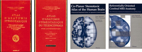
From left to right: the 1957, 1967, 1988, and 1993 Talairach atlas. (Images reproduced with permission from Talairach J et al., eds. Atlas d'anatomie stéréotaxique. Paris: Masson and Cie. 1957; from Talairach J and Szikla G, eds., Atlas of stereotaxic anatomy of telencephalon. Paris: Masson and Cie. 1967; from Talairach J and Tournoux P, eds. Co‐Planar Stereotaxic Atlas of the Human Brain, 3‐Dimensional Proportional System: An Approach to Cerebral Imaging. New York: Thieme Medical Publishers. 1988; and from Talairach J and Tournoux P, eds. Referentially Oriented Cerebral MRI Anatomy: An Atlas for Stereotaxic Anatomical Correlations for Gray and White Matter. New York: Thieme Medical Publishers. 1993.)
It took Talairach 10 years to extend the stereotaxic approach to the entire brain. For this he implemented the concept of proportional scaling to account for differences in brain size in the three direction of the AC‐PC system (see Fig. 2). Working with a rare tenacity, he validated the concept in tens of post‐mortem studies and published in 1967 a second stereotaxic atlas devoted to the telecephalon [Talairach and Szikla, 1967, Fig. 3], which also became a reference and a key for the advent of stereo encephalography (he invented with Jean Bancaud) and a new method for the surgery of epilepsy.
As often for pioneers, academic recognition came late to Talairach. In 1966, at last, he was named Associate professor of neurosurgery, and later full professor in 1975, three years before his official retirement in 1978. But retirement made no sense for such a man and Talairach pursued very actively his research work, bringing all pieces together. In 1988 he published his third stereotaxic atlas [Talairach and Tournoux, 1988, Fig. 3], the one that most brain mappers have in their library. Actually, the 1988 Talairach atlas came at the very beginning of the brain mapping domain and has largely contributed to its expansion. Since then, this emblematic book has cumulated over 7,600 citations, making it one of the most highly cited neuroscientific work. But even this was not enough for Talairach who at the age of 82 published a fourth atlas [Talairach and Tournoux, 1993, Fig. 3], building upon the advent of MRI to describe gray white matter correlations.
Such exceptional scientific curiosity, creativity, productivity, and longevity clearly place Jean Talairach among the scientific giants of the XXth century. But this should not make us forget that he was also a surgeon devoted to his patients, a teacher dedicated to his students, a leader who remained very close to his collaborators, and above all an exceptional human being.
REFERENCES
- Talairach J,Hecaen H,David M,Monnier M,De Ajuriaguerra J ( 1949): Recherches sur la coagulation thérapeutique des structures sous‐corticales chez l'homme. III. Techniques de stimulation et coagulation électriques des centres sous‐corticaux chez l'Homme. Rev Neurol 81: 4–24. [Google Scholar]
- Talairach J,David M,Tournoux P,Corredor H,Kvasina T ( 1957): Atlas d'anatomie stéréotaxique. Repérage radiologique indirect des noyaux gris centraux des régions mésencephalo‐sous‐optique et hypothalamique de l'homme. Paris: Masson & Cie; 294 p. [Google Scholar]
- Talairach J,Szikla G ( 1967): Atlas of Stereotaxic Anatomy of the Telencephalon. Anatomo‐Radiological Studies. Paris: Masson & Cie; 326 p. [Google Scholar]
- Talairach J,Tournoux P ( 1988): Co‐planar Stereotaxic Atlas of the Human Brain. 3‐Dimensional Proportional System: An Approach to Cerebral Imaging. Stuttgart: Georg Thieme Verlag; 122 p. [Google Scholar]
- Talairach J,Tournoux P ( 1993): Referentially Oriented Cerebral MRI Anatomy. Stuttgart: Georg Thieme Verlag; 234 p. [Google Scholar]
- Talairach J ( 2007): Souvenirs des études stéréotaxiques du cerveau humain. Une vie, une équipe, une méthodologie. L'école de Sainte‐Anne. Montrouge: John Libbey Eurotext; 131 p. [Google Scholar]